41(7), 907~913(2012) http://dx.doi.org/10.3746/jkfn.2012.41.7.907
Piceatannol에 의한 AGS 인체 위암세포의 G1 Arrest 및 Prostaglandin E 2 생성의 억제
최 영 현
동의대학교 한의과대학 생화학교실, 대학원 바이오물질제어학과(BK21 Program), 항노화연구소 및 블루바이오소재개발센터
Piceatannol-Induced G1 Arrest of the Cell Cycle is Associated with Inhibition of Prostaglandin E 2 Production in Human Gastric Cancer AGS Cells
Yung Hyun Choi
Dept. of Biochemistry, Dongeui University College of Oriental Medicine, Busan 614-052, Dept. of Biomaterial Control (BK21 Program), and Anti-Aging Research Center & Blue-Bio Industry
Regional Innovation Center, Dongeui University, Busan 614-714, Korea
Abstract
Piceatannol (trans-3,4,3',5'-tetrahydroxystilbene) is a polyphenol detected in grapes, rhubarb, and sugarcane.
Although recent experimental data revealed that this compound is known to exhibit immunosuppressive and antitumorigenic activities in several cell lines, the molecular mechanisms underlying anticancer activity are poor- ly understood. In the present study, we investigated possible further mechanisms by which piceatannol exerts its anti-proliferative action in cultured human gastric cancer AGS cells. Piceatannol treatment resulted in the inhibition of growth and G1 arrest of the cell cycle in a concentration-dependent manner, as determined by MTT assay and flow cytometry analysis. The induction of G1 arrest by piceatannol was associated with the modulation of cyclin-dependent kinases (Cdks) and cyclins, up-regulation of the expression of Cdk inhibitor p21 (WAF1/CIP1) in both transcriptional and translational levels, and the inhibition of phosphorylation of retino- blastoma proteins and E2F1 expression. In addition, piceatannol treatment caused a progressive decrease in the expression levels of cyclooxygenase (COX)-2 without significant changes in the levels of COX-1, which was correlated with a decrease in prostaglandin E2 synthesis.
Key words: piceatannol, G1 arrest, p21, COX-2
E-mail: [email protected]
Phone: 82-51-850-7413, Fax: 82-51-853-4036
서 론
최근 cyclooxgenase-2(COX-2)의 발현 증가가 다양한 암 발생과 연관성이 있음이 보고된 이후, COX-2가 정상세포의 초기 암화 과정에서 매우 중요한 역할을 하는 것은 명백한 현상으로 인식되어지고 있다. 이는 COX-2의 발현 증가와 동반된 지속적인 염증성 자극은 정상세포의 비정상적인 증 식 촉진을 유래하기 때문에 COX-2가 암 예방 및 암의 증식 을 막을 수 있는 매우 중요한 표적인자가 될 수 있음을 의미 한다(1,2). COXs는 prostaglandin(PG) endoperoxide syn- thase로서 arachidonic acid를 PGs 및 다양한 eicosanoids로 의 전환에 관여하는 rate-limiting enzyme이다. 그중 체내에 서 일정 비율로 생성되는 COX-1은 항상성 유지에 관여하는 반면, COX-2는 neoplastic transformation, 세포증식, 혈관 신생, 침윤 및 전이활성 증가와 연관된 immediate-early gene으로서 작용한다(2,3). 따라서 COX-2의 선택적 발현 억
제에 의한 과다한 PGs의 생성을 저하시킬 수 있는 신물질은 화학적 암 예방제로의 개발 가능성이 매우 높다.
등대풀속 식물인Euphorbia lagascae에서 처음 분리 동 정된 piceatannol(3,3',4,5'-tetrahydroxy-trans-stilbene)은 (4) resveratrol 유도체의 하나로서 포도, 대황, 사탕수수 등 에 많이 함유되어 있는 hydroxystilbene의 일종이다(5,6).
Piceatannol의 생리활성에 관한 초기 연구 중, piceatannol은 protein-tyrosine kinase와(7) nuclear factor kappa B(NF-κ B)의 활성을 강력하게 억제함으로써(8) 면역 조절 및 항염증 효능이 있다고 보고된 바 있다. 이러한 효능에 관한 구체적 인 기전 연구로서 piceatannol이 림프구에서 interferon-α에 의해 유도된 signal transducer and activator of tran- scription 3(STAT3)의 signal transducer 및 활성자의 기능 을 억제함이 밝혀졌는데(9-11), 이는 piceatannol이 다양한 세포 내 표적인자의 발현을 조절함으로써 면역 조절 및 항염 증 효능을 나타낼 수 있는 대표적인 예에 해당된다.
Table 1. Oligonucleotides used in RT-PCR
Name Sequence of primers
Cdk2 Cdk4 Cdk6 cyclin D1 cyclin E p53 p21 p27 COX-1 COX-2 GAPDH
sence antisence sence antisence sence antisence sence antisence sence antisence sence antisence sence antisence sence antisence sence antisence sence antisence sence antisence
5'-GCT-TTC-TGC-CAT-TCT-CAT-CG-3' 5'-GTC-CCC-AGA-GTC-CGA-AAG-AT-3' 5'-ACG-GGT-GTA-AGT-GCC-ATC-TG-3' 5'-TGG-TGT-CGG-TGC-CTA-TGG-GA-3' 5'-CGA-ATG-CGT-GGC-GGA-GAT-C-3' 5'-CCA-CTG-AGG-TTA-GAG-CCA-TC-3' 5'-TGG-ATG-CTG-GAG-GTC-TGC-GAG-GAA-3' 5'-GGC-TTC-GAT-CTG-CTC-CTG-GCA-GGC-3' 5'-AGT-TCT-CGG-CTC-GCT-CCA-GGA-AGA-3' 5'-TCT-TGT-GTC-GCC-ATA-TAC-CGG-TCA-3' 5'-GCT-CTG-ACT-GTA-CCA-CCA-TCC-3' 5'-CTC-TCG-GAA-CAT-CTC-GAA-GCG-3' 5'-CTC-AGA-GGA-GGC-GCC-ATG-3' 5'-GGG-CGG-ATT-AGG-GCT-TCC-3' 5'-AAG-CAC-TGC-CGG-GAT-ATG-GA-3' 5'-AAC-CCA-GCC-TGA-TTG-TCT-GAC-3' 5'-TGC-CCA-GCT-CCT-GGC-CCG-CCG-CTT-3' 5'-GTG-CAT-CAA-CAC-AGG-CGC-CTC-TTC-3' 5'-TTC-AAA-TGA-GAT-TGT-GGG-AAA-AT-3' 5'-AGA-TCA-TCT-CTG-CCT-GAG-TAT-CTT-3' 5'-CGG-AGT-CAA-CGG-ATT-TGG-TCG-TAT-3' 5'-AGC-CTT-CTC-CAT-GGT-GGT-GAA-GAC-3'
최근 연구에 의하면 piceatannol은 암세포의 세포주기 교란 및 apoptosis 유도를 통하여 암세포의 증식을 억제할 수 있는 것으로 알려져 있으나(10,12-17), 새로운 암예방 및 항 암 작용 기전의 표적이 되고 있는 COXs의 발현과 연관된 prostaglandin E2(PGE2) 생성 조절에 관한 연구는 거의 이루 어진 바가 없다. 본 연구실에서는 그동안 piceatannol의 생리 활성에 관한 다양한 연구를 수행한 바 있는데, piceatannol은 신경교세포에서 lipopolysaccharide(LPS)에 의한 COX-2의 발현 및 PGE2생성을 저하시켰으며, 이는 NF-κB 활성 억제 와 연관성이 있었다(18). 또한 piceatannol에 의한 인체 혈구 암세포의 증식억제는 telomerase 활성 저하와 연관성이 있 었으며(19), 혈구암세포의 apoptosis 유도는 anti-apoptotic 단백질인 Bcl-2의 발현 저하와 caspase의 활성 증가에 의한 것이었으며(20), tumor necrosis factor-related apopto- sis-inducing ligand(TRAIL) 저항성 극복을 위한 활용 가능 성도 높음을 제시한 바 있다(21). 본 연구에서는 piceatannol 의 항암 효능에 관한 부가적인 자료를 얻기 위하여 picea- tannol에 의한 인체 위암세포의 세포주기 조절 측면에서의 증식억제 현상 및 이와 연관된 COXs의 발현과 그들에 의해 조절 받는 PGE2의 생성에 미치는 영향을 조사하였다.
재료 및 방법
세포배양, piceatannol의 처리 및 생존율 측정
본 연구에 사용된 AGS 위암세포(American Type Culture Collection, Rockville, MD, USA)는 10% fetal bovine se- rum(FBS)에 1%의 penicillin 및 streptomycin이 포함된 90
%의 DMEM 배지(Gibco BRL, Grand Island, NY, USA)를
사용하여 37oC, 5% CO2조건 하에서 배양하였다. Piceatan- nol은 Sigma Chemical Co.(St. Louis, MO, USA)에서 구입 하여 dimethyl sulfoxide(DMSO)에 10 mM의 stock solution 으로 만든 후, 적정농도로 배지에 희석하여 처리하였다.
AGS 세포의 증식에 미치는 piceatannol의 영향을 조사하기 위하여 6 well plate에 AGS 세포를 1×10³개로 분주하고 24 시간 동안 안정화시킨 후, piceatannol을 48시간 동안 처리한 세포를 대상으로 3-(4,5-dimetylthiazol-2-yl)-2,5-diphenyl- tetrazolium(MTT) assay를 실시하였다.
Flow cytometry를 이용한 세포주기 분석
적정농도의 piceatannol이 48시간 처리된 AGS 세포들을 모아 Cycle TEST PLUS(Becton Dickinson, San Jose, CA, USA) solution A 및 B를 상온에서 각각 10분씩 처리한 후, Cycle TEST PLUS solution C를 처리하여 4oC에서 10분간 염색하였다. 그 후 DNA flow cytometer(BD FACSCalibur) 를 사용하여 세포주기의 각각에 해당되는 histogram을 분석 하였다.
Reverse transcription-polymerase chain reaction (RT-PCR) 분석
동일한 조건에서 배양된 AGS 세포들에 TRIzol reagent (Invitrogen Co., Carlsbad, CA, USA)를 4oC에서 1시간 동안 처리하여 RNA를 분리하였다. 분리된 RNA를 정량한 후, 각 각의 primer(Table 1), DEPC water 그리고 ONE-STEP RT-PCR PreMix kit(Intron, Seoul, Korea)를 넣고 Master- cycler gradient(Eppendorf, Hamburg, Germany)를 이용하 여 증폭하였다. 각 PCR 산물들의 양적 차이를 확인하기 위 하여 1×TAE buffer로 1% agarose gel을 만들고 well당 각
0 20 40 60 80 100 120
0 25 50 75 100
Piceatannol (μM)
C e ll v ia b il it y ( % ) .
*
*
*
(A)
(B) Piceatannolo (μM)
Fig. 1. Inhibition of the cell growth and morphological changes by piceatannol in AGS human gastric cancer cells.
(A) Cells were plated at 1×10
3cells per 60-mm plate, and in- cubated for 24 hr. The cells were treated with variable concen- trations of piceatannol for 48 hr and the cell viability was meas- ured by the metabolic-dye-based MTT assay. Results are ex- pressed as the means±SD of three independent experiments. The significance was determined by a Student’s t-test (
*p<0.05, com- pared with control). (B) AGS cells were incubated with variable concentrations of piceatannol. After 48 hr incubation, the cells were sampled and examined under light microscopy. Magnification,
×200.
각의 primer에 해당하는 PCR 산물에 DNA gel loading sol- ution을 섞어서 loading 한 후 50 V에서 전기영동을 행하였 다. 전기영동으로 DNA 분리가 끝난 gel을 ethidium bro- mide(EtBr)를 이용하여 염색한 후 UV 하에서 발현의 차이 를 확인하였으며, glyceraldehyde-3-phosphate dehydro- genase(GAPDH)를 internal control로 사용하였다.
단백질의 분리, 전기영동 및 Western blot 분석 Piceatannol이 처리된 세포들에 적정량의 lysis buffer[25 mM Tris-Cl(pH 7.5), 250 mM NaCl, 5 mM EDTA, 1%
NP-40, 1 mM phenylmethylsulfonyfluoride, 5 mM dithio- threitol]를 첨가하여 4oC에서 1시간 반응시킨 후, 14,000 rpm 으로 30분 원심분리 하여 그 상층액을 취하였다. 상층액에서 얻은 단백질을 sodium dodecyl sulphate(SDS)-polyacryl- amide gel을 이용하여 전기영동으로 분리하였다. 분리된 단 백질을 함유한 acrylamide gel을 nitrocellulose membrane (Schleicher and Schuell, Keene, NH, USA)으로 전이시킨 후, 적정 항체를 처리하여 enhanced cheiluminoscence(ECL) 용액(Amersham Life Science Corp., Arlignton Heights, IL, USA)을 적용시킨 X-ray film에 감광시켜 특정단백질의 양 을 분석하였다. 본 실험에 사용된 항체들은 Santa Cruz Bio- technology Inc.(Santa Cruz, CA, USA)에서 구입하였으며, immunoblotting을 위한 peroxidase-labeled 항체는 Amer- sham에서 구입하였다.
Prostaglandin E2의 측정
PGE2생성량 측정을 위해 사용한 PGE2 EIA kit는 Cay- man Chemicals(Ann Arbor, MI, USA)에서 구입하였다.
AGS 세포에 다양한 농도의 piceatannol을 48시간 동안 처리 후, 상층액만 이용하여 PGE2EIA kit에 제시된 방법에 따라 처리한 다음 ELISA reader를 이용한 420 nm의 흡광도로 반응의 정도를 측정하였다.
통계처리
모든 실험결과는 평균±표준편차로 표시하였고 Sigma- Plot을 이용하여 Studentt-test를 이용하여 통계적 유의성 을 얻었다.
결과 및 고찰
Piceatannol에 의한 AGS 세포의 증식 억제
AGS 위암세포의 증식에 미치는 piceatannol의 영향을 조 사하기 위하여 AGS 세포에 piceatannol을 48시간 동안 처리 한 후, MTT assay를 실시한 결과는 Fig. 1A에 나타낸 바와 같다. Fig. 1A의 결과에서 알 수 있듯이 25 μM 이하의 저농 도 처리군서는 대조군에 비하여 큰 차이가 없었으나, picea- tannol 처리 농도가 증가될수록 AGS 위암세포의 생존율은 점차 감소되어 75 μM 및 100 μM 처리군에서는 대조군에 비하여 약 50% 및 75% 정도 감소되었다. 즉 piceatannol이
적정농도 이상 처리된 배지에서 자란 AGS 세포는 picea- tannol 처리농도 의존적으로 세포증식이 억제되었음을 알 수 있었다. 아울러 piceatannol 처리의 농도가 증가할수록 AGS 세포의 다양한 형태적 변형도 동반되었다(Fig. 1B).
Piceatannol에 의한 세포주기 G1 arrest 유도
세포주기 조절의 관점에서 암세포는 세포주기의 비정상 화에 기인된 질병으로 정의될 수 있으며, 특정 시기의 세포 주기 억제는 세포주기 조절 양성인자의 발현 저하 또는 음성 조절 인자의 과발현에 의한 것으로 요약될 수 있다. 진핵세 포의 세포주기 조절은 각 주기별 관여하는 다양한 유전자들 에 의해 조절되는데, 기본적으로 세포주기 checkpoint 각 시 기에 요구되어지는 양성 조절인자인 cyclins에 의하여 cy- clin-dependent kinases(Cdks)의 연속적인 활성과 불활성 및 Cdk 저해제의 발현 정도에 따라 결정되어진다(22). 따라 서 piceatannol의 처리에 의한 AGS 세포의 증식억제가 세포 주기 특정 시기의 진행억제와 연관성을 지니는지의 여부를 조사하기 위하여 AGS 세포의 세포주기 분포에 미치는 pi- ceatannol의 영향을 조사하였다. 이를 위하여 정상 및 다양
Table 2. Effects of piceatannol on cell cycle distribution of AGS cells
Piceatannol
(μM) % of cells
G1 S G2/M
25 0 50 75 100
41.27 49.71 50.94 52.09 66.70
27.06 26.89 25.31 25.09 17.00
31.67 23.40 23.75 22.82 16.30 Exponentially growing cells were treated for 48 hr with in- creasing concentration of piceatannol. The cells were collected and stained with PI for flow cytometry analysis. Results are expressed as average from two separate experiments.
(A)
(B)
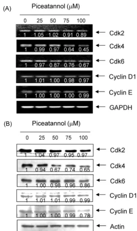
Fig. 2. Effects of piceatannol on the expression of Cdks and cyclins in AGS cells. (A) After 48 hr incubation with picea- tannol, total RNAs were isolated and reverse-transcribed. The resulting cDNAs were subjected to PCR with the indicated pri- mers and the reaction products were subjected to electrophoresis in a 1% agarose gel and visualized by EtBr staining. GAPDH was used as an internal control. (B) The cells grown under the same conditions as (A) were lysed and then cellular proteins were separated by SDS-polyacrylamide gels and transferred onto ni- trocellulose membranes. The membranes were probed with the indicated antibodies. Proteins were visualized using an ECL de- tection system. Actin was used as an internal control. The num- bers represent the average densitometric analyses as compared with GAPDH and actin, respectively.
한 농도의 piceatannol이 처리된 배지에서 AGS 세포를 48시 간 배양한 후 flow cytometry를 이용하여 분석하였다. Table 2에 나타낸 바와 같이, piceatannol이 함유되지 않은 정상 배지에서 자란 암세포의 경우 G1, S 및 G2/M기에 해당되는 세포의 빈도는 약 41%, 27% 및 23% 정도였다. 그러나 pi- ceatannol이 처리된 배지에서 자란 AGS 세포의 경우 특히 G1기에 속하는 세포의 빈도가 piceatannol 처리농도가 증가 할수록 상승되어 75 μM 및 100 μM 처리군에서 각각 약 52%
및 67%를 차지하였다. 이러한 G1기에 해당되는 세포의 빈 도 증가에 따라 S 및 G2/M기에 속하는 세포의 빈도는 상대 적으로 감소되어 piceatannol 처리에 AGS 세포의 증식억제 는 세포주기 G1 arrest와 연관이 있음을 알 수 있었다. 이는 인체 전립선암세포(23), 방광암세포(13) 및 혈관 평활근(16) 등에서 관찰된 경우와 잘 일치되는 결과였으나, SK-Mel-28 흑색종세포에서는 piceatannol 처리에 의하여 S기 arrest가 관찰되었으며(14), HL-60 혈구암세포에서는 G2/M arrest 가 유발되었음이 보고되어(24), 실험 대상 세포의 종류에 따 른 세포주기 조절에 차이가 있음을 알 수 있었다.
세포주기 주요 조절인자들의 발현에 미치는 piceatannol 의 영향
세포주기 진행의 조절에 관여하는 인자 중, D-type cyclin 은 G1기로 접어들면서 발현이 증가되어 Cdk4 및 Cdk6과 복합체를 형성하면서 활성화되어 종양억제인자인 retino- blastoma 단백질(pRB)을 인산화 시킨다(22). pRB는 인산화 의 정도에 따라 전사조절인자 E2F family에 속하는 단백질 들과의 결합을 통해 E2F 인자들의 활성을 조절하는데, pRB 가 인산화 되면 E2F가 유리되어 S기 특이적 유전자를 포함 한 DNA 합성과 연관된 많은 유전자들의 발현을 촉진시킨 다. 비록 pRB에 대한 인산화 장소는 다르지만 cyclin E/
Cdk2 복합체에 의한 pRB의 인산화도 이와 유사한 결과를 낳는데, DNA 손상이나 분화와 같은 증식 억제 신호는 Cdk inhibitor의 발현을 증가시켜 cyclins/Cdks의 활성을 억제하 여 S기로의 진입이 되지 못하게 된다. 부가적으로 cyclin A 는 G1 후기에 발현을 시작하여 S기와 G2기 동안 발현이 증 가되며 cyclin B1은 G2기에서 M기로의 전이를 조절하며, 각각은 Cdk2 및 Cdc2의 kinase 활성을 위한 양성 조절자로
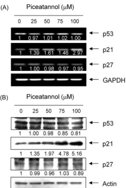
서 중요한 역할을 한다(25,26). 한편 Cdks는 다양한 세포증 식 억제 신호들에 의해 유도되는 Cdk inhibitor에 의하여 그 활성이 조절되는, 크게 두 가지의 family로 분류(INK4 및 CIP/KIP family)되고 있다. 그중 CIP/KIP family에 속하는 p21 및 p27은 DNA 손상에 의한 종양 억제인자인 p53에 의 해 조절을 받는다고 알려져 있다(27,28). 상기 결과에 의하면 piceatannol에 의한 AGS 세포의 증식억제가 G1 arrest와 연 관이 있었기에, 이와 연관된 기전 해석을 위하여 몇 가지 유전자들의 발현에 미치는 piceatannol의 영향을 조사하였 다. Fig. 2에 나타낸 것처럼 조사된 3가지 Cdks의 발현이 piceatannol 처리 농도 증가에 따라 점차 감소되었으며, cy- clin E 및 D1의 발현도 특히 단백질 수준에서 고농도 처리군 에서 약간 감소되었는데, 이러한 현상은 대장암세포에서 관 찰된 경우와 유사한 결과였다(23). 그리고 Cdk inhibitor 중, p27의 발현은 큰 변화가 없었으나, p21의 발현이 piceatannol
(A)
(B)
Fig. 3. Induction of p21 expression by piceatannol in AGS cells. (A) After 48 hr incubation with piceatannol, total RNAs were isolated and reverse-transcribed. The resulting cDNAs were subjected to PCR with the p53, p21 and p27 primers and the reaction products were subjected to electrophoresis in a 1%
agarose gel and visualized by EtBr staining. GAPDH was used as an internal control. (B) The cells were lysed and then cellular proteins were separated by SDS-polyacrylamide gels and trans- ferred onto nitrocellulose membranes. The membranes were pro- bed with the anti-p53, anti-p21 and anti-p27 antibodies. Proteins were visualized using an ECL detection system. Actin was used as an internal control. The numbers represent the average densi- tometric analyses as compared with GAPDH and actin, respec- tively.
Fig. 4. Effects of piceatannol on the expression of pRB and E2Fs in AGS cells. AGS cells were treated with the indicated concentrations of piceatannol for 48 hr and collected. The cells were lysed and then cellular proteins were separated by SDS- polyacrylamide gels and transferred onto nitrocellulose membranes.
The membranes were probed with anti-pRB, anti-E2F1 and an- ti-E2F4 antibodies. Proteins were visualized using an ECL de- tection system. Actin was used as an internal control. The num- bers represent the average densitometric analyses as compared with actin.
처리에 의하여 매우 증가되었다(Fig. 3). 아울러 pRB는 다소 탈인산화 되는 경향성을 보였으며, E2F4의 발현은 큰 변화 가 없었으나 E2F1의 발현은 piceatannol 처리 농도 의존적 으로 감소되었다(Fig. 4). 특히 p53의 발현은 변화가 없음에 도 불구하고 p21의 발현이 증가된 것은 piceatannol 처리에 의한 p21의 발현이 p53 비의존적으로 이루어지고 있음을 의 미하며, 이러한 p53 비의존적 p21의 발현 증가는 방광암세포 에서 관찰된 경우와 매우 일치한 결과였다(13). 따라서 본 연구의 결과는 비록 piceatannol이 cyclin이나 Cdk 등의 발 현에는 큰 변화를 주지 못하였지만, p21의 발현을 증가시킴 으로써 pRB의 인산화를 억제하여 cyclins/Cdks의 활성을 저해하였을 것이며, 아울러 pRB의 인산화 억제 및 부분적은 E2F 인자들의 발현 저하는 S기 진입에 요구되는 인자들의 발현을 억제하였을 것으로 추정된다.
Piceatannol에 의한 COX-2의 발현 및 PGE2의 생성 억제 본 연구실의 선행연구 결과에 의하면 신경교세포에서 pi- ceatannol은 NF-κB의 활성 억제를 통하여 LPS에 의한 COX-2의 발현을 차단함으로써 PGE2의 생성을 억제하였으
며(18), 유사한 결과들이 유방암세포(29), 상피세포(30) 및 대식세포(31) 등에서도 보고된 바 있다. 이러한 선행연구 결 과를 바탕으로 piceatannol에 의한 AGS 위암세포의 G1 ar- rest 유도과정에서 COX-2의 발현저해에 따른 PGE2의 생성 억제가 동반되는지를 조사하였다. Fig. 5A 및 B의 결과에서 알 수 있듯이 COX-1의 mRNA 및 단백질 발현에는 picea- tannol이 별다른 영향을 미치지 못하였으나, COX-2의 경우 전사 및 번역 수준에서 piceatannol의 처리 농도 증가에 따라 점차적인 발현의 감소를 보여주었다. 이러한 piceatannol에 의한 COX-2의 선택적 발현 저하가 PGE2의 생성 저하와 연 관성이 있는지를 조사하기 위하여 PGE2의 생성에 미치는 piceatannol의 영향을 조사한 결과, piceatannol의 처리 농도 가 증가할수록 PGE2의 생성이 유의적으로 감소되었으며, PGE2의 생성 감소는 COX-2의 발현 저하와 유사한 경향성 을 보여주었다. 따라서 piceatannol 처리에 의한 AGS 위암 세포의 G1 arrest 유도에는 COX-2의 선택적 발현 억제에 따른 PGE2의 생성 저해가 동반되어 있음을 알 수 있었다.
요 약
포도, 대황, 사탕수수 등을 포함한 다양한 식물에서 발견 되는 hydroxystilbene의 일종인 piceatannol은 암세포의 증 식을 억제하고 apoptosis를 유발하는 것으로 알려져 있다.
본 연구에서는 AGS 인체위암세포를 대상으로 piceatannol 에 의한 암세포 증식억제 과정에서 나타나는 또 다른 현상들 을 조사하기 위하여 실시되었다. Piceatannol이 처리된 AGS 위암세포는 piceatannol의 처리 농도의 증가에 따라 생존율 이 감소되었으며, 이는 세포주기 G1 arrest 유발과 연관이 있음을 MTT assay와 flow cytometry 분석을 통하여 확인
(A)
(B)
0 20 40 60 80 100 120
0 25 50 75 100
Piceatannol (μM) R e la ti ve P G E
2p ro d u ct io n .
*
*
(C)
Fig. 5. Down-regulation of COX-2 expression and inhibition of PGE
2production in AGS cells after exposure to picea- tannol. (A) AGS cells were treated with the indicated concen- trations of piceatannol for 48 hr and collected. The cells were lysed and total RNAs were isolated and reverse-transcribed. The resulting cDNAs were subjected to PCR with COX-1 and COX-2 primers and the reaction products were subjected to electro- phoresis in a 1% agarose gel and visualized by EtBr staining.
GAPDH was used as an internal control. (B) After 48 hr incubation with piceatannol, the cells were lysed and then cellular proteins were separated by 10% SDS-polyacrylamide gels. Proteins were visualized by Western blotting using anti-COX-1 and anti- COX-2 antibodies, and ECL detection. Actin was used as an in- ternal control. The numbers represent the average densitometric analyses as compared with GAPDH and actin, respectively. (C) Cells were treated with the indicated concentrations of picea- tannol for 48 hr and collected. The PGE
2accumulation in the me- dium was determined by an EIA kit described in materials and methods. Results are expressed as the means±SD of three in- dependent experiments. The significance was determined by a Student’s t-test (
*p<0.05, compared with control).
하였다. Piceatannol에 의한 AGS 세포의 G1 arrest는 Cdks 및 cyclins의 발현 변화 및 Cdk 저해제인 p21의 발현을 전사 및 번역 수준에서 증가시켰으며, pRB 단백질의 인산화 감소 및 E2F1의 발현 억제와 연관성이 있었다. 아울러 picea- tannol은 COX-2의 mRNA 및 단백질의 발현을 억제하였으 나 COX-1의 발현에는 영향을 미치지 않았으며, piceatannol 에 의한 COX-2의 발현억제는 PGE2의 생성 저하와 관련이
있었다. 본 연구의 결과는 piceatannol에 의한 세포주기 G1 arrest 유발이 COX-2의 선택적 발현 차단과 연관이 있음을 보여 주는 것이다.
감사의 글
본 연구는 지식경제부·부산광역시 지원 지역혁신센터사 업 동의대학교 블루바이오 소재 개발 및 실용화 지원 센터 (RIC 08-06-07)의 지원에 의하여 이루어진 결과의 일부입 니다.
문 헌
1. Anderson WF, Umar A, Viner JL, Hawk ET. 2002. The role of cyclooxygenase inhibitors in cancer prevention.
Curr Pharm8: 1035-1062.
2. Gately S, Kerbel R. 2003. Therapeutic potential of selective cyclooxygenase-2 inhibitors in the management of tumor angiogenesis.
Prog Exp Tumor Res37: 179-192.
3. Fujita H, Koshida K, Keller ET, Takahashi Y, Yoshimito T, Namiki M, Mizokami A. 2002. Cyclooxygenase-2 pro- motes prostate cancer progression.
Prostate53: 232-240.
4. Ferrigni NR, McLaughlin JL, Powell RG, Smith Jr CR. 1984.
Use of potato disc and brine shrimp bioassays to detect ac- tivity and isolate piceatannol as the antileukemic principle from the seeds of
Euphorbia lagascae.
J Nat Prod47:
347-352.
5. Bavaresco L, Fregoni C, Cantu E, Trevisan M. 1999. Stilbene compounds: from the grapevine to wine.
Drugs Exp Clin Res25: 57-63.
6. Roupe K, Teng XW, Fu X, Meadows GG, Davies NM. 2004.
Determination of piceatannol in rat serum and liver micro- somes: pharmacokinetics and phase I and II biotransforma- tion.
Biomed Chromatogr18: 486-491.
7. Geahlen RL, McLaughlin JL. 1989. Piceatannol (3,4,3',5'- tetrahydroxy-trans-stilbene) is a naturally occurring pro- tein-tyrosine kinase inhibitor.
Biochem Biophys Res Com- mun165: 241-245.
8. Ashikawa K, Majumdar S, Banerjee S, Bharti AC, Shishodia S, Aggarwal BB. 2002. Piceatannol inhibits TNF-induced NF-κB activation and NF-κB-mediated gene expression through suppression of IκBα kinase and p65 phosphor- ylation.
J Immunol169: 6490-6497.
9. Kumari AL, Ali AM, Das S, Pardhasaradhi BV, Varalakshmi CH, Khar A. 2005. Role of STAT3 and NF-κB signaling in the serum factor-induced apoptosis in AK-5 cells.
Bio- chem Biophys Res Commun336: 860-867.
10. Barton BE, Karras JG, Murphy TF, Barton A, Huang HF.
2004. Signal transducer and activator of transcription 3 (STAT3) activation in prostate cancer: direct STAT3 in- hibition induces apoptosis in prostate cancer lines.
Mol Cancer Ther3: 11-20.
11. Su L, David M. 2000. Distinct mechanisms of STAT phos- phorylation via the interferon-α/β receptor. Selective in- hibition of STAT3 and STAT5 by piceatannol.
J Biol Chem275: 12661-12666.
12. Ferreira MJ, Duarte N, Gyemant N, Radics R, Cherepnev G, Varga A, Molnar J. 2006. Interaction between doxor- ubicin and the resistance modifier stilbene on multidrug re- sistant mouse lymphoma and human breast cancer cells.
Anticancer Res
26: 3541-3546.
13. Kuo PL, Hsu YL. 2008. The grape and wine constituent piceatannol inhibits proliferation of human bladder cancer cells via blocking cell cycle progression and inducing Fas/membrane bound Fas ligand-mediated apoptotic path- way.
Mol Nutr Food Res52: 408-418.
14. Larrosa M, Tomás-Barberán FA, Espín JC. 2003. Grape polyphenol resveratrol and the related molecule 4-hydrox- ystilbene induce growth inhibition, apoptosis, S-phase ar- rest, and upregulation of cyclins A, E, and B1 in human SK-Mel-28 melanoma cells.
J Agric Food Chem51: 4576- 4584.
15. Larrosa M, Tomás-Barberán FA, Espín JC. 2004. The grape and wine polyphenol piceatannol is a potent inducer of apoptosis in human SK-Mel-28 melanoma cells.
Eur J Nutr43: 275-284.
16. Lee B, Lee EJ, Kim DI, Park SK, Kim WJ, Moon SK. 2009.
Inhibition of proliferation and migration by piceatannol in vascular smooth muscle cells.
Toxicol In Vitro23: 1284- 1291.
17. Wieder T, Prokop A, Bagci B, Essmann F, Bernicke D, Schulze-Osthoff K, Dorken B, Schmalz HG, Daniel PT, Henze G. 2001. Piceatannol, a hydroxylated analog of the chemopreventive agent resveratrol, is a potent inducer of apoptosis in the lymphoma cell line BJAB and in primary, leukemic lymphoblasts.
Leukemia15: 1735-1742.
18. Jin CY, Moon DO, Lee KJ, Kim MO, Lee JD, Choi YH, Park YM, Kim GY. 2006. Piceatannol attenuates lipopoly- saccharide-induced NF-κB activation and NF-κB-related proinflammatory mediators in BV2 microglia.
Pharmacol Res54: 461-467.
19. Choi YH. 2008. Induction of S phase arrest of the cell cycle by piceatannol is associated with inhibition of telomerase activity in human leukemic U937 cells.
J Life Sci18: 96-102.
20. Kim YH, Park C, Lee JO, Kim GY, Lee WH, Choi YH, Ryu CH. 2008. Induction of apoptosis by piceatannol in human leukemic U937 cells through down-regulation of Bcl-2 and activation of caspases.
Oncol Rep19: 961-967.
21. Kang CH, Moon DO, Choi YH, Choi IW, Moon SK, Kim WJ, Kim GY. 2011. Piceatannol enhances TRAIL-induced apoptosis in human leukemia THP-1 cells through Sp1-
and ERK-dependent DR5 up-regulation.
Toxicol in Vitro25: 605-612.
22. Sherr CJ. 2000. The Pezcoller lecture: cancer cell cycles revisited.
Cancer Res60: 3689-3695.
23. Lee YM, Lim DY, Cho HJ, Seon MR, Kim JK, Lee BY, Park JH. 2009. Piceatannol, a natural stilbene from grapes, in- duces G1 cell cycle arrest in androgen-insensitive DU145 human prostate cancer cells via the inhibition of CDK activity.
Cancer Lett285: 166-173.
24. Fritzer-Szekeres M, Savinc I, Horvath Z, Saiko P, Pem- berger M, Graser G, Bernhaus A, Ozsvar-Kozma M, Grusch M, Jaeger W, Szekeres T. 2008. Biochemical effects of pi- ceatannol in human HL-60 promyelocytic leukemia cells- synergism with Ara-C.
Int J Oncol33: 887-892.
25. Girard F, Strausfeld U, Fernandez A, Lamb NJ. 1991. Cyclin A is required for the onset of DNA replication in mamma- lian fibroblasts.
Cell67: 1169-1179.
26. Brandeis M, Hunt T. 1996. The proteolysis of mitotic cy- clins in mammalian cells persists from the end of mitosi until the onset of S phase.
EMBO J15: 5280-5289.
27. Elledge SJ, Harper JW. 1994. Cdk inhibitors: on the thresh- old of checkpoints and development.
Curr Opin Cell Biol6: 847-852.
28. Morgan DO. 1995. Principles of CDK regulation.
Nature374: 131-134.
29. Son PS, Park SA, Na HK, Jue DM, Kim S, Surh YJ. 2010.
Piceatannol, a catechol-type polyphenol, inhibits phorbol ester-induced NF-κB activation and cyclooxygenase-2 ex- pression in human breast epithelial cells: cysteine 179 of IKKβ as a potential target.
Carcinogenesis31: 1442-1449.
30. Liu D, Kim DH, Park JM, Na HK, Surh YJ. 2009. Piceatannol inhibits phorbol ester-induced NF-kB activation and COX- 2 expression in cultured human mammary epithelial cells.
Nutr Cancer