Copyright © 2021 Korean Neurological Association 487
Multiple Cranial Neuropathies Due to a Mixed Infection in Skull Base Osteomyelitis: A Nanopore Sequencing Study
Dear Editor,
Skull base osteomyelitis (SBO) has substantial mortality and its incidence is increasing due to the growing prevalence of immunodeficiency diseases such as AIDS, diabetes melli- tus, chemotherapy, and immune suppressants.1,2 Staphylococcus aureus and Pseudomonas aeruginosa are reported as the two most common causative pathogens in SBO.2 However, identifying the offending pathogen in SBO is challenging due to the vagueness of its clinical symptoms, and tissue cultures and imaging modalities may only reveal the tip of the ice- berg.2-4 Nanopore technology is a new technique for sequencing that involves measuring the current flow through nanoscale molecular holes. Nanopore sequencing provides many ad- vantages for metagenomics research, including simple and rapid library preparation, and real-time analysis of the reads.5 Here we report a case of multiple cranial neuropathies caused by SBO of mixed infection in order to raise awareness of this condition.
A 70-year-old male presented to the neurology clinic with right ocular pain and facial pal- sy. He had been taking cephalosporins since visiting the otolaryngology clinic 5 days pre- viously, but facial pain and diplopia had developed over the preceding 2 days. In addition, total right-eye blindness occurred, which prompted him to visit the neurology clinic. He had no previous illness other than hypertension. The examination at the neurology clinic revealed multiple unilateral cranial nerve (CN) palsy, including of CNs 2, 3, 4, 5, 6, and 7.
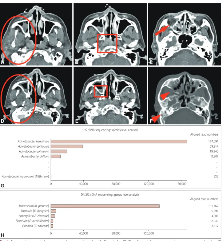
He had right facial pain and headache, but was alert without fever or neck stiffness. The ini- tial laboratory test revealed leukocytosis at 16,070/mm3 with left shifting (neutrophils at 79.6%) and elevated C-reactive protein at 11.2 mg/dL. Serum glucose and HbA1C were 262 mg/dL and 14.9%, respectively. Paranasal sinus computed tomography (PNS-CT) revealed fatty infiltrations in the inferomedial extraconal space of the right orbit, and retrobulbar and retromaxillary fat, suggesting inflammation around the right sinus (Fig. 1A-C). Since endo- scopic sinus surgery (ESS) led to methicillin-resistant Staphylococcus aureus (MRSA) be- ing identified in tissue cultures, we administered vancomycin. After 2 weeks the CN palsy remained unchanged, but the facial pain had gradually improved. Follow-up PNS-CT showed progression of inflammation involving the pterygoid muscle, sinus wall erosion, and soft- tissue invasion into the right middle ear (Fig. 1D-F). With clinical suspicion of invasive fun- gal infection, the patient underwent a second ESS with debridement and tissue culture. We performed 16S rDNA PCR for bacterial identification (Fig. 1G) and PCR of the D1/D2/D3 region of the large-subunit rDNA (Fig. 1H) for fungal detection in tissue specimens. Nano- pore sequencing (Oxford Nanopore Technologies, Oxford, UK) was performed from the PCR products to identify Acinetobacter bereziniae and multiple fungal strains, including Malassezia globosa, Yarrowia lipolytica, Aspergillus clavatus, Fusarium verticillioides, and Candida albicans.
The facial edema and headache improved after initiating liposomal amphotericin B at 5.0 mg/kg. The extents of soft tissue swelling and necrosis were decreased in follow-up images.
Keun Tae Kima Yu Hun Jeonga Seon-Jae Ahnb Jangsup Moonb,c Yong Won Choa Kon Chub,d
a Department of Neurology,
Keimyung University School of Medicine, Daegu, Korea
b Department of Neurology, Seoul National University Hospital, Seoul, Korea
c Department of Genomic Medicine, Seoul National University Hospital, Seoul, Korea
d Comprehensive Epilepsy Center, Biomedical Research Institute, Seoul National University Hospital, Seoul, Korea
pISSN 1738-6586 / eISSN 2005-5013 / J Clin Neurol 2021;17(3):487-489 / https://doi.org/10.3988/jcn.2021.17.3.487
Received January 22, 2021 Revised March 24, 2021 Accepted March 25, 2021 Correspondence Kon Chu, MD, PhD Department of Neurology, Comprehensive Epilepsy Center, Biomedical Research Institute, Seoul National University Hospital, 101 Daehak-ro, Jongno-gu, Seoul 03080, Korea Tel +82-2-2072-1878 Fax +82-2-3672-7553
E-mail [email protected]
cc This is an Open Access article distributed under the terms of the Creative Commons Attribution Non-Com- mercial License (https://creativecommons.org/licenses/by-nc/4.0) which permits unrestricted non-commercial use, distribution, and reproduction in any medium, provided the original work is properly cited.
JCN Open Access LETTER TO THE EDITOR
488 J Clin Neurol 2021;17(3):487-489
Multiple Cranial Neuropathies in SBO
JCN
Based on the clinical response and diagnostic evaluations, the neuromicrobiological manifestations were compatible with SBO involving a mixed infection of bacteria and fungi. Can-
dida albicans, Fusarium proliferatum, and Acinetobacter bau- mannii were identified in the tissue culture at 1 week after ini- tiating the antifungal agent.
Acinetobacter bereziniae Acinetobacter guillouiae Acinetobacter johnsonii Acinetobacter defluvii
…
…
… Acinetobacter baumannii (15th rank)
Malassezia (M. globosa) Yarrowia (Y. lipolytica) Aspergillus (A. clavatus) Fusarium (F. verticillioides) Candida (C. albicans)
167,091 39,217 19,940 11,907
…
…
… 533
131,762 5,991 4,901 2,636 2,517 0
0
40,000
40,000
16S rDNA sequencing: species level analysis
D1/2/3 rDNA sequencing: genus level analysis 80,000
80,000
120,000
120,000
160,000
G
H
Fig. 1. Enhanced paranasal sinus computed tomography before (A–C) and after (D–F) antibacterial treatment, and nanopore sequencing data (G and H). A: Fat infiltration, and loss of fat plain of pterygoid space and fat infiltration with swelling and enhancement (ellipse). B: Swelling and en- hancement of the nasopharynx (square). C: Fat infiltration in the inferior portion of the orbit (arrow). D: Nondifferentiation of the pterygoid space due to the progression of inflammation (ellipse). E: Penetration of the posterior wall of the maxilla by fat infiltration (square). F: Bone erosion in the posterior wall of the maxilla (upper arrowhead), and soft tissue invasion into the right middle ear (lower arrowhead). G: 16S rDNA sequencing for species-level analysis. The most-closely aligned species are considered pathogen species. The result indicates that Acinetobacter baumannii is not the causative agent. H: D1/D2/D3 rDNA sequencing for genus-level analysis. The species in parentheses is the most-closely aligned for the corresponding genus.
Aligned read numbers
Aligned read numbers
A
D
B
E
C
F
www.thejcn.com 489
Kim KT et al.
JCN
Tissue culture is usually considered to confirm the caus- ative strain; however, it has a low sensitivity and may miss a mixed infection.1,4 In our case, antifungal treatment was de- layed since only MRSA was identified in the first tissue cul- ture and as the causative agent. However, a reassessment was needed since clinical discrepancy was suspected after 2 weeks of the appropriate antibiotic treatment. MRSA is believed to be a cofactor rather than coexisting independently, because Staphylococcus aureus has the potential to cause inflamma- tion, tissue barrier disruption, and mucociliary dysfunction.6 A mixed infection of bacteria and fungi should be consid- ered in SBO, since approximately 50% of cases might be neg- ative in tissue cultures.7 Clinicians should be aware of this protean condition to differentiate it from treatment failure or an insufficient treatment period. Nanopore technology can contribute to the early identification of multiple pathogens.
However, it should still be considered as a complementary di- agnostic approach rather than as providing a definitive diag- nosis, since the possibility of sequencing errors and misalign- ments are limitations of sequencing-based diagnostics.
Author Contributions
Conceptualization: Keun Tae Kim, Kon Chu. Data curation: Yu Hun Jeong, Seon-Jae Ahn, Jangsup Moon. Writing—original draft: Keun Tae Kim.
Writing—review & editing: all authors.
ORCID iDs
Keun Tae Kim https://orcid.org/0000-0002-7124-0736 Yu Hun Jeong https://orcid.org/0000-0001-6220-7953 Seon-Jae Ahn https://orcid.org/0000-0003-0520-7812
Jangsup Moon https://orcid.org/0000-0003-1282-4528 Yong Won Cho https://orcid.org/0000-0002-6127-1045 Kon Chu https://orcid.org/0000-0001-5863-0302 Conflicts of Interest
The authors have no potential conflicts of interest to disclose.
Acknowledgements
This work was supported by the National Research Foundation of Korea (NRF) grant funded by the Ministry of Science and ICT, Republic of Ko- rea (NRF-2019R1A2C4070284).
REFERENCES
1. Ridder GJ, Breunig C, Kaminsky J, Pfeiffer J. Central skull base osteo- myelitis: new insights and implications for diagnosis and treatment.
Eur Arch Otorhinolaryngol 2015;272:1269-1276.
2. Johnson AK, Batra PS. Central skull base osteomyelitis: an emerging clinical entity. Laryngoscope 2014;124:1083-1087.
3. Mejzlik J, Cerny M, Zeinerova L, Dedkova J, Kopriva J, Zadrobilek K, et al. The routes of infection spread in central skull-base osteomyeli- tis and the diagnostic role of CT and MRI scans. BMC Med Imaging 2019;19:60.
4. Turner JH, Soudry E, Nayak JV, Hwang PH. Survival outcomes in acute invasive fungal sinusitis: a systematic review and quantitative synthesis of published evidence. Laryngoscope 2013;123:1112-1118.
5. Moon J, Kim N, Kim TJ, Jun JS, Lee HS, Shin HR, et al. Rapid diag- nosis of bacterial meningitis by nanopore 16S amplicon sequencing:
a pilot study. Int J Med Microbiol 2019;309:151338.
6. Vickery TW, Ramakrishnan VR, Suh JD. The role of Staphylococcus aureus in patients with chronic sinusitis and nasal polyposis. Curr Al- lergy Asthma Rep 2019;19:21.
7. Clancy CJ, Nguyen MH. Finding the “missing 50%” of invasive can- didiasis: how nonculture diagnostics will improve understanding of disease spectrum and transform patient care. Clin Infect Dis 2013;56:
1284-1292.