• 검색 결과가 없습니다.

Cefepime-Induced Encephalopathy in a Tertiary Medical Center in Korea

N/A
N/A
Protected

Academic year: 2021

Share "Cefepime-Induced Encephalopathy in a Tertiary Medical Center in Korea"

Copied!
8
0
0

로드 중.... (전체 텍스트 보기)

전체 글

(1)

Cefepime-Induced Encephalopathy in a Tertiary Medical Center in Korea

INTRODUCTION

Cefepime is a widely used fourth-generation cephalosporin. It is commonly used as a first-line antibiotic to treat various infectious diseases such as neutropenic fever, urinary tract infections, hospital-acquired pneumonia, bacterial meningitis, and skin, intra-ab- dominal, and gynecological infections.
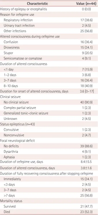
1

The clinical features of cefepime-induced en- cephalopathy (CIE) are confusion, agitation, myoclonic jerks, epileptic seizures, coma, and convulsive status epilepticus.

2

CIE was recently classified as a type-1 antibiotic-asso- ciated encephalopathy (AAE).
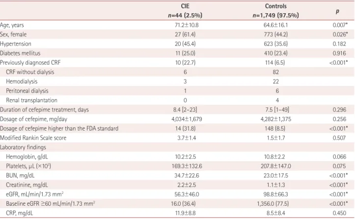
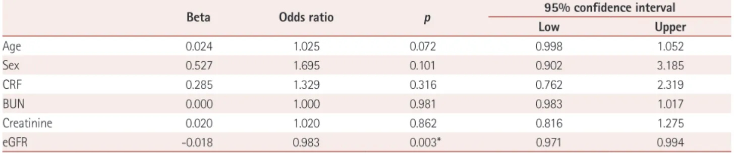
3

In type-1 AAE, seizures are observed more frequently than in psychosis. Predisposing factors for CIE include a high cefepime dosage, reduced drug renal clearance, and older age.

4-6

The reported incidence of CIE has ranged from 0.2% to 7.0%.

2,7-9

Appa et al.

2

reported an incidence at their center of 1 (0.2%) in 480 courses of cefepime. Naeije et al.

7

reported Ji-Ye Jeon

a,b

Yong Won Cho

b

Hye-Jin Moon

b,c

a

Department of Neurology, Kyungpook National University Chilgok Hospital, School of Medicine, Kyungpook National University, Daegu, Korea

b

Department of Neurology, Dongsan Medical Center,

Keimyung University School of Medicine, Daegu, Korea

c

Department of Neurology, Soonchunhyang University Bucheon Hospital, Soonchunhyang University College of Medicine, Bucheon, Korea

pISSN 1738-6586 / eISSN 2005-5013 / J Clin Neurol 2020;16(3):408-415 / https://doi.org/10.3988/jcn.2020.16.3.408

Received May 23, 2019 Revised March 12, 2020 Accepted March 16, 2020 Correspondence Hye-Jin Moon, MD Department of Neurology, Soonchunhyang University Bucheon Hospital, Soonchunhyang University College of Medicine,

170 Jomaru-ro, Bucheon 14584, Korea Tel +82-32-621-6569

Fax +82-32-621-5018

E-mail [email protected]

cc

This is an Open Access article distributed under the terms of the Creative Commons Attribution Non-Com- mercial License (https://creativecommons.org/licenses/by-nc/4.0) which permits unrestricted non-commercial use, distribution, and reproduction in any medium, provided the original work is properly cited.

JCN Open Access ORIGINAL ARTICLE

Background and Purpose Cefepime is a widely used fourth-generation cephalosporin. It is commonly used as a first-line antibiotic to treat various infectious diseases such as hospi- tal-acquired pneumonia, urinary tract infections, and bacterial meningitis. The primary out- come of this study was the development of cefepime-induced encephalopathy (CIE) at a ter- tiary medical center in Korea. We also aimed to describe the clinical features of CIE.

Methods We enrolled 1,793 consecutive patients treated with cefepime. The CIE group in- cluded 44 patients who experienced altered consciousness after receiving cefepime without any other obvious cause and showed full recovery after stopping cefepime. This study collect- ed demographic data, laboratory findings, and clinical data including the cause of infection, duration for onset of altered consciousness, duration of cefepime treatment, dosage of ce- fepime, duration of recovering consciousness after stopping cefepime, sequelae after enceph- alopathy, and electroencephalography data.

Results Forty-four (2.5%) patients were included in the CIE group. The age was significantly higher in the CIE group than in the control group (71.2±10.8 years vs. 64.7±16.1 years, mean±

standard deviation; p=0.007), and females constituted a significantly large proportion in the CIE group (27 of 44, 61.4%). The CIE group had higher blood urea nitrogen (34.7±22.6 mg/dL vs. 23.0±17.5 mg/dL, p<0.001) and creatinine (2.2±2.5 mg/dL vs. 1.1±1.3 mg/dL, p<0.001), and lower estimated glomerular filtration rate (eGFR) (56.3±46.0 mL/min/1.73 mm

2

vs. 98.8±66.3 mL/min/1.73 mm

2

, p<0.001). Multivariate analysis showed that only eGFR was significantly re- lated to CIE.

Conclusions The incidence of CIE was 2.5% in this study. It is essential to consider the possi- bility of CIE occurring, especially in patients with lower values of eGFR and dialysis.

Key Words encephalopathy, cefepime, epilepsy, incidence, electroencephalography.

(2)

Jeon JY et al. JCN

that 14 (1.25%) out of 1,120 patients who received cefepime showed continuous or intermittent generalized periodic epileptiform discharges. Fugate et al.

9

found that 7 of 100 patients in an intensive care unit (ICU) setting had definite CIE. In Korea, Kang and Kim

8

reported that 21 (0.85%) of 2,461 patients were diagnosed with CIE. This wide range of the reported incidence rates may be expected given both the heterogeneity in diagnostic criteria for CIE and the di- versity of the populations analyzed in these studies.

In this study we aimed to 1) determine the single-center incidence of CIE in Korea, 2) describe the clinical charac- teristics of CIE, and 3) identify the potential risk factors for CIE.

METHODS

Subjects

We selected consecutive patients diagnosed with an infec- tious disease and treated with cefepime at the Dongsan Med- ical Center in Korea from January 2013 to March 2015. The included patients were aged 18–80 years and first-time users of cefepime for an infectious condition. The following exclu- sion criteria were applied: 1) altered consciousness at the start of cefepime treatment, 2) neurological deficits or psy- chiatric disorders, 3) development of an altered conscious- ness due to other conditions such as mental retardation or alcohol or drug abuse, or 4) received antiepileptic drug treat- ment for epilepsy or seizure within the previous year. After applying the exclusion criteria, 1,802 patients were enrolled.

The study was approved by the Institutional Review Board of the Dongsan Medical Center (IRB No. 2015-11-020).

Data collection

We defined the CIE group as patients who fulfilled the follow- ing criteria: 1) altered consciousness, hallucinations, behavior change, myoclonus, or seizure after receiving cefepime, 2) neurological alteration not due to any other cause such as central nervous system deficit, delirium, psychosis, or meta- bolic cause, 3) experienced neurological alterations but recov- ered within a few days after stopping cefepime and subse- quently fully recovered their consciousness to the premorbid state, and 4) all of the aforementioned criteria confirmed by a neurologist or their own physician. Neurologists reviewed all medical records to include patients with newly developed acute neurological deterioration or expressed altered levels of consciousness during cefepime treatment. Patients who died during cefepime treatment or after stopping cefepime were excluded.

Demographic and laboratory findings were collected at the beginning of cefepime treatment. The demographic data

included age, sex, and laboratory findings such as white blood cells, red blood cells, platelets, hemoglobin, electro- lytes, blood sugar, liver enzymes, bilirubin, albumin, blood urea nitrogen (BUN), creatinine, C-reactive protein (CRP), and erythrocyte sedimentation rate. The data related to clin- ical mental status included the score on the modified Rankin Scale during cefepime treatment, whether patients developed an altered consciousness after using cefepime, the duration for onset of altered consciousness after using ce- fepime, the duration of recovering consciousness after stop- ping cefepime, the severity of the altered consciousness ac- cording to the Glasgow Coma Scale score, and the presence or absence of seizures. The data related to infection included the duration of cefepime treatment, the dosage of cefepime, the duration of recovering consciousness after stopping ce- fepime, and the reasons for cefepime treatment such as community-acquired pneumonia, hospital-acquired pneu- monia, urinary tract infection, colitis, or neutropenic fever.

If seizures occurred, data including the type of seizures, the number of seizures, and the presence of generalized tonic–

clonic seizure were collected. Additionally, outcome-related data such as survival or death and sequelae of CIE were col- lected. The electroencephalography (EEG) data of the CIE group recorded during the encephalopathy episode were re- viewed by an epilepsy specialist.

Statistical analyses

Continuous variables are presented as number and fre- quency or mean±standard-deviation values. We also con- ducted a series of internal subgroup comparisons of nomi- nal variables using the chi-squared test, and analyzed the continuous variables using analysis of variance or the inde- pendent t-test. A probability value of p<0.05 was consid- ered statistically significant.

To identify the factors contributing to CIE development, data analysis was performed for score derivation based on univariate and multivariate logistic regressions. All statisti- cal analyses were performed using IBM SPSS Statistics (version 21.0, IBM Corp., Armonk, NY, USA).

RESULTS

Subject characteristics and factors correlated with the CIE group

Nine of the 1,802 recruited participants were excluded. Eight

out of the nine excluded patients showed altered conscious-

ness after cefepime use but did not recover within a few days

after stopping cefepime. An underlying severe infectious

conditions or metabolic encephalopathy were possible expla-

nations for the altered consciousness. One patient died of an

(3)

Cefepime-Induced Encephalopathy in Korea

JCN

unknown cause during cefepime use. Forty-four (2.5%) of the 1,793 finally enrolled patients showed an altered con- sciousness during cefepime treatment and recovered after stopping cefepime, and were thus included in the CIE group.

The remaining 1,749 (97.5%) patients showed no alteration of their consciousness during cefepime treatment.

The age was significantly higher in the CIE group (71.2±10.8 years, p=0.007) than in the control group (64.6±16.1 years). Fe- males constituted a significantly larger proportion of patients in the CIE group (27 of 44, 61.4%) than in the control group (773 of 1,749, 44.2%). The daily dosage of cefepime and the duration of cefepime treatment did not differ significantly between the two groups. However, the proportion of subjects receiving ce- fepime doses that were higher than that specified in the Food and Drug Administration standard was higher in the CIE group than in the control group (31.8% vs. 8.5%, p<0.001).

The laboratory findings revealed that the CIE group had higher BUN (34.7±22.6 mg/dL vs. 23.0±17.5 mg/dL, p<

0.001), higher creatinine (2.2±2.5 mg/dL vs. 1.1±1.3 mg/dL, p<0.001), and lower estimated glomerular filtration rate (eGFR) (56.3±46.0 mL/min/1.73 mm

2

vs. 98.8±66.3 mL/min/

1.73 mm

2

, p<0.001) than the control group. The proportion

of patients with eGFR above 60 mL/min/1.73 mm

2

was sig- nificantly higher in the control group than in the CIE group (77.5% vs. 36.4%, p<0.001). The CRP, hemoglobin count, and platelet count did not differ significantly between the groups (Table 1).

In a univariate analysis and a reanalysis using multivari- ate logistic regression for investigating the factors contrib- uting to the development of CIE, only eGFR showed a sig- nificant correlation with CIE (p=0.003). The other variables of age, sex, and CRP, BUN, and creatinine levels showed no significant correlation with CIE in the multivariate logistic regression analysis (Table 2).

Clinical characteristics in the CIE group

Among 44 (2.5%) of the 1,793 patients in the CIE group, 20 (45.4%) had hypertension and 11 (25.0%) had diabetes melli- tus. Ten (22.7%) patients suffered from chronic renal failure, of which three underwent hemodialysis, one underwent perito- neal dialysis, and the others had not received dialysis when the study was conducted. No patient had a previous diagnosis of epilepsy or encephalitis. Seventeen (38.6%) and two (4.5%) pa- tients received cefepime to treat respiratory and urinary tract

Table 1. Comparison of the CIE and control groups

CIE n=44 (2.5%)

Controls

n=1,749 (97.5%) p

Age, years 71.2±10.8 64.6±16.1 0.007*

Sex, female 27 (61.4) 773 (44.2) 0.026*

Hypertension 20 (45.4) 623 (35.6) 0.182

Diabetes mellitus 11 (25.0) 410 (23.4) 0.916

Previously diagnosed CRF 10 (22.7) 114 (6.5) <0.001*

CRF without dialysis 6 82

Hemodialysis 3 22

Peritoneal dialysis 1 6

Renal transplantation 0 4

Duration of cefepime treatment, days 8.4 [2–23] 7.5 [1–49] 0.296

Dosage of cefepime, mg/day 4,034±1,679 4,282±1,375 0.256

Dosage of cefepime higher than the FDA standard 14 (31.8) 148 (8.5) <0.001*

Modified Rankin Scale score 3.7±1.4 1.5±1.7 0.507

Laboratory findings

Hemoglobin, g/dL 10.2±2.5 10.8±2.2 0.066

Platelets, μL (×10

3

) 169.3±132.6 207.8±147.0 0.075

BUN, mg/dL 34.7±22.6 23.0±17.5 <0.001*

Creatinine, mg/dL 2.2±2.5 1.1±1.3 <0.001*

eGFR, mL/min/1.73 mm

2

56.3±46.0 98.8±66.3 <0.001*

Baseline eGFR ≥60 mL/min/1.73 mm

2

16.0 (36.4) 1,356.0 (77.5) <0.001*

CRP, mg/dL 11.9±8.8 8.5±8.4 0.450

Data are n (%), n, mean±standard-deviation, or mean [range] values.

*Statistical significance (p<0.05).

BUN: blood urea nitrogen, CIE: cefepime-induced encephalopathy, CRF: chronic renal failure, CRP: C-reactive protein, eGFR: estimated glomerular filtra-

tion rate, FDA: Food and Drug Administration.

(4)

Jeon JY et al. JCN

Table 2. Results from the multivariate logistic regression analysis of cefepime-induced encephalopathy

Beta Odds ratio p 95% confidence interval

Low Upper

Age 0.024 1.025 0.072 0.998 1.052

Sex 0.527 1.695 0.101 0.902 3.185

CRF 0.285 1.329 0.316 0.762 2.319

BUN 0.000 1.000 0.981 0.983 1.017

Creatinine 0.020 1.020 0.862 0.816 1.275

eGFR -0.018 0.983 0.003* 0.971 0.994

Age, Sex, eGFR and CRF were considered independent variables.

*Statistical significance (p<0.05).

BUN: blood urea nitrogen, CRF: chronic renal failure, eGFR: estimated glomerular filtration rate.

infections, respectively.

During cefepime treatment, 16 (36.4%) patients suffered from confusion, 15 (34.1%) from drowsiness, 9 (20.5%) from stupor, and 4 (9.1%) were semicomatose or comatose. Pa- tients began to show symptoms of altered consciousness within a mean of 3.6 days after antibiotic administration. The duration of an altered consciousness was 3.8±1.6 days, and the duration of cefepime treatment was 8.4±5.5 days (range=2–23 days). Four (9.1%) patients suffered clinical sei- zures and three were diagnosed with status epilepticus. Dur- ing encephalopathy, four (9.1%) patients experienced dysar- thria and one (2.3%) suffered from aphasia. The recovery of consciousness to premorbid levels took more than 1 week in 56.8% of the patients, whereas this happened immediately af- ter stopping cefepime in 34.1% of them. Twenty-one (47.7%) patients survived, while 25 (52.3%) died due to infections or comorbidities (Table 3).

The overall incidence of CIE was 2.5%, but this was much higher in the dialysis patients (12.5%).

CIE occurred in about 1.1% of patients who did not receive dialysis, and eGFR was within the normal range (>60 mL/min/

1.73 mm

2

). The incidence of CIE among subjects with eGFR below 60 mL/min/1.73 mm

2

was 63.6%. CIE developed in 12.4% of the patients with eGFR below 30 mL/min/1.73 mm

2

but more than 11 mL/min/1.73 mm

2

, in 4.8% of the patients with eGFR below 11 mL/min/1.73 mm

2

, and in 4.0% of the patients with eGFR not more than 60 mL/min/1.73 mm

2

but 30 mL/min/1.73 mm

2

or more (Table 4).

EEG findings in the CIE group

Thirty of the 44 CIE patients underwent an EEG examina- tion at that time of altered consciousness. Eight of these pa- tients showed 2-Hz generalized periodic discharges (GPDs) with triphasic morphology, two of which had continuous 2- to 3-Hz GPDs with triphasic morphology and were diag- nosed with nonconvulsive status epilepticus. The remaining six patients had occasional GPDs with triphasic morpholo-

gy. Ten out of 30 patients who underwent EEG showed con- tinuous slow theta activity in the background, while 8 showed a slowing of delta to theta activity with triphasic morphology.

Two patients had normal EEG findings, one showed back- ground suppression without reactivity, and one showed fron- tal intermittent rhythmic delta activity. Below we describe the clinical features and EEG characteristics of two represen- tative cases.

Representative case 1

A 66-year-old woman who had chronic kidney disease with peritoneal dialysis was diagnosed with peritoneal dialysis-re- lated peritonitis and admitted to the department of nephrol- ogy. She was treated with 1-g cefepime once daily, and the peritoneal device was removed. After 4 days she developed confusion, disorientation, and irritability. A neurological ex- amination revealed that the patient was confused but fully oriented. EEG showed continuous frontally predominant 1-Hz GPDs with triphasic morphology and background the- ta slowing (Fig. 1A).

The neurologist immediately instructed the physician to stop cefepime. She recovered consciousness at 6 hours after stopping cefepime, and returned to her normal appearance after 1 day. In the follow-up EEG, the continuous theta slow- ing persisted but the epileptiform discharges had disappeared (Fig. 1B), and so the diagnosis of CIE was confirmed. All of the symptoms improved after replacing cefepime with ceftri- axone and clindamycin, and the patient was discharged with- out any complications.

Representative case 2

A 74-year-old woman with chronic kidney disease visited the

emergency room with cough, dyspnea, and palpitation. She

had been receiving on hemodialysis for 19 years. She was di-

agnosed with pneumonia and treated with empirical antibiot-

ics (2-g cefepime once after hemodialysis). After 3 days she

developed confusion, insomnia, and irritability, and 2 days

(5)

Cefepime-Induced Encephalopathy in Korea

JCN

Table 3. Clinical characteristics in the cefepime-induced encepha- lopathy group

Characteristic Value (n=44) History of epilepsy or encephalitis 0 (0.0) Reason for cefepime use

Respiratory infection 17 (38.6)

Urinary tract infection 2 (4.5)

Other infections 25 (56.8)

Altered consciousness during cefepime use

Confusion 16 (36.4)

Drowsiness 15 (34.1)

Stupor 9 (20.5)

Semicomatose or comatose 4 (9.1)

Duration of altered consciousness

<1 day 7 (15.9)

1–2 days 3 (6.8)

3–7 days 16 (36.4)

8–10 days 18 (40.9)

Duration for onset of altered consciousness, days 3.6 [0–17]

Clinical seizure

No clinical seizure 40 (90.9)

Complex partial seizure 1 (2.3)

Generalized tonic–clonic seizure 1 (2.3)

Unknown 2 (4.5)

Status epilepticus (n=43)

Convulsive 1 (2.3)

Nonconvulsive 2 (4.7)

Focal neurological deficit

No deficits 39 (88.6)

Dysarthria 4 (9.1)

Aphasia 1 (2.3)

Duration of cefepime use, days 8.4±5.5 Duration of altered consciousness, days 3.8±1.6 Duration of fully recovering consciousness after stopping cefepime

Immediately 15 (34.1)

<3 days 2 (4.5)

3–7 days 2 (4.5)

>7 days 25 (56.8)

Mortality status

Survived 21 (47.7)

Died 23 (52.3)

Data are n (%), mean±standard-deviation, or mean [range] values.

Table 4. Incidence of CIE according to eGFR eGFR, mL/min/1.73 mm

2

CIE

(n=44)

Controls (n=1,749) p

Dialysis 4 (12.5) 28 (87.5) -

0 ≤eGFR <11 2 (4.8) 40 (95.2) <0.001*

11 ≤eGFR <30 13 (12.4) 92 (87.6)

30 ≤eGFR ≤60 10 (4.0) 240 (96.0)

eGFR >60 15 (1.1) 1,349 (98.9)

Data are n (%) values.

*Statistical significance (p<0.05).

CIE: cefepime-induced encephalopathy, eGFR: estimated glomerular filtration rate.

later her treating physicians made an emergency call to a neu- rologist after she fell into a stupor. A neurological examina- tion revealed that she exhibited stupor with intact oculoce- phalic and pupil reflexes. Her power of all major joints were 3/5 of medical research council grade, and her sensory func- tion for painful stimulation was intact all over the body. Brain magnetic resonance imaging did not reveal any acute destruc- tive lesion. EEG showed continuous 3-Hz GPDs (Fig. 2A).

The neurologist diagnosed her with nonconvulsive status epilepticus due to CIE. The patient was administered 2 mg of lorazepam, but the GPDs persisted (Fig. 2B). This was fol- lowed by 3,000 mg of intravenous levetiracetam, and ce- fepime was replaced with levofloxacin. She was transferred to the department of neurology and EEG monitoring was start- ed in the ICU. The epileptiform discharges disappeared after the levetiracetam infusion. She gradually recovered her con- sciousness and returned to her normal appearance after 5 days. At the end of the EEG monitoring, slow background ac- tivity without epileptiform discharges was observed (Fig. 2C).

The patient recovered her usual appearance and was dis- charged on levetiracetam at 1,500 mg twice daily.

DISCUSSION

The present study found that the overall incidence of CIE in patients receiving cefepime in a single tertiary hospital was 2.5%. This is higher than in previously reported studies, with the exception of Fugate et al.

9

reporting that CIE was diag- nosed in about 7% of patients in the ICU. Most studies have found the incidence of CIE to be around 1%.

2,7,8

Naeije et al.

7

and Kang and Kim

8

reported incidence rates 1.25% and 0.85%, respectively. These discrepancies between studies might be due to the differences in the diagnostic criteria for CIE. The two large case–control studies used specific EEG character- istics as important diagnostic criteria. However, since about half of the patients in the present retrospective study did not undergo EEG while symptoms persisted, we decided to ex- clude EEG findings from the diagnostic criteria for CIE.

Cefepime is a fourth-generation cephalosporin that is one of the beta-lactam antibiotics with a broad antimicrobial spectrum.

10

Beta-lactam antibiotics enhance glutamate-medi- ated excitatory neurotransmission and the properties of gam- ma-aminobutyric acid, resulting in convulsions.

11

Bhattacha- ryya et al.

3

reported three distinct clinical phenotypes of AAE.

Encephalopathy due to cephalosporins and penicillin are

commonly accompanied by seizures or myoclonus arising

(6)

Jeon JY et al. JCN

Fig. 2. EEG findings in a cefepime-induced encephalopathy patient with nonconvulsive status epilepticus. A: Continuous 3-Hz GPDs and its evo- lution.* B: Despite injecting 2 mg of lorazepam, the GPDs persisted.* C: After administering 3,000 mg of levetiracetam intravenously, and followed by cefepime replacement with levofloxacin, background theta-to-alpha slow activity without epileptiform discharges was observed. *The EEG re- cords contain large numbers of artifacts since the electrodes were attached by inexperienced residents of neurology under emergency situations.

EEG: electroencephalography, GPDs: generalized periodic discharges.

A

C

B

Fig. 1. EEG findings in a patient with cefepime-induced encephalopathy without clinical seizures. A: Continuous frontally predominant 1-Hz generalized periodic discharges with triphasic morphology and background theta slowing were evident during cefepime treatment. B: Fol- low-up EEG after stopping cefepime showed that continuous theta slowing persisted but epileptiform discharges had disappeared. EEG:

electroencephalography.

A B

(7)

Cefepime-Induced Encephalopathy in Korea

JCN

within days after administration. In this study, 9.1% of the CIE patients suffered clinical seizures, of which 8% were diag- nosed as status epilepticus. EEG in the CIE group revealed that 26.7% showed GPDs with a triphasic morphology. These EEG findings were similar to those of several previous case reports. Many case reports have found that the EEG patterns in patients with CIE included rhythmic triphasic waves that were nonreactive to stimulation,

5,6,12,13

nonconvulsive status epilepticus with generalized spikes, and slow-wave or sharp and slow-wave activity.

6,14

Johnson et al.

15

reported a case having normal renal func- tion with CIE showing stimulus-induced rhythmic, periodic, or ictal discharges (SIRPIDs). However, our patients with CIE did not show SIRPIDs. Though many studies have found GPDs in EEG during CIE, some have also found vari- ous EEG patterns including nonspecific findings or diffuse triphasic waves with generalized slowing only in CIE pa- tients.

16

EEG patterns vary according to the severity of en- cephalopathy and the timing of the EEG study. In the present retrospective study, we did not include the EEG characteris- tics in the diagnostic criteria of CIE due to the low rate and varying time points of EEG being performed. Nevertheless, we believe that identifying and reporting various EEG pat- terns in CIE is valuable, and so consider that further pro- spective studies are needed.

Cefepime is largely excreted via the kidney, with 85% of the drug excreted in an unchanged form in the urine. The remainder is metabolized by the body to N-methylpyrro- lidine and a 7-epimer isomer. This study found that de- creased eGFR was the most-significant risk factor for CIE.

We showed that lower eGFR levels and dialysis were related to a higher incidence of CIE. We further found that 63.6%

of the CIE patients had an eGFR of below 60 mL/min/1.73 mm

2

, which is similar to previous studies.

8

Among patients with reduced renal function who do not receive dialysis, the incidence of CIE was highest when eGFR was above 11 mL/min/1.73 mm

2

and below 30 mL/min/1.73 mm

2

. Fur- ther studies are needed to explain whether the physician had followed the dosing standard according to the eGFR of each patient, and what the serum level of cefepime was for each patient.

Elderly patients show decreased creatinine clearance with age, with the eGFR possibly decreasing to about half its normal value before the serum creatinine level increases to above the upper limit. It is therefore essential to monitor the renal function in elderly patients with severe diseases and sepsis, and in those who are receiving cefepime. In this study we found that those in the CIE group were on aver- age older than controls. However, multivariate analysis re- vealed that renal function itself is a more-significant risk

factor than age.

The present study was subject to a few limitations. Unlike previous studies,

8

we did not include EEG findings in the diag- nostic criteria for CIE due to the retrospective design of this study. This might have reduced the specificity of the CIE diag- noses. Furthermore, we did not consider the history of previ- ous seizures or encephalopathy, or the family history of epilep- sy. Previous seizures or encephalopathies could significantly influence the EEG patterns of patients and the development of encephalopathy. The estimated incidence of CIE could be low- er than the actual incidence since most of the patients receiv- ing cefepime were admitted to the internal medicine depart- ment rather than to departments related to neurology. Thus, patients suffering from CIE were easily lost since the physi- cians might not have considered their altered mental status, and may have inferred that the encephalopathy was due to their poor general condition such as high fever, poor oral in- take, malignancy, or sepsis. The lack of further analysis of the causes of delayed recovery and the lack of evaluation for the serum level of cefepime were further limitations.

In conclusion, the overall incidence of CIE was 2.5% in our center. Impaired renal function was the only significant risk factor for CIE. The incidence of CIE was highest (exceeding 12%) in patients receiving dialysis and those with eGFR above 11 mL/min/1.73 mm

2

but below 30 mL/min/1.73 mm

2

. Cau- tion is needed while prescribing cefepime to such patients.

Additional long-term follow-up and prospective studies are needed to evaluate the development of CIE over time and to determine the factors contributing to the development of CIE.

Author Contributions

Conceptualization: Hye-Jin Moon, Yong Won Cho. Data curation: Hye-Jin Moon, Ji-Ye Jeon. Formal analysis: Hye-Jin Moon, Ji-Ye Jeon. Funding ac- quisition: Hye-Jin Moon. Investigation: Hye-Jin Moon, Ji-Ye Jeon. Meth- odology: all authors. Project administration: Hye-Jin Moon. Resources:

Hye-Jin Moon, Yong Won Cho. Supervision: Hye-Jin Moon, Yong Won Cho. Validation: Hye-Jin Moon, Ji-Ye Jeon. Visualization: Ji-Ye Jeon. Writ- ing—original draft: Ji-Ye Jeon. Writing—review & editing: Hye-Jin Moon, Yong Won Cho.

ORCID iDs

Ji-Ye Jeon https://orcid.org/0000-0003-1111-3521 Yong Won Cho https://orcid.org/0000-0002-6127-1045 Hye-Jin Moon https://orcid.org/0000-0002-6109-7171 Conflicts of Interest

The authors have no potential conflicts of interest to disclose.

Acknowledgements

This work was supported by the research promoting grant from the Keimyung University Dongsan Medical Center in 2015 and the Soonc- hunhyang University Research Fund.

REFERENCES

1. Martin SI, Kaye KM. Beta-lactam antibiotics: newer formulations

(8)

Jeon JY et al. JCN

and newer agents. Infect Dis Clin North Am 2004;18:603‐619.

2. Appa AA, Jain R, Rakita RM, Hakimian S, Pottinger PS. Characteriz- ing cefepime neurotoxicity: a systematic review. Open Forum Infect Dis 2017;4:ofx170.

3. Bhattacharyya S, Darby RR, Raibagkar P, Gonzalez Castro LN, Berkow- itz AL. Antibiotic-associated encephalopathy. Neurology 2016;

86:963‐971.

4. Lin CJ, Chen SP, Wang SJ, Fuh JL. Cefepime-related encephalopathy in peritoneal dialysis patients. J Chin Med Assoc 2011;74:87‐90.

5. Dixit S, Kurle P, Buyan-Dent L, Sheth RD. Status epilepticus associ- ated with cefepime. Neurology 2000;54:2153‐2155.

6. Thabet F, Al Maghrabi M, Al Barraq A, Tabarki B. Cefepime-induced nonconvulsive status epilepticus: case report and review. Neurocrit Care 2009;10:347‐351.

7. Naeije G, Lorent S, Vincent JL, Legros B. Continuous epileptiform discharges in patients treated with cefepime or meropenem. Arch Neurol 2011;68:1303‐1307.

8. Kang JK, Kim SB. Neurotoxicity by cefepime: case-control study. J Neurocrit Care 2014;7:104-110.

9. Fugate JE, Kalimullah EA, Hocker SE, Clark SL, Wijdicks EF, Rabin- stein AA. Cefepime neurotoxicity in the intensive care unit: a cause

of severe, underappreciated encephalopathy. Crit Care 2013;17:R264.

10. Mine Y, Watanabe Y, Sakamoto H, Hatano K, Kuno K, Higashi Y, et al. In vitro antibacterial activity of FK037, a novel parenteral broad- spectrum cephalosporin. J Antibiot (Tokyo) 1993;46:71‐87.

11. Sugimoto M, Uchida I, Mashimo T, Yamazaki S, Hatano K, Ikeda F, et al. Evidence for the involvement of GABA(A) receptor blockade in convulsions induced by cephalosporins. Neuropharmacology 2003;

45:304‐314.

12. Fishbain JT, Monahan TP, Canonico MM. Cerebral manifestations of cefepime toxicity in a dialysis patient. Neurology 2000;55:1756‐1757.

13. Jallon P, Fankhauser L, Du Pasquier R, Coeytaux A, Picard F, Hefft S, et al. Severe but reversible encephalopathy associated with cefepime.

Neurophysiol Clin 2000;30:383‐386.

14. Coelho FM, Bernstein M, Yokota PK, Coelho RM, Wachemberg M, Sampaio LP, et al. Cefepime-induced encephalopathy in patient without renal failure. Einstein (Sao Paulo) 2010;8:358‐360.

15. Johnson E, Hannawi Y, Martinez NC, Ritzl EK. Cefepime-associated SIRPIDs in a patient with normal renal function. Neurohospitalist 2016;6:167‐169.

16. Schlidt K, Kadlec A, Bhandari S, Jha P. Cefepime-induced neurotox-

icity: five cases reported in a single institution. Cureus 2018;10:e3666.

수치

Table 1. Comparison of the CIE and control groups
Table 2. Results from the multivariate logistic regression analysis of cefepime-induced encephalopathy
Table 3. Clinical characteristics in the cefepime-induced encepha- encepha-lopathy group
Fig. 1. EEG findings in a patient with cefepime-induced encephalopathy without clinical seizures

참조

관련 문서

Statistics for mass loss, duration of failure, and HCl, HCN, HF, and CO emissions are shown in Table 1. This data is taken directly from all of the cell tests. This

It considers the energy use of the different components that are involved in the distribution and viewing of video content: data centres and content delivery networks

After first field tests, we expect electric passenger drones or eVTOL aircraft (short for electric vertical take-off and landing) to start providing commercial mobility

1 John Owen, Justification by Faith Alone, in The Works of John Owen, ed. John Bolt, trans. Scott Clark, &#34;Do This and Live: Christ's Active Obedience as the

Thus, this study has researched the literature study of the current statistic data and study article and field survey in some farm households, analysis

Also, this study used opinions of Poomsae players who engaged in national contests wearing a mask to prevent infection of Covid 19 and preceding research

 Procedure for creating the equipment failure rate data segment of a CPQRA analysis data base.  Define

In this paper, we developed a data management system for shipboard machinery equipment and the monitoring application for the verification of the utility, and we