DOI 10.17480/psk.2015.59.1.17
Acacetin의 HeLa 세포 Apoptosis 유도 효과
한수정 · 김안근# 숙명여자대학교 약학대학
(Received February 3, 2015; Revised February 13, 2015; Accepted February 16, 2015)
Effect of Acacetin on the Apoptosis Induction of HeLa Cells
Su Jung Han and An Keun Kim#
College of Pharmacy, Sookmyung Women’s University, Seoul 140-742, Korea
Abstract — In this study, we examined the effect of acacetin on the apoptosis induction of HeLa human cervical cancer cells. The results showed that acacetin inhibited the cell viability and induced apoptosis, leading to PARP cleavage and acti- vation of caspase-9, -3, and -7. Moreover, acacetin-induced apoptosis was attenuated by a broad caspase inhibitor, z-VAD- fmk. Also, acacetin resulted in a loss of mitochondria membrane potential. Taken together, our results demonstrate that the induction of apoptosis by acacetin in HeLa cells is associated with caspase activation via the mitochondria pathway.
Keywords □ HeLa cells, acacetin, apoptosis
우리나라에서 자궁경부암은 지난 10년 사이에 발생률와 사망 률이 감소하는 추세를 보이고 있으나, 연령 15~33세군에서 세 번째로 발생빈도가 높은 것으로 나타나고 있다.1)현재 자궁경부 암의 치료는 주로 수술, 방사선, 화학요법을 통해 이루지고 있다.
이 중 화학요법은 강한 항암작용을 가지고 있으나 암세포뿐만 아 니라 정상세포도 함께 손상을 입히고, 반복 치료를 하는 과정에 서 고농도의 용량을 사용하게 되면서 여러 가지 부작용을 초래 한다. 뿐만 아니라 항암제에 대한 내성으로 인해 기존의 방법으 로 암세포를 치료하는데는 한계가 있다.2,3)따라서 이러한 문제 점을 해결할 수 있는 새로운 항암제의 개발이 요구되고 있는 실 정이다.
Apoptosis는 세포가 스스로 죽음을 결정해 분해되는 과정으로 programmed cell death라고도 한다. 세포손상이 일어나면 세포 가 수축하여 염색체가 응축하고 핵분해과정을 거쳐 세포내부의 물질들이 apoptosis 사멸체를 형성하는 형태적 변화를 볼 수 있 다.4)또한 세포질쪽에 위치한 포스파티딜 세린이 세포막으로 노
출하게 되는데 이것은 대식세포를 불러일으켜 형성된 사멸체가 식세포작용으로 처리되어 염증반응을 일으키지 않고 사멸하는 특징을 갖고 있다.5)이와 관련하여 정상세포에는 영향을 주지 않 으면서 암세포에만 특이적으로 apoptosis 유도 효과를 나타내는 물질을 탐색하고 그와 관련된 기전을 밝히는 연구가 계속해서 추 진되고 있는 중이다.6-8)
최근 천연물질이 암 예방과 치료에 효과가 있다고 알려져 있으 며, 특히 기존연구들을 통해 천연물질에서 추출한 플라보이드류 의 항산화, 항염증 및 항암효과가 보고되어 주목을 받고 있다.9-11) Acacetin은 식물에 함유되어 있는 플라보노이드 계열 중 하나이 며 다양한 생리활성을 가지고 있다고 알려져 있다(Fig. 1).12,13) 또한 acacetin이 위암세포에서 caspase를 활성화하여 apoptosis 를 일으킨다고 보고되었으며,14)전립선암에서는 G1 및 G2/M기
#
Corresponding Author An Keun Kim
College of Pharmacy, Sookmyung Women’s University, Seoul 140-742, Korea
Tel.: 02-710-9566 Fax.: 02-710-9871 E-mail: [email protected] Short Report
종설Fig. 1 − Chemical structure of acacetin.
arrest와 apoptosis 유도에 의해 세포증식을 억제한다고 보고된 바 있다.15) 하지만 아직도 acacetin의 항암활성에 대한 연구는 미 미한 실정이다.
따라서 본 연구는 acacetin을 자궁경부암세포주인 HeLa 세포 에 처리하여 세포생존율 및 apoptosis 유도 확인하고 그와 관련 된 기전을 조사하였다.
시약 및 방법
시약
세포배양에 사용한 RPMI 1640 배지, Dulbecco’s phosphate buffered saline(DPBS), fetal bovine serum(FBS), trypsin- EDTA, penicillin/streptomycin 항생제는 WelGENE(Daegu, South Korea)에서 구입하였다. Acacetin, 3-(4,5-dimethylthiazol- 2-yl)-2,5-diphenyl-tetrazoliumbromide(MTT)시약은 Sigma-Aldrich (St. Louis, MO, USA)에서 구입하였고, Annexin V-FITC apoptosis detection Kit는 BD Biosciences(San Jose, CA, USA)제품을 사용하였다. Caspase-9, cleaved caspase-3, cleaved caspase-7, PARP, β-actin antibody는 cell signaling(Danvers, MA, USA)에서 구입하였고, western blotting detection kit(ECL solution)은 Amersham Pharmacia Biotech(Piscataway, NJ, USA)사에서 구입하였다.
세포배양
본 연구에서 사용한 HeLa 세포는 한국 세포주 은행(Korea Cell Line Bank)으로부터 분양받았고 10% FBS, 항생제(10,000 units/ml penicillin G sodium, 10,000 μg/ml streptomycin sulfate)를 포함하는 RPMI 1640 배지에서 37oC, 5% CO2인 환 경에서 배양하였다.
MTT assay
세포생존율은 MTT assay를 이용하여 측정하였다. HeLa 세 포를 96-well plate에 분주하여 안정화시킨 후 조건에 따라 acacetin을 처리하고 24시간 동안 배양하였다. MTT를 첨가하여 4시간 방치한 후, DMSO를 well당 100 μg/ml을 가하여 formazan crystal을 용해시켜 ELISA plate reader로 540 nm에서 흡광도를 측정하였다.
Annexin V-FITC/PI assay
HeLa 세포를 6-well plate에 배양하여 acacetin을 조건에 따라 24시간 처리하였다. Trypsin-EDTA 처리 후 cell을 떼어내 원심 분리하고 PBS로 두 번 세척한다. 생성된 pellet을 1x binding buffer에 풀어주고 annexin V-FITC와 PI 시약으로 염색하였다.
FACSCanto II flow cytometer(BD Biosciences, San jose, CA,
USA)를 이용하여 apoptosis 발생을 확인하였다.
Western blotting
Acacetin을 24시간 동안 cell에 처리한 후, protease inhibitor 가 포함하는 RIPA buffer로 lysis하였다. 4oC에서 1시간 방치한 뒤 원심분리한 다음 단백질정량을 통해 동일한 양의 단백질을 준 비하였다. 이 단백질을 SDS-PAGE gel에서 전기영동으로 분리 하여 PVDF membrane에 transfer 시키고 5% non-fat milk을 함유한 TBS-T에 넣어 실온에서 2시간 동안 blocking하였다. 1 차 항체를 처리하여 4oC에서 하룻밤 동안 반응시킨 후, 2차 항 체와 반응시켰다. 단백질 발현은 ECL용액을 처리하여 LAS- 3000(Fuji, Tokyo, Japan)에서 측정하였다.
Measurement of mitochondria membrane potential 미토콘드리아 막 전위의 변화를 조사하기 위하여 DiOC6염색 을 시행하였다. Acacetin을 0, 1, 2, 4, 6 μg/ml의 농도로 처리하 여 24시간 지난 후 trypsin-EDTA로 cell을 수확하였다. DiOC6 를 첨가해 빛을 차단하여 37oC에서 20분간 방치하고 PBS로 세 척한 후 flow cytometer로 관찰하였다.
통계처리
본 연구의 실험 결과는 평균±SD로 표시하였다. 통계처리는 one-way ANOVA를 이용하여 분석하고 사후검정으로 Turkey test를 이용하였다. p-value가 0.05 미만인 경우를 유의성이 있는 것으로 판정하였다.
결과 및 고찰
세포 증식 억제 효과 확인
HeLa 세포에 acacetin을 처리하였을 때 세포 생존율을 알아보
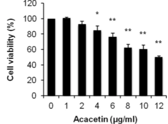
Fig. 2 − Effect of acacetin on HeLa cell viability. Cells were treated
with various concentrations of acacetin for 24 h and cell
viability was evaluated by MTT assay. Data represents the
mean±SD (n=3). *p<0.01, **p<0.001 vs. control.
기 위해 MTT assay를 시행하였다. Acacetin의 농도는 0 μg/ml 에서 12 μg/ml를 범위로 하여 24시간 동안 처리하였다. 실험 결 과 acacetin에 의해 HeLa 세포의 생존율이 억제되었으며, 농도 가 증가함에 따라 세포 증식 억제 효과가 더 크게 나타난 것을 확인할 수 있었다(Fig. 2). 이후의 실험은 70% 이상의 생존율을 나타내는 농도를 택하여 진행하였다.
Apoptosis 발생 확인
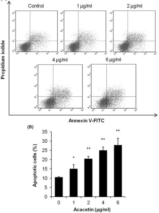
세포 증식 억제 효과가 apoptosis 유도에 의한 것인지 확인하
기 위하여 apoptosis V-FITC/PI 이중 염색 후 유세포분석기를 사용하여 apoptosis의 발생 정도를 측정하였다. 실험결과 acacetin 농도가 1~6 μg/ml로 높아짐에 따라 초기 apoptosis의 발생 비율 이 15~27%로 유의적로 증가한 것이 관찰되었다(Fig. 3).
PARP는 DNA 복구를 돕는 효소로, apoptosis 유도 과정에서 불활성화되어 세포사멸를 유도하기 때문에 PARP cleavage는 apoptosis의 주요 지표 중의 하나이다.16) PARP의 발현양을 western blotting으로 확인한 결과 acacetin을 처리한 경우 대조 군에 비해 cleaved PARP(85 kDa)가 뚜렷하게 증가하였다(Fig.
Fig. 3 − Effect of acacetin on HeLa cell apoptosis. Cells were treated with indicated concentrations of acacetin for 24 h. Apoptosis was
evaluated by annexin V-FITC/PI double staining assay. Representative results are shown in (A). In (B), the percentage of apoptosis
is represented as a bar graph. Data represents the mean±SD (n=4). *p<0.01, **p<0.001 vs. control.
4). 따라서 acacetin이 apoptosis를 일으켜 HeLa 세포의 증식을 억제하는 것을 확인할 수 있었다.
Apoptosis 유도 인자의 발현 확인
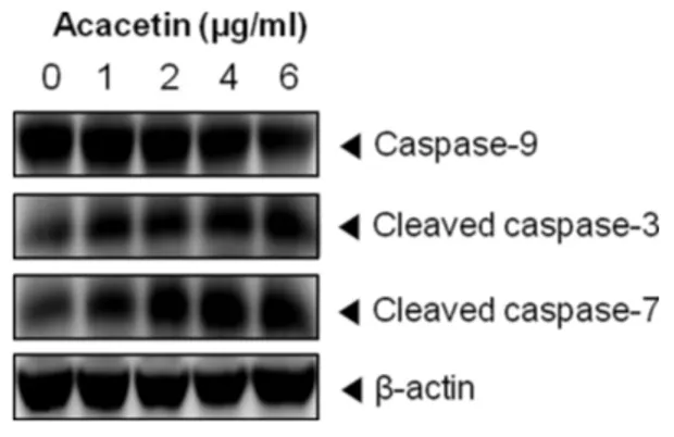
Caspase는 apoptosis가 일어날 때 활성화되는 단백질 분해효 소로 apoptosis 신호전달과정에서 중요한 역할을 한다.17) Caspase 를 크게 두 부류로 나누면 개시역할을 하는 initiator caspase와 실행역할을 하는 effector caspase가 있다. Apoptosis 초기자극에 의해 상위단계의 caspase가 활성화되어 apoptosis를 개시하고 단 계적인 연쇄반응을 통해 하위단계의 caspase를 활성화하여 apoptosis를 일으킨다.18) Acacetin에 의한 apoptosis 유도가 caspase 활성화와 관련되어 일어났는지의 여부는 western blotting을 통하여 확인하였다. 그 결과 acacetin의 농도가 커짐 에 따라 initiator caspase인 caspase-9이 감소하는 반면, effector caspase인 caspase-3, caspase-7은 증가한 것으로 관찰 되었다(Fig. 5).
또한 apoptosis의 유도기전에서 caspase 활성화가 중요한 역 할을 하는 것인지 알아보기 위하여 broad caspase inhibitor인 z-
VAD-fmk를 acacetin 6 μg/ml와 같이 처리한 후 발생한 apoptosis 을 확인하였다. Fig. 6에서 나타낸 바와 같이 초기 apoptosis는 acacetin을 처리하였을 때 26%였으나 z-VAD-fmk에 의해 15%로 유의적인 감소를 보였다. 이를 통해 acacetin에 의한 apoptosis는 caspase 활성화를 통하여 유도된 것임을 확인할 수 있었다.
Mitochondria pathway와의 관련성 확인
Apoptosis 신호전달경로는 death receptor pathway와 mitochondria pathway로 구분지을 수 있다. Death receptors pathway는 death ligand와 세포표면에 존재하는 death receptor 결합으로 caspase-8이 활성화되어 apoptosis를 유도한다.19) Mitochondria pathway는 세포 내의 자극으로 미토콘드리아 막의 투과성이 변화되어 세포질내 apoptosis 인자를 방출해 caspase- 9를 활성화시켜 apoptosis가 시작된다.20)앞선 실험 결과로부터 acacetin에 의한 apoptosis 유도 효과가 caspase-9의 활성화와 관 련있다는 것을 확인했기 때문에 mitochondria pathway에서 나타 나는 특징인 미토콘드리아 내부 전위차 손실 정도를 DiOC6를 사 용해 알아보았다. DiOC6는 미토콘드리아를 염색하는 시약으로 DiOC6의 형광 감소량을 통해 미토콘드리아 막전위 변화를 알 수 있다. DiOC6로 염색한 후 유세포분석기로 분석한 결과, acacetin 이 농도가 증가함에 따라 DiOC6형광 정도가 유의적으로 감소 함을 관찰할 수 있었다(Fig. 7). 따라서 acacetin에 의한 apoptosis 유도 기전은 mitochondria pathway와 관련이 있다는 것을 알 수 있었다. 이러한 결과는 Shim HY 등의 연구에서 acacetin이 MCF- 7 유방암세포에서 미토콘드리아를 매개로 caspase의 활성화를 통 해 apoptosis를 일으킨다는 결과와 일치하며,21) HeLa 자궁경부 암세포에서 acacetin의 apoptosis 유도 효과는 본 연구를 통해 처 음으로 확인되었다.
Fig. 4 − Effect of acacetin on PARP cleavage. Cells were treated with different concentrations of acacetin for 24 h. Whole cell extracts were prepared and analyzed by western blotting using anti-PARP antibody. β-Actin was used as loading control.
Fig. 5 − Effect of acacetin on caspase activation. Cells were treated with acacetin (0~6 μg/ml) for 24 h and cell lysates were prepared. Western blotting were performed for analysis of caspase-9, caspase-3 and caspase-7. β-Actin was used as loading control.
Fig. 6 − Effects of caspase inhibitor on acacetin-induced apoptosis.
Cells were treated with 6 μg/ml acacetin in the presence or
absence of 20 μM pan caspase inhibitor (z-VAD-fmk) for
24 h. Apoptosis (%) was evaluated by determined by annexin
V-FITC/PI double staining analysis. Data represents the
mean±SD (n=3). *p<0.001 vs. control; # p<0.001 vs. cells
treated with 6 μg/ml acacetin.
결 론
본 연구에서는 acacetin에 의해 HeLa 자궁경부암 세포의 세 포생존율이 유의적으로 감소한 것을 확인하고 이는 apoptosis 유
도와 관련이 있다는 것을 초기 apoptosis의 발생 증가와 PARP cleavage 발현을 통해 알 수 있었다. Acacetin에 의한 apoptosis 유도 효과는 caspase-9 및 caspase-3, -7을 활성화시킴으로서 일 어난 것으로 나타났다. 또한 미토콘드리아 막 전위 변화에 의해 mitochondria pathway가 관여한다는 것이 확인되었다.
감사의 말씀
이 논문은 숙명여자대학교 교내연구비 지원에 의해 수행되었 음(1-1303-0050).
References
1) Korea National Statistical Office. : Annual report of cancer statistics in Korea in 2012 (2014).
2) Waggoner, S. E. : Cervical cancer. Lancet 361, 2217 (2003).
3) Fujimoto, J. : Novel strategy of anti-angiogenic therapy for uterine cervical carcinomas. Anticancer Res. 29, 2665 (2009).
4) Van Cruchten, S. and Van Den Broeck, W. : Morphological and biochemical aspects of apoptosis, oncosis and necrosis. Anat.
Histol. Embryol. 31, 214 (2002).
5) Elmore, S. : Apoptosis: a review of programmed cell death.
Toxicol. Pathol. 35, 495 (2007).
6) Fulda, S. and Debatin, K. M. : Extrinsic versus intrinsic apoptosis pathways in anticancer chemotherapy. Oncogene 25, 4798 (2006).
7) Aggarwal, B. B. and Shishodia, S. : Molecular targets of dietary agents for prevention and therapy of cancer. Biochem.
Pharmacol. 71, 1397 (2006).
8) Mahmood, Z. and Shukla, Y. : Death receptors: targets for cancer therapy. Exp. Cell Res. 316, 887 (2010).
9) Amin, A. R., Kucuk, O., Khuri, F. R. and Shin, D. M. : Perspectives for cancer prevention with natural compounds. J.
Clin. Oncol. 27, 2712 (2009).
10) Ramos, S. : Effects of dietary flavonoids on apoptotic pathways related to cancer chemoprevention. J. Nutr. Biochem. 18, 427 (2007).
11) Huang, W. Y., Cai, Y. Z. and Zhang, Y. : Natural phenolic compounds from medicinal herbs and dietary plants: potential use for cancer prevention. Nutr. Cancer 62, 1 (2010).
12) Pan, M. H., Lai, C. S., Wang, Y. J. and Ho, C. T. : Acacetin suppressed LPS-induced up-expression of iNOS and COX-2 in murine macrophages and TPA-induced tumor promotion in mice. Biochem. Pharmacol. 72, 1293 (2006).
13) Miyazawa, M. and Hisama, M. : Antimutagenic activity of flavonoids from Chrysanthemum morifolium. Biosci. Biotechnol.
Biochem. 67, 2091 (2003).
14) Pan, M. H., Lai, C. S., Hsu, P. C. and Wang, Y. J. : Acacetin
Fig. 7 − Effect of acacetin on mitochondria membrane potential.
After incubation with acacetin, cells were stained with DiOC 6 . ΔΨ m was evaluated by flow cytometric analysis.
Representative histograms are shown in (A). In (B), disruption of ΔΨ m (%) is presented as a bar graph. Data represents the mean±SD (n=3). *p<0.01, **p<0.001 vs.
control.
induces apoptosis in human gastric carcinoma cells accompanied by activation of caspase cascades and production of reactive oxygen species. J. Agric. Food Chem. 53, 620 (2005).
15) Singh, R. P., Agrawal, P., Yim, D., Agarwal, C. and Agarwal, R. : Acacetin inhibits cell growth and cell cycle progression, and induces apoptosis in human prostate cancer cells: structure- activity relationship with linarin and linarin acetate.
Carcinogenesis 26, 845 (2005).
16) Li, X. and Darzynkiewicz, Z. : Cleavage of Poly(ADP-ribose) polymerase measured in situ in individual cells: relationship to DNA fragmentation and cell cycle position during apoptosis.
Exp. Cell Res. 255, 125 (2000).
17) Lavrik, I. N., Golks, A. and Krammer, P. H. : Caspases:
pharmacological manipulation of cell death. J. Clin. Invest. 115,
2665 (2005).
18) Kumar, S. : Caspase function in programmed cell death. Cell Death Differ. 14, 32 (2007).
19) Jin, Z. and El-Deiry, W. S. : Overview of cell death signaling pathways. Cancer Biol. Ther. 4, 139 (2005).
20) Belizario, J. E., Alves, J., Occhiucci, J. M., Garay-Malpartida, M. and Sesso, A. : A mechanistic view of mitochondrial death decision pores. Braz. J. Med. Biol. Res. 40, 1011 (2007).
21) Shim, H. Y., Park, J. H., Paik, H. D., Nah, S. Y., Kim, D. S. and Han, Y. S. : Acacetin-induced apoptosis of human breast cancer MCF-7 cells involves caspase cascade, mitochondria-mediated death signaling and SAPK/JNK1/2-c-Jun activation. Mol. Cells 24, 95 (2007).