A Doctoral Dissertation
Influence of basal core promoter and
precore mutations on HBV replication
and sensitivity to antiviral agent in
genotype A and C
Graduate School, Cheju
N
ational
U
niversity
Department of Medicine
Cui, XiuJi
January 2009
삭제됨: n
Influence of basal core promoter and precore mutations
on HBV replication and sensitivity to antiviral agent in
genotype A and C
Cui, Xiu Ji
(Supervised by Prof. Byung-Cheol Song)
A thesis submitted in partial fulfillment of the requirement
for the degree of doctor of philosophy in medicine
2009. 01.
Department of Medicine
GRADUATE SCHOOL
CHEJU NATIONAL UNIVERSITY
삭제됨: This thesis has been
examined and approved.
<sp> <sp> <sp> <sp> <sp> Date Approved 2008.12.12 삭제됨:
B형 간염 바이러스 유전자의 basal
core promoter와 precore부위의 변이가
B형 간염 바이러스의 복제 능력 및
항
바이러스 치료제에 영향
지도교수 송 병 철최
수 길
이
논문을 의학 박사학위 논문으로 제출함
2009년 1월제주대학교
대학원
2009년 1월 삭제됨: 최수길의 의학 박사 논문을 인준함 위 원 장 조문제 부위원장 이근화 위 원 김영리 위 원 김흥업 위 원 송병철Abstract
Hepatitis B virus (HBV) with genotype A and C has different antiviral response and frequency of basal core promoter (BCP) and precore (PC) mutations. In this study, we investigated the influence of these mutations on hepatitis B virus replication and sensitivity to antiviral agents. Genotype A and C recombinant HBV strains with either BCP mutation, PC mutation or BCP+PC mutation were used to study the viral replicating capacity and sensitivity to antiviral agents (lamivudine, entecarvir, and clevudine) in Huh7 cell line. BCP mutation, PC mutation and BCP+PC mutations increased the viral replicating capacity regardless of genotypes. Furthermore, these mutations increased the antiviral resistance to lamivudine, entecarvir and clevudine. In conclusion, BCP and PC mutations increased viral replication regardless of HBV genotypes in vitro. In addition, patients with these mutations might need more amount of antiviral agents. In the future, clinical trials are needed to compare the effectiveness of antiviral agents using current standard dose and higher dose in patients with these mutations.
.
Keywords: HBV, basal core promoter, precore, genotype, antiviral agent, resistance
삭제됨: s
CONTENTS ABSTRACT ……….i CONTENTS……….…ii LIST OF SCHEME………..iv LIST OF TABLES………...v LIST OF FIGURES……….vi 1. INTRODUCTION……….1
2. MATERIALS AND METHODS………..10
1. Construction of plasmid DNA containing HBV DNA -genotype A2 or C2…………...10
2. PCR based Site-directly mutagenesis ……….……….10
3. Cell culture and transfection ………..………..13
4. Isolation of intracellular HBV core particle and southern blot analysis …………...…...14
5. Isolation of extracellular HBV and quantitative real-time PCR analysis …………...….15
3. RESULTS……….17
1. Replication competency of clinical isolates……….17
3. Influence of BCP and PC mutations on extracellular HBV DNA level………...18
4. Sensitivity to antiviral agents………...18
4. DISCUSSION………..32
5 REFERENCES………..36
6. ABSTRACT IN KOREAN………..46
LIST OF SCHEME
Scheme 1. Procedure of the present study………...………20
LIST OF TABLES
Table 1. Primers used in this study………21 Table 2. Sensitivity of wild type- and mutant type-HBV to LMV, ETV and CLV…………..22
LIST OF FIGURES
Figure 1. Replication competency of pHY106-HBV with genotype A2 and genotype C2…….23 Figure 2. Effect of BCP, PC and BCP+PC mutation on intracellular viral replication………...24 Figure 3. Effect of BCP, PC and BCP+PC mutation on HBV secretion……….25 Figure 4. Sensitivity of wild type- and mutant type-HBV to Lamivudine (LMV)
Figure A & B………..26 Figure C & D………..27 Figure 5. Sensitivity of wild type- and mutant type-HBV to Entecarvir (ETV)
Figure A & B……….28 Figure C & D………29 Figure 6. Sensitivity of wild type- and mutant type-HBV to Clevudine (CLV)
Figure A & B………30 Figure C & D………31
1. Introduction
Hepatitis B virus (HBV), a major cause of liver disease, has infected approximately 2 billion people in worldwide and more than 350 million are chronic HBV infection. Annually, there are over 4 million new cases of HBV infection and around 500 0000 to 1.2 million people died from HBV infection (Lavanchy,2004). HBV infection can lead to various clinical outcome such as acute hepatitis B (AHB), inactive hepatitis B surface antigen (HBsAg) carrier, chronic hepatitis B (CHB), liver cirrhosis (LC), and hepatocellular carcinoma (HCC) (Chen,1993, Lok and McMahon,2007).
HBV is a hepadnavirus that contains a 3.2 Kb-length relaxed circular double stranded DNA containing four overlapping open reading frames (ORF) including surface gene (preS1/S2/S gene), polymerase gene (Pol gene), precore/core gene (PreC/C) and X gene that encode surface protein (envelope), polymerase, HBeAg and core protein, and X protein, respectively (Seeger and Mason,2000). Once the HBV enters the hepatocyte, the relaxed circular DNA (RC-DNA) is transferred into nucleus and converted into covalently closed circular DNA (cccDNA) (Tuttleman, et al.,1986), which transcribes pregenomic RNA (pgRNA) and subgenomic RNA by cellular RNA polymerase II(Sells, et al.,1988). The subgenomic RNA includes preS1-mRNA for large surface protein, preS2-mRNA for middle surface protein,
mRNA for small surface protein, precore RNA for e protein and X-mRNA for X protein(Seeger and Mason,2000). The pgRNA, which is controlled by core promoter and enhancer II, plays two roles: (1) serves as mRNA-template for viral polymerase and core protein; (2) serves as template for viral DNA replication. Binding of viral polymerase to the epsilon (ε), a hairpin structure in stem-loop of pgRNA, triggers the encapsidation of polymerase-pgRNA complex into capsid by core protein (Bartenschlager and Schaller,1992, Hirsch, et al.,1990). In the capsid, polymerase initiates RNA intermediated reverse transcription to synthesizing a new RC-DNA. After completion of viral DNA replication, the capsid can either re-transfer the RC-DNA to nucleus for formation of cccDNA or move to the endoplasmic reticulum to be assembled by envelope proteins and exported(Nassal,1999).
Hepatocyte is the major target of HBV and secrets the replicated virions into blood stream. HBV also can be detected in other body fluid such as saliva, semen, vaginal secretions, menstrual blood, perspiration, breast milk, tears, and urine of HBV infected individual(Lavanchy,2004). The HBV infection shows geographical distribution and is divided into high, intermediate and low endemic area basis on the prevalence of HBV infection more than 8%, 2~7 % and less than 2%, respectively(VHPB,1998).
Transmission of HBV infection can be either vertical (perinatal) or horizontal. Vertical transmission is the major route of HBV infection in highly endemic area such as Asia-pacific
region, sub-Saharan Africa. In contrast, most patients are infected by HBV at adulthood through horizontal transmission in low endemic area such as North America, Western and Northern Europe and Australia. For examples, using contaminated syringe or surgical instrument, unprotected sexual activity, blood transfusion and donor organs. Recently, it has been reported that HBV infection also can be transmitted from father to child (Tajiri, et al.,2007).
The clinical outcomes are different between the two routes of transmission. About 90% of individuals infected through vertical transmission likely develop to chronic HBV infection defined as positive for hepatitis B surface antigen (HBsAg) for more than 6 month. On the contrary, less than 5 % of individuals infected during adulthood develop to chronic HBV infection. Among the chronic HBV infections, around 2~6% of HBeAg-positive and 8~10% of HBeAg-negative patients annually progress to LC. Furthermore, the annual incidence of HCC in HBV infection related liver cirrhosis is about 2~3 %(de Franchis, et al.,2003). Chronic HBV infection is a strong risk factor for HCC and LC. Beasley et al. reported that the relative risk ratio in developing HCC between HBsAg-positive men and HBsAg-negative men was 223:1 (Beasley, et al.,1981). Recently, it has been reported the association between serum HBV DNA level and the risk of HCC. Yu MW et al. prospectively followed 4,841 chronic HBV infected Taiwanese men without HCC for 14 years, the adjusted odds ratio of HCC was 7-fold higher in men with serum HBV DNA level more than 5.91 log10 copies/ml than that less than 3.61 log10
copies/ml (Yu, et al.,2005). In another prospective cohort study also reported that the high risk of HCC associated with increasing HBV DNA level (Chen, et al.,2006).
HBV can be classified into eight genotypes (genotype A-H) basis on the divergence of more than 8% of completed HBV genome(Arauz-Ruiz, et al.,2002, Delius, et al.,1983, Norder, et al.,1994, Okamoto, et al.,1988, Stuyver, et al.,2000). Each genotype can be further classified into subgenotype with more than 4% of genetic divergence(Schaefer,2007, Sugauchi, et al.,2004, Sugauchi, et al.,2004, Tanaka, et al.,2005). HBV genotype and subgenotype shows geographic distribution and has correlation with the severity and progression of liver disease(Arauz-Ruiz, et al.,1997, Ding, et al.,2001, Lindh, et al.,1997, Norder, et al.,1993, Orito, et al.,2001). In genotype A, genotype A1 (Aa) is predominantly found in South Africa, genotype A2 (Ae) is mostly found in Western Europe and genotype A3~5 are mostly found in western Africa such as Cabon, Carmeroon, Mali and Nigeria. Genotype B has five subgenotypes (genotype B1~B5): genotype B1 (Bj) is predominant in Japan; genotype B2 (Ba) prevalent in East Asia except Japan; genotype B3~B5 are mostly found in South Asia. Genotype C also has five subgenotypes: genotype C1 (Cs) is common in South East Asia (Vietnam, Myanmar, Thailand, and Southern China); genotype C2 (Ce) is predominant in East Asia (Korea, Japan, and Northern China). Genotype D has five genotypes (genotype D1~D5) and commonly found in the Mongolia, India, South Africa and Australia. Genotype E is predominantly found in Africa;
genotype F (genotype F1~F4) is found in Central and South America, Bolivia and Argentina. Genotype G has been reported in the United States and France and recently reported genotype H is found in Central America(Schaefer,2007) .
The HBV genotype also shows the clinical and virological differences during the natural course of CHB infection. Chronic HBV infection is more frequent in genotype A than D and genotype A is more likely progress to chronic HBV infection after acute HBV infection than genotype B and C(Lin and Kao,2008, Mayerat, et al.,1999). Although genotype A has higher rate of HBeAg positivity than genotype D, the rate of sustained HBeAg seroconversion is higher than genotype D and has better clinical outcome than genotype D(Sanchez-Tapias, et al.,2002). Between genotype B and C, genotype C has higher rate of HBeAg-positivity and histologic activity, later HBeAg seronconverstion, more frequent reactivation after HBeAg seroconversion, less HBsAg seroclearance and possesses higher viral replicating capacity than genotype B(Chu, et al.,2002, Sumi, et al.,2003, Yuen, et al.,2003, Zhang, et al.,2008).
Currently, there are two types of antiviral agents approved by the US Food and Drug Administration (FDA) for CHB therapy. One is the immunomodulator, including interferon alpha and peginterferon alpha; another is the HBV polymerase inhibitor, nucleos(t)ide analogue (lamivudine, adefovir dipivixil, entecarvir and clevudine). The nucleos(t)ide analogues inhibit HBV polymerase in different step of viral replication. Lamivudine, a first FDA approved
nucleoside analogue in 1998, affects HBV reverse transcriptase activity; entecarvir and celuvudine inhibit both minus- and plus-strand DNA synthesis; adefovir affects on the priming of reverse transcription and inhibits minus-strand DNA synthesis(Zoulim and Perrillo,2008).
Recently, several studies reported the different antiviral response in different HBV genotype. Genotype C has poor antiviral response to interferon-based therapy compared with genotype A or B(Kao, et al.,2000, Yu, et al.,2005). Although, the HBV genotype has few affect on the nucleos(t)ide analogues treatment, the durability of lamivudine as well as interferon treatment is low in genotype C than genotype A or B and has been supposed to associate with the high pretreatment serum HBV DNA level and HBV genotypes, which are the important independent predictors in both interferon and nucleos(t)ide analogues s treatment (Chien et al.,2003, Janssen et al.,2005, Song et al.,2004, Song et al.,2000, Wai et al.,2002).
During the life cycle of HBV, the most frequently occurring HBV mutations are the A to T mutation at nucleotide 1762 and/or the G to A mutation at nucleotide 1764 (A1762T/G1764A) in the basal core promoter (BCP)(Okamoto et al.,1994, Song et al.,2006), and the G to A mutation at nucleotide 1896 (G1896A) in the precore (PC) region (Brunetto et al.,1989, Carman et al.,1989, Kidd-Ljunggren et al.,1997). The core promoter region of HBV was mapped between nucleotide 1613 and 1849 (Lo and Ting,1994, Yuh, et al.,1992), and consists of the upper regulatory region, which contains both positive and negative cis-acting elements, and the
BCP, which contains four TATA-like boxes to control the transcription of precore RNA and pregenomic RNA (Chen, et al.,1995, Lo and Ting,1994, Yuh, et al.,1992). The A1762T/G1764A mutation that located at the second TATA-like box in the BCP (nt1742-nt1849) region is frequently detected in chronic HBV infected patients (Okamoto, et al.,1994, Sato, et al.,1995). It has been reported that the A1762T/G1764A double mutations in the BCP suppressed the HBeAg synthesis and enhanced the viral replication (Buckwold, et al.,1996, Buckwold, et al.,1997, Moriyama, et al.,1996). Recently, Sato S et al. reported that the A1762T/G1764A mutations in the BCP were associated with fulminant hepatitis(Sato, et al.,1995). However, subsequent studies have denied this (Laskus, et al.,1995, Liu, et al.,2004, Sterneck, et al.,1996). In addition, some studies reported that the A1762T/G1764A mutation in the BCP were associated with the progression of the liver disease (Lindh, et al.,1999, Shindo, et al.,1999). However, Chun et al. reported that there was no relation between the A1762T/G1764A mutation and liver disease (Chun, et al.,2000). Therefore, the association between the A1762T/G1764A mutation in the BCP and clinical outcomes is still in controversy.
The G to A mutation at nucleotide 1896A (G1896A) in the precore region is known to create a premature stop codon to O prevent the production of HBeAg (Brunetto, et al.,1989, Carman, et al.,1989). HBeAg, known as an accessory protein of HBV is not essential for viral replication, but has been used clinically as a marker of viral replication (Chang, et al.,1987,
Chen, et al.,1992).
The G1896A mutation in the precore was originally hypothesized to a cause of fulminant hepatitis B (Carman, et al.,1991, Carman, et al.,1989, Liang, et al.,1991, Omata, et al.,1991) or severe chronic hepatitis (Brunetto, et al.,1989, Carman, et al.,1989), suggesting that the precore stop codon mutation may be more pathogenic. However, subsequent studies showed that the G1896A mutation in the precore was not associated with fulminant hepatitis (Laskus, et al.,1993) and it also can be found in asymptomatic carriers (Akarca, et al.,1994, Tur-Kaspa, et al.,1992).
Many studies suggested that the G1896A was associated with specific HBV genotype. Because this mutation was restricted to HBV genotype with T at nucleotide1858, which makes base pair with G at nucleotide 1896 in stem-loop structure (Tong, et al.,1993). If HBV genome has T at nucleotide 1858, such as genotype B and C, G at nucleotide 1896 is easily changed by A for stabilizing the stem-loop structure of HBV RNA genome (Li, et al.,1993, Lok, et al.,1994).
Interestingly, the frequency of BCP mutations (A1762T/G1764A) and PC mutation (G1896A) differ by HBV genotypes and subgenotype (Chan, et al.,1999, Kao, et al.,2003, Lindh, et al.,1999, Orito, et al.,2001, Song, et al.,2006, Yuen, et al.,2004). These mutations frequently occur in HBV genotype C2 (Ce), which is the major genotype in Korea, but rare in
genotype A2 (Ae), which is prevalent in European country (Song, et al.,2006, Tanaka, et al.,2006, Yuen, et al.,2004). Thus, we suppose that the mutations (A1762T/G1764A and G1896A) may have impact on the different outcome of antiviral therapy between the HBV genotype through increasing the virus replication.
Therefore, the purpose of the present study is to evaluate the influence of BCP mutations (A1762T/G1764A) and PC mutation (G1896A) on HBV replication capacity and sensitivity to antiviral agents in genotype A and C
2. Materials and Methods
2-1. Construction of plasmid DNA containing HBV DNA -genotype A2 or C2
Full-length HBV DNA was amplified from the serum of a genotype A2 patient and a genotype C2 patient using the primers designed by Günther et al.(Gunther, et al.,1995), which has SapI restriction site. This PCR product was digested with SapI enzyme (New England Biolabs, Beverly, MA, USA) at 37 for at least 16 hours and recovered from 1% agarose gel ℃ and ligated into pHY106 plasmid (a kind gift from Delaneay, Gilead Science) (Yang, et al.,2004), which was digested with SapI enzyme. After transformation into E. coli, the individual recombinant clone was selected and purified using QIAGEN plasmid Midi Kit (QIAGEN, GmBH, Germany). The clones were verified to be replication competent. The serum samples were collected with written informed consent and the study protocol was approved by the Ethics Committees of our institution.
2-2. PCR based site-directed mutagenesis
To evaluate the influence of BCP mutation and PC mutation on viral replication and sensitivity to antiviral agents, the BCP mutation, PC mutation and BCP+PC mutation were introduced into wild-type pHY106-HBV by site-directed mutagenesis method using the
following primer pairs (Quick change II site-directly mutagenesis kit, Stratagene, La Jolla, CA, USA): For genotype A2, BCP (Forward) (5’-GAG GAG ATT AGG TTA ATG ATC TTT GTA TTA GGA GGC-3’) and BCP (Reverse) (5’-GCC TCC TAA TAC AAA GAT CAT TAA CCT AAT CTC CTC-3’); for genotype C2, BCP (Forward) (5’-GAG GAG ATT AGG TTA ATG ATC TTT GTA CTA GGA GGC-3’) and BCP (Reverse) (5’-GCC TCC TAG TAC AAA GAT CAT TAA CCT AAT CTC CTC-3’) (Table 1). In brief, polymerase chain reaction (PCR) was carried out in a tube containing 50 mL, which was composed of the following components: 125 ng of each primer, 0.2mM of each of the four dNTP, 5 mL of 10´reaction buffer, 2.5 unit of Pfu Ultra Hi-Fidelity DNA polymerase (Stratagene, La Jolla, CA, USA) and 50 ng of wild-type pHY106-HBV. The PCR was programmed to the first incubation of the samples at 95°C for 30 seconds, followed by 12 cycles at 95°C for 30 seconds, at 55°C for 30 seconds and then at 68°C for 10 minutes. Then, the PCR product was treated with Dpn I enzyme and incubated at 37 for 2 ℃ hours to remove parental DNA template.
To make clone harboring PC mutation, 1.0–fold HBV genome was amplified with P1/P2 primer pair using the wild type of pHY106-HBV DNA as the template by Takara LA Taq polymerase (Takara, Shiga, Japan) and ligated into pGEM-T Easy vector (Promega, Madison, WI, USA). Briefly, the PCR was carried out in a tube containing 50 mL, which was composed of the following components: 0.2 mM concentration of each of the primer, 0.2mM concentration of
each of the four dNTP, 25 mL of 2´PCR buffer, 0.5 unit of Takara LA taq polymease (Takara LA taq with GC buffer, Japan) and 50 ng of wild type of pHY106-HBV DNA. The PCR was programmed to the first incubation of the samples at 94°C for 5min, followed by 15 cycles at 94°C for 1 minute, at 60°C for 1 minute and then at 72°C for 3.5 minutes, with a 10 minutes extension step at 72°C. The PCR products were purified from 1% of agarose gel using QIAquick Gel Extraction Kit (Qiagen GmbH, Hilden, Germany). Then, the purified PCR produt was ligated into pGEM-T Easy vector (Promega, Madison, WI, USA) according to the manufacture.
The precore mutation (G1896A) was performed with primer pairs using the pGEM-T Easy vector-HBV DNA as template through QuickChange II site-directed mutagenesis kit (Stratagene, La Jolla, CA, USA) and digested with SapI enzyme. Finally, it was ligated into SapI-digested pHY106 vector again. Because the nucleotide at position 1858 is cytosine, which makes base pair with guanine at position 1896 in genotype A2, the cytosine at position nucleotide 1858 was changed with thymine prior to perform precore (G1896A) mutation. Primer pairs used for precore mutation PC were as follows: for genotype A2 and C2, (Forward), 5’-GCC TTG GGT GGC TTT AGG GCA TGG ACA TTG ACC-3’; PC (Reverse), 5’-GGT CAA TGT CCA TGC CCT AAA GCC ACC CAA GGC-3’; For Genotype A2, PC (Forward, C1858T,), 5’-TCT CTT GTA CAT GTC CTA CTG TTC AAG CCT CCA A-3’; and PC (Reverse, C1858T), 5’-TTG
GAG GCT TGA ACA GTA GGA CAT GTA CAA GAG A-3’ (Table 1). Sequences of the plasmid DNA were identified at each step of the cloning process.
2-3. Cell culture and transfection
Huh7 cells were grown in low-glucose DMEM medium supplemented with 10% of (vol/vol) fetal bovine serum (GIBCO, Grand Island, NY, USA) at 37 and 5% CO℃ 2. To determine the
viral replication competency, 8 x 10⁵ Huh7 cells were seeded in 10 cm-culture dish. Twenty-four
hours post-seeding, cells were transfected with 2 mg of each plasmid DNA using the lipofectamine (Invitrogen, Carlsbad, CA, USA) and harvested after 72 hour transfection. Transfection efficiency was measure by co-transfection of 0.1 mg of pSEAP (Clontech, Shiga, Japan) expressing secreted alkaline phosphatase and measured its enzymatic activity in the culture medium. To test sensitivity to antiviral agents, 6 x 10⁵ Huh7 cells were seeded in 10
cm-culture dish. Twenty-four hours post-seeding, cells were transfected with 2 mg of each plasmid DNA using the lipofectamine. From the following day, the transfected cells were fed with fresh medium containing different concentration of lamivudine (LMV) (GlaxoSmithKine, Greenford, Middlesex, UK), Entecavir (ETV) (Bristol-Mayers Squibb, NY, USA) and Clevudine (CLV) (Bukwang, Seoul, Korea) every 24 hours up to 3 days, and then harvested. The antiviral agents were dissolved in sterilized and deinoized-water and then filtered with 0.20 mmsyringe filter
(Advantec, Tokyo, Japan).
2-4. Isolation of intracellular HBV core particle and southern blot analysis
Intracellular HBV DNA in core particle was isolated according to manufacture introduced by Gunther S et al..(Gunther, et al.,1995) In brief, the cells were washed twice with cold phosphate-buffered saline and lysed with 900 ul of lysis buffer (50 mM Tris Hcl pH=7.4, 1 mM EDTA, 1% Nonidet P40 (Roche Applied Science, Indianapolis, IN, USA) per 10 cm-culture dish. The lysate was transferred to a 1.5 ml microcentrifuge tube and incubate on ice for 30 minutes after mix thoroughly, then, centrifuge at 14,000 rpm for 10 minutes at 4 . After then, ℃ the supernatants were transfer to a new microcentrifuge tube and was adjusted to final concentration of 10 mM MgCl and 2mM CaCl2 andtreated with 20 units of DNase I (Takara,
Shiga, Japan) and for 1 hour at 37 . The enzyme was stopped by addition of EDTA to final ℃ concentration of 25mM. HBV capsid was digested with final concentration of 0.5 mg/ml proteinase K (Amresco, Cleveland, OH, USA) and 1 % of SDS for 4 hours at 55 . Then, the ℃ nucleic acid was purified by phenol-chloroform extraction and ethanol precipitation method.
The amount of intracellular HBV DNA replicative intermediate, immature DNA forms, was analyzed by 1% agarose gel electrophoresis at low voltage (50 Volt) and blot onto nylon membrane (Whatman, UK) and hybridized with biotin-labeled random primer generated from
full-length HBV DNA using random prime labeling kit (Pierce, Rockford, IL, USA) and visualized by exposing to an X ray film. The amount of intracellular HBV DNA intermediate was quantified by Southern blot using North2South Chemiluminescent Hybridization and Detection kit (Pierce, Rockford, IL, USA) according to manufacture’s instruction. The efficiency of transfection was normalized by cotransfecting 0.1 mg secreted form of alkaline phosphatase expression plasmd, pSEAP. Great EscAPeTM SEAP Chemiluminescence Kit 2.0
(Clontech, Shiga, Japan) was used to measure the secreted form of alkaline phosphatase according to manufacture’s instruction. The density of signals was measure by Image J software (http://rsbweb.nih.gov/ij/). The 50% inhibitory concentration (IC50) was calculated with nonlinear regression by GrapPad Prism 4 software.
2-5. Isolation of extracellular HBV and quantitative real-time PCR analysis
The supernatant from each culture dish was briefly centrifuged and HBV DNA was isolated from 200㎕ of the supernatant using QIAamp MiniElute virus spin kit (QIAamp, GmBH, Germany). Quantification of extracellular HBV DNA was performed by real-time PCR (Biorad, Hercules, CA, USA) using Taq-man probe: (Forward: 5'-ACA TCA GGA TTC CTA GGA CC-3'; Reverse: 5'-GGT GAG TGA TTG GAG GTT G-CC-3'; Probe: 5'-CAG AGT CTA GAC TCG TGG TGG ACT TC-3') (Table 1). For generation standard curve, first, the pHY106-HBV
plasmid DNA were measured by spectrometer at 260 nm and roughly adjusted to 1x10⁹
copies/ml. Then, the exact amount of plasmid DNA was quantified as 3.4 x 10⁸ copies/ml by
COBAS® AMPLICOR HBV MONITOR Test (Roche Molecular System, Branchburg, NJ, USA) and diluted serially in 10-fold ranging from 3.4 x 10⁸ to 3.4 x 10³ copies/ml,
3. Results
3-1. Replication competency of clinical isolates
Full-length HBV DNA was successfully amplified and introduced into pHY106 vector. The viral replication competency of HBV-genotype A2- and C2- was determined by Southern blot. Of the strains, the well replicated strain (Genotype A2, Lane 1; Genotype C2, Lane 1) from each genotype was selected and used for subsequent study (figure 1).
3-2. Influence of BCP mutations and PC mutations on intracellular viral replication
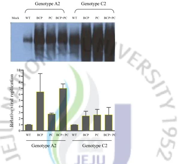
To evaluate the influence of BCP, PC, and BCP+PC mutation on intracellular viral replication in genotype A2 and C2, pHY106-HBV DNA containing these mutations were transiently transfected into Huh7 cell line and the replicating intermediates were detected by Southern blot (figure 2A). The signals of density were normalized to wild type of each HBV genotype. BCP mutation, PC mutation and BCP+PC mutations were all increase the intracellular viral replication regardless of HBV genotypes. In genotype A2, the BCP mutation and BCP+PC mutation increased the viral replication 6.5 times and 7.0 times compared with wild-type, respectively, and PC mutation alone increased the viral replication of 2.8 times. In genotype C2, all the mutant types had similar effect (BCP mutation. 2.5 times; BCP+PC
mutation, 2.7 times, PC mutation, 2.6 times vs. wildtype) on the intracellular viral replication compared with wild-type. Interestingly, the BCP mutation was seemed to be more effective in HBV with genotype A2 than genotype C2. (Figure 2B)
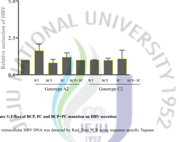
3-3. Influence of BCP and PC mutations on extracellular HBV DNA level
In contrast to the influence of mutations on the intracellular viral replication, there were no remarkable differences between wild type and mutant type. Only in genotype A2, the BCP mutation slightly increased the extracellular HBV DNA level (Figure 3). The relative fold-change in viral secretion is as followed: in genotype A2, BCP mutation. 1.64 times; BCP+PC mutation, 1.2 times, PC mutation, 0.82 times compared with wild-type; in genotype C2, BCP mutation. 1.01 times; BCP+PC mutation, 1.1times, PC mutation, 0.99 times compared with wild-type.
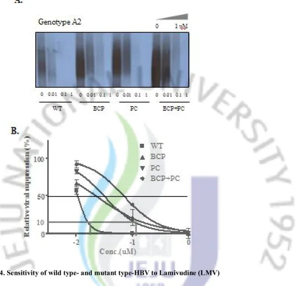
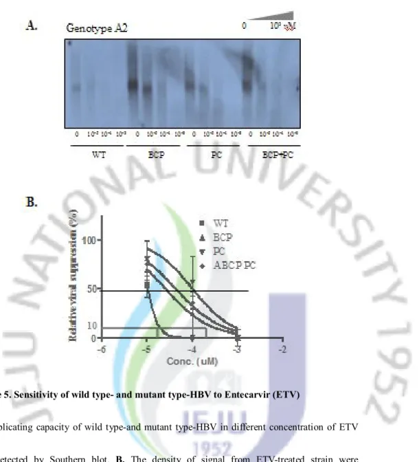
3-4. Sensitivity to antiviral agents
Sensitivity of wild type- and mutant-type of HBV to antiviral agents was evaluated in different concentration of LMV (Final concentration: 0, 0.01, 0.1, 1㎛), ETV (Final concentration: 0, 0.00001, 0.0001, 0.001 ㎛) and CLV (Final concentration: 0, 0.001, 0.01, 0.1 ㎛) and the IC50 value was used to compare the relative sensitivity. The BCP mutation, PC
mutation and BCP+PC mutation either in genotype A2 or genotype C2 all decreased the sensitivity to antiviral agents at various levels (Figure 4~6 and Table 2). Importantly, in HBV strains with these mutations, several folds of antiviral agents were needed to suppress the HBV replication intensively (example: IC90).
Scheme 1. Procedure of the present study
Isolation of HBV DNA from CHB patientsFull-length PCR (primer pair, P1/P2) Sequencing Analysis Choosing of HBV with Genotype A2 & C2
Sap I enzyme
T4 ligase pHY106-HBV DNA with genotype A2 & C2
Replication Competency
Site-directly mutagenesis (BCP, PC, BCP+PC mutation)
Viral replicating capacity (HBV with BCP, PC, BCP+PC mutation)
pHY106 vector
Sensitivity to antiviral agents (lamivudine, entecavir, clevudine) Transfection into Huh7 cell
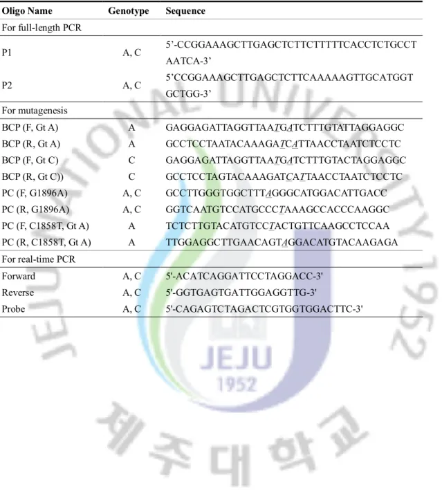
Table 1. Primers used in present study
Oligo Name Genotype Sequence
For full-length PCR P1 A, C 5’-CCGGAAAGCTTGAGCTCTTCTTTTTCACCTCTGCCT AATCA-3’ P2 A, C 5’CCGGAAAGCTTGAGCTCTTCAAAAAGTTGCATGGT GCTGG-3’ For mutagenesis BCP (F, Gt A) A GAGGAGATTAGGTTAATGATCTTTGTATTAGGAGGC BCP (R, Gt A) A GCCTCCTAATACAAAGATCATTAACCTAATCTCCTC BCP (F, Gt C) C GAGGAGATTAGGTTAATGATCTTTGTACTAGGAGGC BCP (R, Gt C)) C GCCTCCTAGTACAAAGATCATTAACCTAATCTCCTC PC (F, G1896A) A, C GCCTTGGGTGGCTTTAGGGCATGGACATTGACC PC (R, G1896A) A, C GGTCAATGTCCATGCCCTAAAGCCACCCAAGGC PC (F, C1858T, Gt A) A TCTCTTGTACATGTCCTACTGTTCAAGCCTCCAA PC (R, C1858T, Gt A) A TTGGAGGCTTGAACAGTAGGACATGTACAAGAGA For real-time PCR Forward A, C 5'-ACATCAGGATTCCTAGGACC-3' Reverse A, C 5'-GGTGAGTGATTGGAGGTTG-3' Probe A, C 5'-CAGAGTCTAGACTCGTGGTGGACTTC-3'
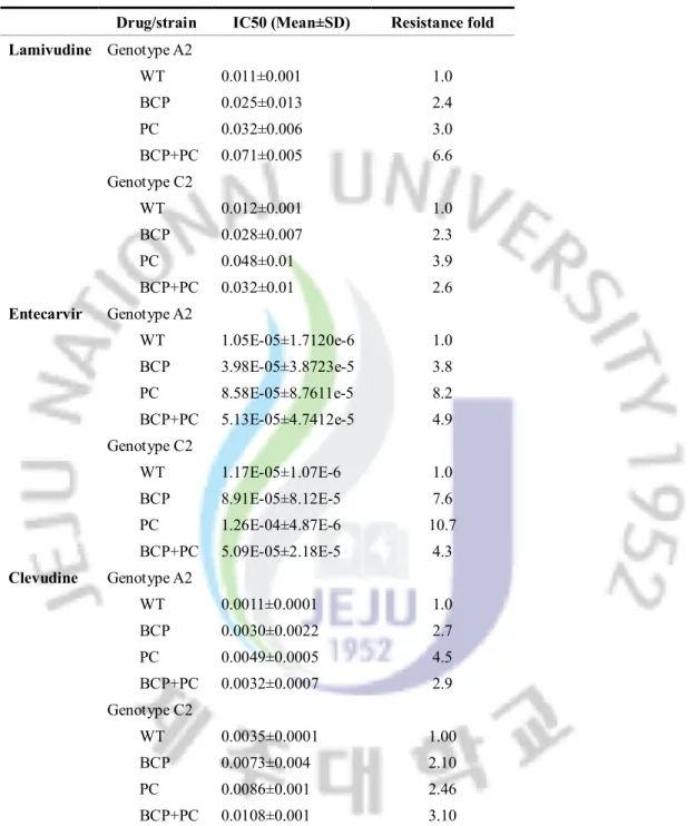
Table 2. Sensitivity of wild type- and mutant type-HBV to LMV, ETV and CLV
Drug/strain IC50 (Mean±SD) Resistance fold Lamivudine Genotype A2 WT 0.011±0.001 1.0 BCP 0.025±0.013 2.4 PC 0.032±0.006 3.0 BCP+PC 0.071±0.005 6.6 Genotype C2 WT 0.012±0.001 1.0 BCP 0.028±0.007 2.3 PC 0.048±0.01 3.9 BCP+PC 0.032±0.01 2.6 Entecarvir Genotype A2 WT 1.05E-05±1.7120e-6 1.0 BCP 3.98E-05±3.8723e-5 3.8 PC 8.58E-05±8.7611e-5 8.2 BCP+PC 5.13E-05±4.7412e-5 4.9 Genotype C2 WT 1.17E-05±1.07E-6 1.0 BCP 8.91E-05±8.12E-5 7.6 PC 1.26E-04±4.87E-6 10.7 BCP+PC 5.09E-05±2.18E-5 4.3 Clevudine Genotype A2 WT 0.0011±0.0001 1.0 BCP 0.0030±0.0022 2.7 PC 0.0049±0.0005 4.5 BCP+PC 0.0032±0.0007 2.9 Genotype C2 WT 0.0035±0.0001 1.00 BCP 0.0073±0.004 2.10 PC 0.0086±0.001 2.46 BCP+PC 0.0108±0.001 3.10
Note. Results are from duplicated experiment. The resistance fold is the IC50 ratio mutant/WT and present as Mean±SD. Abbreviation: WT, wild-type; BCP, Basal Core Promoter; PC, Precore.
Figure 1. Replication competency of pHY106-HBV with genotype A2 and genotype C2
Five clones from each genotype were transfected into Huh7 cell and the replication competency were detected by Southern blot.
Figure 2. Effect of BCP, PC and BCP+PC mutation on intracellular viral replication A.pHY106-HBV with wild- or mutant-type were transfected into 8x10⁵ Huh7 cells and the
intracellular HBV DNA were detected by Southern blot. B. The relative intracellular viral replication is represented as fold changes compared with wild-type strain. Abbreviation: WT, wild-type; BCP, Basal Core promoter; PC, Precore.
0 1 2 3 4 5 6 7 8 9 10
R
el
at
iv
e
vi
ra
l r
ep
li
ca
ti
on
WT BCP PC BCP+ PC WT BCP PC BCP+ PC Mock WT BCP PC BCP+PC WT BCP PC BCP+PC Genotype A2 Genotype C2 Genotype C2 Genotype A2Figure 3. Effect of BCP, PC and BCP+PC mutation on HBV secretion
The extracellular HBV DNA was detected by Real Time PCR using sequence specific Taqman probe and the result were compared with wild type-HBV of each genotype. Abbreviation: WT, wild-type; BCP, Basal Core promoter; PC, Precore
Genotype A2 Genotype C2
0.0
2.5
5.0
R
el
at
iv
e
se
cr
ec
tio
n
of
H
B
V
WT BCP PC BCP+ PC WT BCP PC BCP+ PCFigure 4. Sensitivity of wild type- and mutant type-HBV to Lamivudine (LMV)
A. Replicating capacity of wild type-and mutant type-HBV in different concentration of LMV
was detected by Southern blot. B. The density of signal from LMV-treated strain were normalized to non-treated strain either in wild type or mutant type of HBV, and the data were used for analyzing IC50 value by non linear regression method. Abbreviation:WT, wild type;
Figure 4. Sensitivity of wild type- and mutant type-HBV to Lamivudine (LMV)
C. Replicating capacity of wild type-and mutant type-HBV in different concentration of LMV
was detected by Southern blot. D. The density of signal from LMV-treated strain were normalized to non-treated strain either in wild type or mutant type of HBV, and the data were used for analyzing IC50 value by non linear regression method. Abbreviation: WT, wild type;
Figure 5. Sensitivity of wild type- and mutant type-HBV to Entecarvir (ETV)
A. Replicating capacity of wild type-and mutant type-HBV in different concentration of ETV
was detected by Southern blot. B. The density of signal from ETV-treated strain were normalized to non-treated strain either in wild type or mutant type of HBV, and the data were used for analyzing IC50 value by non linear regression method. Abbreviation: WT, wild type; BCP, Basal Core promoter; PC, Precore
Figure 5. Sensitivity of wild type- and mutant type-HBV to Entecarvir (ETV)
C. Replicating capacity of wild type-and mutant type-HBV in different concentration of ETV
was detected by Southern blot. D. The density of signal from ETV-treated strain were normalized to non-treated strain either in wild type or mutant type of HBV, and the data were used for analyzing IC50 value by non linear regression method. Abbreviation: WT, wild type; BCP, Basal Core promoter; PC, Precore
Figure 6. Sensitivity of wild type- and mutant type-HBV to Clevudine (CLV)
A. Replicating capacity of wild type-and mutant type-HBV in different concentration of CLV
was detected by Southern blot. B. The density of signal from CLV-treated strain were normalized to non-treated strain either in wild type or mutant type of HBV, and the data were used for analyzing IC50 value by non linear regression method. Abbreviation: WT, wild type; BCP, Basal Core promoter; PC, Precore
Figure 6. Sensitivity of wild type- and mutant type-HBV to Clevudine (CLV)
C. Replicating capacity of wild type-and mutant type-HBV in different concentration of CLV
was detected by Southern blot. D. The density of signal from CLV-treated strain were normalized to non-treated strain either in wild type or mutant type of HBV, and the data were used for analyzing IC50 value by non linear regression method. Abbreviation: WT, wild type;
4.Discussion
In the present study, we evaluated the BCP mutation (A1762T/G1764A) and PC mutation (G1896A) on the impact of viral replication and sensitivity to nucleoside analogues (lamivudine, entecavir and clevudine) either in genotype A2 or in genotype C2.
Recently, it has been reported that elevated serum HBV DNA level and HBV genotype C are strong independent risk factor of HCC (Chen, et al.,2006, Yang, et al.,2008). HBV is a DNA virus that replicates via reverse transcription of RNA intermediate and can be increased by the naturally occurring nucleotide mutations in virus genome (Baumert, et al.,1998, Buckwold, et
al.,1996, Delius, et al.,1983, Moriyama, et al.,1996).
Among them, BCP mutation is the frequently occurring mutation during viral replication(Okamoto, et al.,1994). In the present study, BCP mutation increased the intracellular viral replication regardless of genotypes and this result is consistent with the previous in vitro studies(Baumert, et al.,1998, Buckwold, et al.,1996, Moriyama, et al.,1996). BCP, which is mapped between nucleotide 1742 and 1849 and contains four TATA-like boxes, has central role in precore mRNA and pregenomic mRNA transcription during HBV replication (Chen, et
al.,1995, Yuh, et al.,1992). In vitro studies have shown that the BCP mutation enhanced viral
increased pregenomic mRNA transcription. HBeAg has been reported to inhibit HBV DNA synthesis, thus the reduction of HBeAg production by BCP mutation increase viral replication. Moriyama K et al. reported that the BCP mutation increased pregenomic mRNA, a template of viral genome replication, transcription to enhance viral replication(Moriyama, et al.,1996). But, there still exist controversy on the effect of BCP on increasing pregenomic mRNA (Baumert, et
al.,1998, Buckwold, et al.,1996, Moriyama, et al.,1996, Scaglioni, et al.,1997). Interestingly, in
our study, BCP mutation more effectively increased viral replication in genotype A2 than in genotype C2. Generally, BCP mutation was more frequent in genotype C2 than in genotype A2, thus we supposed it might be more effective in HBV-genotype C2 replication. The mechanism of different effect of BCP mutation on viral replication is not clear, however, it might be associated with the viral quasi-species. In vitro studies showed the various viral replication levels of clinical HBV isolates from the same patients (Parekh, et al.,2003, Yang, et al.,2004). Furthermore, even in the same genotype, BCP mutation increased the viral replication in different level (Parekh, et al.,2003, Scaglioni, et al.,1997, Tacke, et al.,2004).
Another frequent mutation is PC mutation, which creates a stop codon at codon 28 to abolish the HBeAg synthesis. PC mutation also has been reported to increase viral replication via prevention of HBeAg production and stabilized the stem-loop in pregenomic mRNA in vitro (Brunetto, et al.,1991, Carman, et al.,1989, Lok, et al.,1994). In our study, PC mutation
increased the viral replication both in genotype A2 and C2. In contrast to BCP mutation, PC mutation had similar effect on the viral replication of both genotypes. Overall, BCP and PC mutations increased the intracellular viral replication regardless of genotypes.
In addition, influence of BCP and PC mutations on the secretion of HBV were also evaluated. In result, these mutations slightly increased the extracellular HBV DNA level in HBV-genotype A2, but had no remarkable effect on the secretion of HBV in genotype C2. It might be explained by the minor effect of BCP and PC mutations on the secretion of HBsAg (Tacke, et al.,2004, Wang, et al.,2007).
To date, there exist two types of antiviral agents, immunomodulator (interferon) and viral polymerase inhibitor (nucleos(t)ide analogue). Successful antiviral therapy can improve hepatic inflammation and fibrosis, suppress viral replication, and even improve survival(Dienstag, et al.,1999, Hadziyannis, et al.,2005, Marcellin, et al.,2003, Niederau, et al.,1996, van Zonneveld, et al.,2004). The efficacy of antiviral therapy is related with the baseline serum HBV DNA level, which represents viral replication, both in immunomodulator and nucloes(t)ide analogue treatment(Kau, et al.,2008, Mommeja-Marin, et al.,2003, Perrillo, et al.,1990, Song, et al.,2004, Song, et al.,2000, van Zonneveld, et al.,2004). In present study, IC50 value of lamivudine, entecarvir and clevudine were higher in BCP- and PC-mutant HBV strains compared with wild-type HBV strain. Because these antiviral agents are viral polymerase inhibitor, the amount of
inhibited virus might be same both in wild-type and mutant type. In other words, it needs higher concentration of antiviral agents to effectively suppress the virus containing BCP mutation and PC mutation equal to the suppression level in wild-type strain.
In Korea, most of the HBV is genotype C2 and more than approximately 90% and 30% of them possessed BCP mutation and PC mutation, respectively(Song, et al.,2006, Yoo, et al.,2003). In contrast, the frequency of BCP mutation and PC mutations in HBV-genotype A2 is less than 20% (Sugauchi, et al.,2004, Tanaka, et al.,2004). It means that most Korean patients might need more amount of antiviral agent to suppress complete viral replication
In conclusion, BCP and PC mutations increased viral replication regardless of HBV genotypes in vitro. In addition, patients with theses mutations might need more amount of antiviral agents. In the future, clinical trials are needed to compare the effectiveness of antiviral agents using current standard dose and higher dose in patients with these mutations.
5.References
Akarca, U S, Greene, S, Lok, A S.Detection of precore hepatitis B virus mutants in asymptomatic HBsAg-positive family members. Hepatology, 19, 1366-1370 (1994).
Arauz-Ruiz, P, Norder, H, Robertson, B H, Magnius, L O.Genotype H: a new Amerindian genotype of hepatitis B virus revealed in Central America. J Gen Virol, 83, 2059-2073 (2002).
Arauz-Ruiz, P, Norder, H, Visona, K A, Magnius, L O.Molecular epidemiology of hepatitis B virus in Central America reflected in the genetic variability of the small S gene. J Infect Dis, 176, 851-858 (1997).
Bartenschlager, R, Schaller, H.Hepadnaviral assembly is initiated by polymerase binding to the encapsidation signal in the viral RNA genome. Embo J, 11, 3413-3420 (1992).
Baumert, T F, Marrone, A, Vergalla, J, Liang, T J.Naturally occurring mutations define a novel function of the hepatitis B virus core promoter in core protein expression. J Virol, 72, 6785-6795 (1998).
Beasley, R P, Hwang, L Y, Lin, C C, Chien, C S.Hepatocellular carcinoma and hepatitis B virus. A prospective study of 22 707 men in Taiwan. Lancet, 2, 1129-1133 (1981).
Brunetto, M R, Giarin, M M, Oliveri, F, Chiaberge, E, Baldi, M, Alfarano, A, Serra, A, Saracco, G, Verme, G, Will, H, et al.Wild-type and e antigen-minus hepatitis B viruses and course of chronic hepatitis. Proc Natl Acad Sci U S A, 88, 4186-4190 (1991).
Brunetto, M R, Stemler, M, Schodel, F, Will, H, Ottobrelli, A, Rizzetto, M, Verme, G, Bonino, F.Identificaion of HBV variants which cannot produce precore derived HBeAg and may be responsible for severe hepatitis. Ital J Gastroenterol, 21, 151-154 (1989).
Buckwold, V E, Xu, Z, Chen, M, Yen, T S, Ou, J H.Effects of a naturally occurring mutation in the hepatitis B virus basal core promoter on precore gene expression and viral replication. J Virol, 70, 5845-5851 (1996).
Buckwold, V E, Xu, Z, Yen, T S, Ou, J H.Effects of a frequent double-nucleotide basal core promoter mutation and its putative single-nucleotide precursor mutations on hepatitis B virus gene expression and replication. J Gen Virol, 78 ( Pt 8), 2055-2065 (1997).
Carman, W F, Fagan, E A, Hadziyannis, S, Karayiannis, P, Tassopoulos, N C, Williams, R, Thomas, H C.Association of a precore genomic variant of hepatitis B virus with fulminant hepatitis.
Hepatology, 14, 219-222 (1991).
Carman, W F, Jacyna, M R, Hadziyannis, S, Karayiannis, P, McGarvey, M J, Makris, A, Thomas, H C.Mutation preventing formation of hepatitis B e antigen in patients with chronic hepatitis B infection. Lancet, 2, 588-591 (1989).
Chan, H L, Hussain, M, Lok, A S.Different hepatitis B virus genotypes are associated with different mutations in the core promoter and precore regions during hepatitis B e antigen seroconversion.
Hepatology, 29, 976-984 (1999).
Chang, C, Enders, G, Sprengel, R, Peters, N, Varmus, H E, Ganem, D.Expression of the precore region of an avian hepatitis B virus is not required for viral replication. J Virol, 61, 3322-3325 (1987). Chen, C J, Yang, H I, Su, J, Jen, C L, You, S L, Lu, S N, Huang, G T, Iloeje, U H.Risk of hepatocellular
carcinoma across a biological gradient of serum hepatitis B virus DNA level. JAMA, 295, 65-73 (2006).
Chen, D S.From hepatitis to hepatoma: lessons from type B viral hepatitis. Science, 262, 369-370 (1993). Chen, H S, Kew, M C, Hornbuckle, W E, Tennant, B C, Cote, P J, Gerin, J L, Purcell, R H, Miller, R
H.The precore gene of the woodchuck hepatitis virus genome is not essential for viral replication in the natural host. J Virol, 66, 5682-5684 (1992).
Chen, I H, Huang, C J, Ting, L P.Overlapping initiator and TATA box functions in the basal core promoter of hepatitis B virus. J Virol, 69, 3647-3657 (1995).
Chien, R N, Yeh, C T, Tsai, S L, Chu, C M, Liaw, Y F.Determinants for sustained HBeAg response to lamivudine therapy. Hepatology, 38, 1267-1273 (2003).
Chu, C J, Hussain, M, Lok, A S.Hepatitis B virus genotype B is associated with earlier HBeAg seroconversion compared with hepatitis B virus genotype C. Gastroenterology, 122, 1756-1762 (2002).
Chun, Y K, Kim, J Y, Woo, H J, Oh, S M, Kang, I, Ha, J, Kim, S S.No significant correlation exists between core promoter mutations, viral replication, and liver damage in chronic hepatitis B infection. Hepatology, 32, 1154-1162 (2000).
de Franchis, R, Hadengue, A, Lau, G, Lavanchy, D, Lok, A, McIntyre, N, Mele, A, Paumgartner, G, Pietrangelo, A, Rodes, J, Rosenberg, W, Valla, D.EASL International Consensus Conference on Hepatitis B. 13-14 September, 2002 Geneva, Switzerland. Consensus statement (long version). J
Hepatol, 39 Suppl 1, S3-25 (2003).
Delius, H, Gough, N M, Cameron, C H, Murray, K.Structure of the hepatitis B virus genome. J Virol, 47, 337-343 (1983).
Dienstag, J L, Schiff, E R, Wright, T L, Perrillo, R P, Hann, H W, Goodman, Z, Crowther, L, Condreay, L D, Woessner, M, Rubin, M, Brown, N A.Lamivudine as initial treatment for chronic hepatitis B in the United States. N Engl J Med, 341, 1256-1263 (1999).
Ding, X, Mizokami, M, Yao, G, Xu, B, Orito, E, Ueda, R, Nakanishi, M.Hepatitis B virus genotype distribution among chronic hepatitis B virus carriers in Shanghai, China. Intervirology, 44, 43-47 (2001).
Gunther, S, Li, B C, Miska, S, Kruger, D H, Meisel, H, Will, H.A novel method for efficient amplification of whole hepatitis B virus genomes permits rapid functional analysis and reveals deletion mutants in immunosuppressed patients. J Virol, 69, 5437-5444 (1995).
Hadziyannis, S J, Tassopoulos, N C, Heathcote, E J, Chang, T T, Kitis, G, Rizzetto, M, Marcellin, P, Lim, S G, Goodman, Z, Ma, J, Arterburn, S, Xiong, S, Currie, G, Brosgart, C L.Long-term therapy with adefovir dipivoxil for HBeAg-negative chronic hepatitis B. N Engl J Med, 352, 2673-2681 (2005).
Hirsch, R C, Lavine, J E, Chang, L J, Varmus, H E, Ganem, D.Polymerase gene products of hepatitis B viruses are required for genomic RNA packaging as wel as for reverse transcription. Nature, 344, 552-555 (1990).
Janssen, H L, van Zonneveld, M, Senturk, H, Zeuzem, S, Akarca, U S, Cakaloglu, Y, Simon, C, So, T M, Gerken, G, de Man, R A, Niesters, H G, Zondervan, P, Hansen, B, Schalm, S W.Pegylated interferon alfa-2b alone or in combination with lamivudine for HBeAg-positive chronic hepatitis B: a randomised trial. Lancet, 365, 123-129 (2005).
Kao, J H, Chen, P J, Lai, M Y, Chen, D S.Basal core promoter mutations of hepatitis B virus increase the risk of hepatocellular carcinoma in hepatitis B carriers. Gastroenterology, 124, 327-334 (2003).
Kao, J H, Wu, N H, Chen, P J, Lai, M Y, Chen, D S.Hepatitis B genotypes and the response to interferon therapy. J Hepatol, 33, 998-1002 (2000).
Kau, A, Vermehren, J, Sarrazin, C.Treatment predictors of a sustained virologic response in hepatitis B and C. J Hepatol, 49, 634-651 (2008).
Kidd-Ljunggren, K, Oberg, M, Kidd, A H.Hepatitis B virus X gene 1751 to 1764 mutations: implications for HBeAg status and disease. J Gen Virol, 78, 1469-1478 (1997).
Laskus, T, Persing, D H, Nowicki, M J, Mosley, J W, Rakela, J.Nucleotide sequence analysis of the precore region in patients with fulminant hepatitis B in the United States. Gastroenterology, 105, 1173-1178 (1993).
Laskus, T, Rakela, J, Nowicki, M J, Persing, D H.Hepatitis B virus core promoter sequence analysis in fulminant and chronic hepatitis B. Gastroenterology, 109, 1618-1623 (1995).
Lavanchy, D.Hepatitis B virus epidemiology, disease burden, treatment, and current and emerging prevention and control measures. J Viral Hepat, 11, 97-107 (2004).
Li, J S, Tong, S P, Wen, Y M, Vitvitski, L, Zhang, Q, Trepo, C.Hepatitis B virus genotype A rarely circulates as an HBe-minus mutant: possible contribution of a single nucleotide in the precore region. J Virol, 67, 5402-5410 (1993).
Liang, T J, Hasegawa, K, Rimon, N, Wands, J R, Ben-Porath, E.A hepatitis B virus mutant associated with an epidemic of fulminant hepatitis. N Engl J Med, 324, 1705-1709 (1991).
Lin, C L, Kao, J H.Hepatitis B viral factors and clinical outcomes of chronic hepatitis B. J Biomed Sci, 15, 137-145 (2008).
Lindh, M, Andersson, A S, Gusdal, A.Genotypes, nt 1858 variants, and geographic origin of hepatitis B virus--large-scale analysis using a new genotyping method. J Infect Dis, 175, 1285-1293 (1997).
Lindh, M, Hannoun, C, Dhillon, A P, Norkrans, G, Horal, P.Core promoter mutations and genotypes in relation to viral replication and liver damage in East Asian hepatitis B virus carriers. J Infect Dis, 179, 775-782 (1999).
Liu, C J, Kao, J H, Lai, M Y, Chen, P J, Chen, D S.Precore/core promoter mutations and genotypes of hepatitis B virus in chronic hepatitis B patients with fulminant or subfulminant hepatitis. J Med
Lo, W Y, Ting, L P.Repression of enhancer II activity by a negative regulatory element in the hepatitis B virus genome. J Virol, 68, 1758-1764 (1994).
Lok, A S, Akarca, U, Greene, S.Mutations in the pre-core region of hepatitis B virus serve to enhance the stability of the secondary structure of the pre-genome encapsidation signal. Proc Natl Acad Sci
U S A, 91, 4077-4081 (1994).
Lok, A S, McMahon, B J.Chronic hepatitis B. Hepatology, 45, 507-539 (2007).
Marcellin, P, Chang, T T, Lim, S G, Tong, M J, Sievert, W, Shiffman, M L, Jeffers, L, Goodman, Z, Wulfsohn, M S, Xiong, S, Fry, J, Brosgart, C L.Adefovir dipivoxil for the treatment of hepatitis B e antigen-positive chronic hepatitis B. N Engl J Med, 348, 808-816 (2003).
Mayerat, C, Mantegani, A, Spertini, F, Frei, P C.Mutations in the basal core promoter and precore/core gene of hepatitis B virus in patients with chronic active but not acute hepatitis B. Eur J Clin
Microbiol Infect Dis, 18, 871-878 (1999).
Mommeja-Marin, H, Mondou, E, Blum, M R, Rousseau, F.Serum HBV DNA as a marker of efficacy during therapy for chronic HBV infection: analysis and review of the literature. Hepatology, 37, 1309-1319 (2003).
Moriyama, K, Okamoto, H, Tsuda, F, Mayumi, M.Reduced precore transcription and enhanced core-pregenome transcription of hepatitis B virus DNA after replacement of the precore-core promoter with sequences associated with e antigen-seronegative persistent infections. Virology, 226, 269-280 (1996).
Nassal, M.Hepatitis B virus replication: novel roles for virus-host interactions. Intervirology, 42, 100-116 (1999).
Niederau, C, Heintges, T, Lange, S, Goldmann, G, Niederau, C M, Mohr, L, Haussinger, D.Long-term follow-up of HBeAg-positive patients treated with interferon alfa for chronic hepatitis B. N Engl
J Med, 334, 1422-1427 (1996).
Norder, H, Courouce, A M, Magnius, L O.Complete genomes, phylogenetic relatedness, and structural proteins of six strains of the hepatitis B virus, four of which represent two new genotypes.
Norder, H, Hammas, B, Lee, S D, Bile, K, Courouce, A M, Mushahwar, I K, Magnius, L O.Genetic relatedness of hepatitis B viral strains of diverse geographical origin and natural variations in the primary structure of the surface antigen. J Gen Virol, 74 ( Pt 7), 1341-1348 (1993).
Okamoto, H, Tsuda, F, Akahane, Y, Sugai, Y, Yoshiba, M, Moriyama, K, Tanaka, T, Miyakawa, Y, Mayumi, M.Hepatitis B virus with mutations in the core promoter for an e antigen-negative phenotype in carriers with antibody to e antigen. J Virol, 68, 8102-8110 (1994).
Okamoto, H, Tsuda, F, Sakugawa, H, Sastrosoewignjo, R I, Imai, M, Miyakawa, Y, Mayumi, M.Typing hepatitis B virus by homology in nucleotide sequence: comparison of surface antigen subtypes. J
Gen Virol, 69 ( Pt 10), 2575-2583 (1988).
Omata, M, Ehata, T, Yokosuka, O, Hosoda, K, Ohto, M.Mutations in the precore region of hepatitis B virus DNA in patients with fulminant and severe hepatitis. N Engl J Med, 324, 1699-1704 (1991). Orito, E, Ichida, T, Sakugawa, H, Sata, M, Horiike, N, Hino, K, Okita, K, Okanoue, T, Iino, S, Tanaka, E,
Suzuki, K, Watanabe, H, Hige, S, Mizokami, M.Geographic distribution of hepatitis B virus (HBV) genotype in patients with chronic HBV infection in Japan. Hepatology, 34, 590-594 (2001).
Parekh, S, Zoulim, F, Ahn, S H, Tsai, A, Li, J, Kawai, S, Khan, N, Trepo, C, Wands, J, Tong, S.Genome replication, virion secretion, and e antigen expression of naturally occurring hepatitis B virus core promoter mutants. J Virol, 77, 6601-6612 (2003).
Perrillo, R P, Schiff, E R, Davis, G L, Bodenheimer, H C, Jr., Lindsay, K, Payne, J, Dienstag, J L, O'Brien, C, Tamburro, C, Jacobson, I M, et al.A randomized, controlled trial of interferon alfa-2b alone and after prednisone withdrawal for the treatment of chronic hepatitis B. The Hepatitis Interventional Therapy Group. N Engl J Med, 323, 295-301 (1990).
Sanchez-Tapias, J M, Costa, J, Mas, A, Bruguera, M, Rodes, J.Influence of hepatitis B virus genotype on the long-term outcome of chronic hepatitis B in western patients. Gastroenterology, 123, 1848-1856 (2002).
Sato, S, Suzuki, K, Akahane, Y, Akamatsu, K, Akiyama, K, Yunomura, K, Tsuda, F, Tanaka, T, Okamoto, H, Miyakawa, Y, Mayumi, M.Hepatitis B virus strains with mutations in the core promoter in patients with fulminant hepatitis. Ann Intern Med, 122, 241-248 (1995).
Scaglioni, P P, Melegari, M, Wands, J R.Biologic properties of hepatitis B viral genomes with mutations in the precore promoter and precore open reading frame. Virology, 233, 374-381 (1997). Schaefer, S.Hepatitis B virus taxonomy and hepatitis B virus genotypes. World J Gastroenterol, 13,
14-21 (2007).
Seeger, C, Mason, W S.Hepatitis B virus biology. Microbiol Mol Biol Rev, 64, 51-68 (2000).
Sells, M A, Zelent, A Z, Shvartsman, M, Acs, G.Replicative intermediates of hepatitis B virus in HepG2 cells that produce infectious virions. J Virol, 62, 2836-2844 (1988).
Shindo, M, Hamada, K, Koya, S, Sokawa, Y, Okuno, T.The clinical significance of core promoter and precore mutations during the natural course and interferon therapy in patients with chronic hepatitis B. Am J Gastroenterol, 94, 2237-2245 (1999).
Song, B C, Cui, X J, Kim, H U, Cho, Y K.Sequential accumulation of the basal core promoter and the precore mutations in the progression of hepatitis B virus-related chronic liver disease.
Intervirology, 49, 266-273 (2006).
Song, B C, Ghany, M, Lee, H C, Chung, Y H, Lee, Y S.Which patients with chronic hepatitis B are more likely to relapse after interferon alpha-induced hepatitis B e antigen loss in Korea? J Clin
Gastroenterol, 38, 124-129 (2004).
Song, B C, Suh, D J, Lee, H C, Chung, Y H, Lee, Y S.Hepatitis B e antigen seroconversion after lamivudine therapy is not durable in patients with chronic hepatitis B in Korea. Hepatology, 32, 803-806 (2000).
Sterneck, M, Gunther, S, Santantonio, T, Fischer, L, Broelsch, C E, Greten, H, Will, H.Hepatitis B virus genomes of patients with fulminant hepatitis do not share a specific mutation. Hepatology, 24, 300-306 (1996).
Stuyver, L, De Gendt, S, Van Geyt, C, Zoulim, F, Fried, M, Schinazi, R F, Rossau, R.A new genotype of hepatitis B virus: complete genome and phylogenetic relatedness. J Gen Virol, 81, 67-74 (2000). Sugauchi, F, Kumada, H, Acharya, S A, Shrestha, S M, Gamutan, M T, Khan, M, Gish, R G, Tanaka, Y,
Kato, T, Orito, E, Ueda, R, Miyakawa, Y, Mizokami, M.Epidemiological and sequence differences between two subtypes (Ae and Aa) of hepatitis B virus genotype A. J Gen Virol, 85, 811-820 (2004).
Sugauchi, F, Kumada, H, Sakugawa, H, Komatsu, M, Niitsuma, H, Watanabe, H, Akahane, Y, Tokita, H, Kato, T, Tanaka, Y, Orito, E, Ueda, R, Miyakawa, Y, Mizokami, M.Two subtypes of genotype B (Ba and Bj) of hepatitis B virus in Japan. Clin Infect Dis, 38, 1222-1228 (2004).
Sumi, H, Yokosuka, O, Seki, N, Arai, M, Imazeki, F, Kurihara, T, Kanda, T, Fukai, K, Kato, M, Saisho, H.Influence of hepatitis B virus genotypes on the progression of chronic type B liver disease.
Hepatology, 37, 19-26 (2003).
Tacke, F, Gehrke, C, Luedde, T, Heim, A, Manns, M P, Trautwein, C.Basal core promoter and precore mutations in the hepatitis B virus genome enhance replication efficacy of Lamivudine-resistant mutants. J Virol, 78, 8524-8535 (2004).
Tajiri, H, Tanaka, Y, Kagimoto, S, Murakami, J, Tokuhara, D, Mizokami, M.Molecular evidence of father-to-child transmission of hepatitis B virus. J Med Virol, 79, 922-926 (2007).
Tanaka, Y, Hasegawa, I, Kato, T, Orito, E, Hirashima, N, Acharya, S K, Gish, R G, Kramvis, A, Kew, M C, Yoshihara, N, Shrestha, S M, Khan, M, Miyakawa, Y, Mizokami, M.A case-control study for differences among hepatitis B virus infections of genotypes A (subtypes Aa and Ae) and D.
Hepatology, 40, 747-755 (2004).
Tanaka, Y, Mukaide, M, Orito, E, Yuen, M F, Ito, K, Kurbanov, F, Sugauchi, F, Asahina, Y, Izumi, N, Kato, M, Lai, C L, Ueda, R, Mizokami, M.Specific mutations in enhancer II/core promoter of hepatitis B virus subgenotypes C1/C2 increase the risk of hepatocellular carcinoma. J Hepatol, 45, 646-653 (2006).
Tanaka, Y, Orito, E, Yuen, M F, Mukaide, M, Sugauchi, F, Ito, K, Ozasa, A, Sakamoto, T, Kurbanov, F, Lai, C L, Mizokami, M.Two subtypes (subgenotypes) of hepatitis B virus genotype C: A novel subtyping assay based on restriction fragment length polymorphism. Hepatol Res, 33, 216-224 (2005).
Tong, S P, Li, J S, Vitvitski, L, Kay, A, Treepo, C.Evidence for a base-paired region of hepatitis B virus pregenome encapsidation signal which influences the patterns of precore mutations abolishing HBe protein expression. J Virol, 67, 5651-5655 (1993).
Tur-Kaspa, R, Klein, A, Aharonson, S.Hepatitis B virus precore mutants are identical in carriers from various ethnic origins and are associated with a range of liver disease severity. Hepatology, 16, 1338-1342 (1992).
Tuttleman, J S, Pourcel, C, Summers, J.Formation of the pool of covalently closed circular viral DNA in hepadnavirus-infected cells. Cell, 47, 451-460 (1986).
van Zonneveld, M, Honkoop, P, Hansen, B E, Niesters, H G, Murad, S D, de Man, R A, Schalm, S W, Janssen, H L.Long-term follow-up of alpha-interferon treatment of patients with chronic hepatitis B. Hepatology, 39, 804-810 (2004).
VHPB 1998 Universal HB immunization by 1997: where are we now? In: Viral Hepatitis Prevention Board; http://hgins.uia.ac.be/esoc/VHPB/vhfs2.html
Wai, C T, Chu, C J, Hussain, M, Lok, A S.HBV genotype B is associated with better response to interferon therapy in HBeAg(+) chronic hepatitis than genotype C. Hepatology, 36, 1425-1430 (2002).
Wang, Y, Wei, L, Jiang, D, Cong, X, Fei, R, Chen, H, Xiao, J, Wang, Y.In vitro resistance to interferon-alpha of hepatitis B virus with basic core promoter double mutation. Antiviral Res, 75, 139-145 (2007).
Yang, H, Westland, C, Xiong, S, Delaney, W E t.In vitro antiviral susceptibility of full-length clinical hepatitis B virus isolates cloned with a novel expression vector. Antiviral Res, 61, 27-36 (2004). Yang, H I, Yeh, S H, Chen, P J, Iloeje, U H, Jen, C L, Su, J, Wang, L Y, Lu, S N, You, S L, Chen, D S,
Liaw, Y F, Chen, C J.Associations between hepatitis B virus genotype and mutants and the risk of hepatocellular carcinoma. J Natl Cancer Inst, 100, 1134-1143 (2008).
Yoo, B C, Park, J W, Kim, H J, Lee, D H, Cha, Y J, Park, S M.Precore and core promoter mutations of hepatitis B virus and hepatitis B e antigen-negative chronic hepatitis B in Korea. J Hepatol, 38, 98-103 (2003).
Yu, M W, Yeh, S H, Chen, P J, Liaw, Y F, Lin, C L, Liu, C J, Shih, W L, Kao, J H, Chen, D S, Chen, C J.Hepatitis B virus genotype and DNA level and hepatocellular carcinoma: a prospective study in men. J Natl Cancer Inst, 97, 265-272 (2005).
Yuen, M F, Sablon, E, Wong, D K, Yuan, H J, Wong, B C, Chan, A O, Lai, C L.Role of hepatitis B virus genotypes in chronic hepatitis B exacerbation. Clin Infect Dis, 37, 593-597 (2003).
Yuen, M F, Tanaka, Y, Mizokami, M, Yuen, J C, Wong, D K, Yuan, H J, Sum, S M, Chan, A O, Wong, B C, Lai, C L.Role of hepatitis B virus genotypes Ba and C, core promoter and precore
mutations on hepatocellular carcinoma: a case control study. Carcinogenesis, 25, 1593-1598 (2004).
Yuh, C H, Chang, Y L, Ting, L P.Transcriptional regulation of precore and pregenomic RNAs of hepatitis B virus. J Virol, 66, 4073-4084 (1992).
Zhang, H W, Yin, J H, Li, Y T, Li, C Z, Ren, H, Gu, C Y, Wu, H Y, Liang, X S, Zhang, P, Zhao, J F, Tan, X J, Lu, W, Schaefer, S, Cao, G W.Risk factors for acute hepatitis B and its progression to chronic hepatitis in Shanghai, China. Gut, 57, 1713-1720 (2008).
Zoulim, F, Perrillo, R.Hepatitis B: reflections on the current approach to antiviral therapy. J Hepatol, 48 Suppl 1, S2-19 (2008).
6. Abstract in Korean
연구배경: 만성B형 간염의 치료에 있어서 viral factor인 치료 전 혈청 HBV DNA
의 수준과 B형 간염바이러스의 유전자형이 중요한 예측인자로 작용한다. 혈청 HBV DNA level은 바이러스의 복제능력과 연관 있는데 바이러스의 복제능력에 영향을 주 는 주요한 인자로는 바이러스 유전자의 basal core promoter (BCP) 와 precore (PC)부위 의 변이이며 주로 BCP mutation (A1762T/G1764A) 또는 PC mutation (G1896A) 가 있다. 이러한 변이는 바이러스의 genotype 및 subgenotype에 따라 발생빈도의 차이가 있다. 한국에서의 주요 유전자형인 Genotype C2에서 약 80% 이상의 환자에서 BCP와 PC 변이가 발생하는 반면에 서양인의 주요 유전자형인 genotype A2에서는 발생빈도는 20% 미만이다. 따라서 Genotype C가 다른 유전자형에 비해 항 바이러스 치료효과가 낮은데 이는 바이러스의 복제를 증가시키는 BCP 및 PC변이와 연관이 있을 것으로 생각된다. 또한 현재까지 대부분의 in vitro 항 바이러스 치료 효능 실험은 genotype A에서 진행 되여 genotype C를 모델로 한 in vitro 실험이 필요하다고 생간 된다.
연구목적: genotype C2를 이용한 in vitro 실험모델을 만들고 BCP및 PC변이가 바
이러스의 증식에 대한 영향 및 항바이러스제에 대한 영향을 연구하고자 한다. 재료 및 방법: B형 간염 환자의 혈청에서 HBV DNA를 추출한 다음 염기서열분
석으로 Genotype A2와 C2인 바이러스를 선택한 다음 바이러스의 전체유전자를 PCR 방법으로 증폭하였다. 다음 SapI enzyme으로 자르고 gene expression vector 인 pHY106 에 연결시켰다. genotype A2와 C2인 바이러스 유전자를 가지고 있는 plasmid DNA를 Huh7 cell에 transfection시켜 바이러스 증식능력을 확인한 다음 증식능력을 우수한 plasmid DNA를 wild type로 지정하였다. 또한 BCP변이, PC변이 및 BCP+PC변이가 바 이러스의 증식에 대한 영향을 연구하기 위하여 site-directly mutagenesis 방법으로 BCP, PC 및 BCP+PC변이를 가지는 plasmid DNA를 만들었다. 다음 이들을 Huh7 cell
에 transfection하여 바이러스의 증식능력 및 항 바이러스제에 대한 민감성을 southern blot으로 확인하였다.
결과: BCP, PC 그리고 BCP+PC변이들은 B형 간염 바이러스의 genotype에 연관
없이 모두 wild type에 비해 세포 내 바이러스의 복제를 증가시켰다. 바이러스의 세 포 밖으로의 분비에 대한 영향을 확인하기 위하여 배양 상층 액에서 바이러스 DNA를 추출한 다음 real time PCR로 바이러스 DNA의 양을 측정하였다. 결과 이러 한 변이가 바이러스의 세포 밖으로의 분비에는 큰 영향을 주지 않는 것을 확인하였 다. 다음으로 항바이러스제에 대한 변이의 영향을 확인 하기 위하여 현재 임상에서 사용되는 항바이러스 제제인 lamivudine entecarvir 그리고 clevudine를 농도별로 처리 하여 각각 50%의 바이러스를 억제하는 IC50값을 구하여 비교하였다. 결과, 이러한 변이들은 대체로 유전자 형에 관계없이 모든 mutant type의 IC50값이 wild type 에 비 해 높을 것을 확인 하였다. 특히 중요한 것은 초기 바이러스의 양이 적으면 적을 수록 바이러스 억제 효율이 높아 적은 양의 약물로도 바이러스를 효과적으로 억제 할 수 있었다. 반대로 바이러스의 양이 많을 수록 동일한 수준의 바이러스 억제효 과를 나타내기 위해서 더 높은 농도의 약물이 필요하다는 것을 확인할 수 있습니다. 결론: 첫째, BCP 및 PC 변이는 유전자형에 관계없이 세포 내 바이러스의 복제 능력을 증가 시키지만 바이러스의 분비엔 큰 영향을 미치지 않는다. 둘째, 바이러스의 복제가 증가하면 할 수록 항바이러스제제의 효능이 낮아진다. 따라서 이러한 원인 때문에 BCP/PC mutation 빈번하게 일어나는 genotype C가 다른 유전자형에 비해 항v바이러스 치료효능이 낮을 거라고 생각된다.