143
-Vol. 11, No. 2(December), 2014 The Journal of Medicine and Life Science
Immunoglobulin G4 (IgG4)–related disease is an inflammatory disorder that is characterized by hyper-IgG4-γ-globulinemia and IgG4-producing plasma cell infiltration in affected organs with fibrotic or sclerotic changes1). It can affect various organs, including the pancreas, bile duct, salivary glands, and retroperitoneum2). Recent studies suggest that the cardiovascular system can also be a target of IgG4-related disease (IgG4-RD). However, in most cases, they were presented as periaortitis or periarteritis involving larger arteries, and cardiac and coronary involvement of the disease is rare2-4). Only a few reports have been described as coronary periarteritis-related angina and cardiac mass causing conduction abnormalities5-9). In this study, we report a case of presumed IgG4-related periarteritis manifested as a focal tumorous lesion surrounding the right coronary artery (RCA) and multifocal stenoses in the left coronary artery, which showed a dramatic response to corticosteroid therapy. And to the best of our knowledge, this is the first case report of a presumed IgG4-RD presented as coronary periarteritis in Korea.
A 54-year-old man underwent a coronary CT angiogram for a health check-up, and a soft tissue mass surrounding the RCA was observed in the right atrioventricular (AV) groove (Fig. 1A). Multifocal moderate discrete stenoses, up to 50%, due to soft plaques with positive remodelling were also noted in the left main coronary artery, the left anterior descending (LAD) artery, and the first diagonal artery (Fig. 1B). A subsequent 18fluorine fluorodeoxyglucose (18F FDG) positron emission tomography/computed tomography (PET/CT) was performed because thyroid cancer was detected during the health check-up. The right AV groove mass showed mild FDG uptake with a maximum standardized uptake value (SUV) of 2.8. In contrast-enhanced cardiac magnetic resonance imaging (MRI), the soft tissue mass in the right AV groove showed marked enhancement (Fig. 1C). We checked the serum IgG4 level, and it was markedly elevated at 1569mg/dl. Because of the dangerous location of the mass, we couldn’t have a pathologic confirmation. Instead, he was treated with prednisolone 35mg/day for 4 weeks and after that tapered to 5mg/day in suspicion of the diagnosis of IgG4-related periarteritis involving coronary arteries. Two months after the steroid therapy, follow-up coronary CT angiography showed dramatically decreased size of the right AV groove mass and improvement of multifocal stenoses in the left coronary artery (Fig. 1D and E).
Presumed IgG4-Related Disease Presented as Coronary Periarteritis: A Case
Report Showing Dramatic Response to Steroid Therapy
Jeong Jae Kim
1, Song Yi Kim
2, Sun Young Jeong
1, Seung Hyung Kim
1, Im Kyung Hwang
1Departments of 1Radiology and 2Internal Medicine, Jeju National University School of Medicine, Jeju, Korea (Received October 22, 2014; Revised October 29, 2014; Accepted November 5, 2014)
A 54-year-old man underwent a coronary computed tomographic (CT) angiogram for a health check-up, and a soft tissue mass surrounding the right coronary artery was observed in the right atrioventricular groove. Moderate degree of multifocal stenoses were also noted in the left coronary artery. The serum concentration of immunoglobulin G4 was significantly increased at 1569 mg/dl. In suspicion of IgG4-related periarteritis, he was treated with steroids. Two months after steroid therapy, coronary CT angiography revealed interval improvement of coronary periarteritis.(J Med Life Sci 2014;11(2):143-146)
Key Words
: IgG4-related disease, coronary artery, periarteritis, steroidsINTRODUCTION
Correspondence to : Sun Young Jeong
Department of Radiology, Jeju National University School of Medicine, Aran 13gil 15, Jeju-si, Jeju Special Self-governing Province, Republic of Korea, 690-767
E-mail : [email protected]
Abstract
CASE REPORT
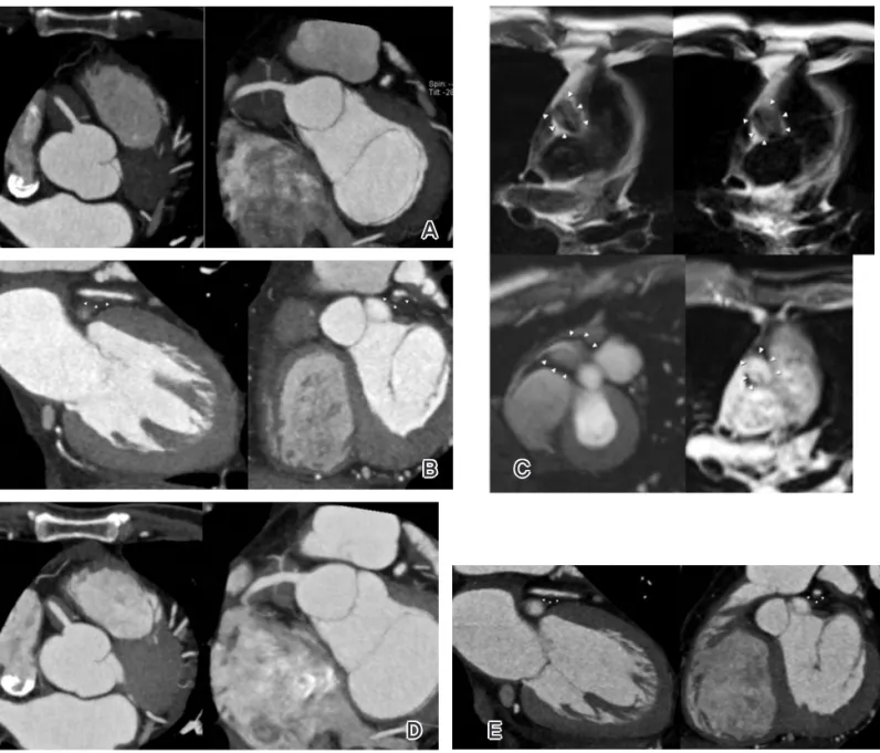
Figure 1. A 54-year-old man with presumed IgG4-related coronary periarteritis.
A & B. Initial coronary CTA reveals a soft tissue mass in the right AV groove, surrounding the proximal RCA (A), and focal perivascular soft tissue density or eccentric wall thickening with positive remodelling in the left main coronary artery (B).
C. In cardiac MRI, a soft tissue mass in the right AV groove (arrowheads) shows iso- to slightly high signal intensity on T1 & T2 WI (upper images) and enhancement in the first-pass perfusion and delayed enhancement scans (lower images). D & E. Two months follow-up coronary CTA shows dramatically decreased size of the right AV groove mass (D) and focal periarterial soft tissue density or eccentric wall thickening in the left main coronary artery (E).
A
A
A
A
A
A
A
B
B
B
B
B
B
B
C
C
C
C
C
C
C
D
D
D
D
D
D
D
E
E
E
E
E
E
E
Jeong Jae Kim, Song Yi Kim, Sun Young Jeong, Seung Hyung Kim, Im Kyung Hwang
144
Recently, there have been several case reports of IgG4-related periarteritis presenting a tumorous lesion surrounding coronary arteries5,8,9). The radiological finding of our case is very similar to those reports. So we checked the serum IgG4 level in suspicion of IgG4-related periarteritis, and it was markedly elevated. According to the comprehensive diagnostic criteria for IgG4-RD by Umehara et al., a diagnosis is definitive in patients with: (i) organ enlargement, mass or nodular lesions, or organ dysfunction; (ii) a serum IgG4 concentration >135 mg/dl; and (iii) histopathological findings of >10 IgG4 cells/HPF and an IgG4+/IgG+ cell ratio >40% (10). Our case fulfills criteria (i) and (ii), but without pathologic examination. The criteria notes that patients having disease in organs difficult to biopsy and who respond to steroids, like our case, may possibly have IgG4-RD. Therefore, our case was clinically diagnosed with IgG4-RD.
Before steroid treatment, we thought multifocal wall thickening and stenoses in the left coronary artery as atherosclerotic change - soft plaques with positive remodelling. Because he was asymptomatic and the degree of stenosis was less than 50%, an invasive coronary angiogram was not performed. Instead, he was treated with steroids for the right AV groove mass. Two months later, not only the mass-like lesion surrounding the RCA but also multifocal wall thickening and stenoses in the left coronary artery were improved. So we could conclude that it is coronary periarteritis associated with IgG4-RD, not atherosclerotic plaques. This implies that IgG4-RD involving coronary arteries can mimick atherosclerotic wall thickening in addition to mass-like lesions.
Although many patients with IgG4-RD have lesions in several organs, either synchronously or metachronously, others show involvement of a single organ10). In the present case, we couldn’t find evidence of other organ involvement other than coronary artery by the disease.
Most of the previous coronary periarteritis cases were treated with surgery or intervention due to complications such as significant stenosis and aneurysm5,8,9). In our case, it was an incidentally detected lesion in a health check-up. He was asymptomatic and there was no evidence of complications such as significant stenosis and aneurysm. He was treated with steroids, and the periarterial lesions were
dramatically decreased. Histologically, IgG4-related disease is characterized by inflammatory cell infiltration and fibrotic or sclerotic change in affected organs1). It is well known that inflammation has a greater likelihood of reponse to corticosteroid therapy and a better prognosis than fibrosis11). Thus, responsiveness to steroid therapy might depend on the proportion of inflammation and fibrosis within the lesion. If IgG4-related coronary periarteritis is not treated, coronary event due to disease progression could be occurred. Fibrotic component within the lesion might be increased in the chronic phase, and that means irreversible change. Awareness of coronary periarteritis and treatment with steroids in inflammation-dominant early phase might prevent future coronary events.
In summary, IgG4-RD can involve coronary artery with surrounding soft tissue mass or atherosclerosis-mimicking lesion. Because it can be effectively treated with corticosteroids, awareness of this disease entity and appropriate diagnosis are important.
1) Masaki Y, Kurose N, Umehara H. IgG4-related disease: a novel lymphoproliferative disorder discovered and established in Japan in the 21st century. J Clin Exp Hematopathol 2011; 51(1):13-20
2) Khosroshahi A, Stone JH. A clinical overview of IgG4-related systemic disease. Curr Opin Rheumatol 2011;23:57-66
3) Van Moerkercke W, Verhamme M, Doubel P, Meeus G, Oyen R, Van Steenbergen W. Autoimmune pancreatitis and extrapancreatic manifestations of IgG4-related sclerosing disease. Acta Gastroenterol Belg 2010;73:239-46. 4) Inoue D, Zen Y, Abo H, Gabata T, Demachi H,
Yoshikawa J, et al. Immunoglobulin G4-related periaortitis and periarteritis: CT findings in 17 patients. Radiology 2011;261:625-33
5) Tanigawa J, Daimon M, Murai M, Katsumata T, Tsuji M, Ishizaka N. Immunoglobulin G4-related coronary periarteritis in a patient presenting with myocardial ischemia. Human pathology 2012;43:1131-4
6) Carbajal H, Waters L, Popovich J, Boniuk M, Chevez-Barrios P, Marcus D, et al. IgG4 related cardiac disease. Methodist DeBakey cardiovascular journal 2013;9(4):230-2 7) Song C, Koh MJ, Yoon YN, Joung B, Kim SH. IgG4-related sclerosing disease involving the superior vena cava and the atrial septum of the heart. Yonsei Med J
REFERENCES
DISCUSSION
Presumed IgG4-Related Disease Presented as Coronary Periarteritis: A Case Report Showing Dramatic Response to Steroid Therapy
145
2013;54(5):1285-8
8) Ikutomi M, Matsumura T, Iwata H, Nishimura G, Ishizaka N, Hirata Y, et al. Giant tumorous lesions surrounding the right coronary artery associated with immunoglobulin-G4-related systemic disease. Cardiology 2011;120(1):22-6
9) Matsumoto Y, Kasashima S, Kawashima A, Sasaki H, Endo M, Kawakami K, et al. A case of multiple immunoglobulin G4-related periarteritis: a tumorous lesion of the coronary artery and abdominal aortic
aneurysm. Human pathology 2008;39(6):975-80
10) Umehara H, Okazaki K, Masaki Y, Kawano M, Yamamoto M, Saeki T, et al. Comprehensive diagnostic criteria for IgG4-related disease (IgG4-RD), 2011. Mod Rheumatol 2012;22(1):21-30
11) Travis WD, Matsui K, Moss J, Ferrans VJ. Idiopathic nonspecific interstitial pneumonia: prognostic significance of cellular and fibrosing patterns: survival comparison with usual interstitial pneumonia and desquamative interstitial pneumonia. Am J Surg Pathol 2000;24:19-33
146
-Jeong Jae Kim, Song Yi Kim, Sun Young -Jeong, Seung Hyung Kim, Im Kyung Hwang