|
J
BD
Journal of Breast DiseaseClinicopathological Significance of SMAD4 Expression in Breast
Cancer
Jun-Seok Woo, M.D.
1, Min Sung Chung, M.D., Ph.D.
1, Seung Sam Paik, M.D., Ph.D.
21Departments of Surgery, 2Pathology, College of Medicine, Hanyang University, Seoul, Korea
Purpose: SMAD4 is a member of the SMAD family and acts as a central mediator of transforming growth factor beta signaling. Little is known about SMAD4 expression and its prognostic significance in breast cancer. We evaluated the clinicopathological and prognostic significance of SMAD4 expression in breast cancer. Methods: Two hundred and fifty-five patients with invasive ductal carcinoma of the breast from 2000 to 2008 were retrospectively analyzed. We investigated SMAD4 expression using a tissue microarray-based immunohistochemical assay and evaluated the association between SMAD4 and prognosis of breast cancer. Results: High SMAD4 expression was positively associated with early stage (p=0.009), estrogen receptor positivity (p=0.026), and human epidermal growth factor receptor 2 negativity (p=0.001). A significant difference in overall survival (OS) was associated with high SMAD4 expression in patients with T1 stage tumors (hazard ratio: 0.459, p=0.024). Conclusion: High SMAD4 expression was correlated with several favorable prognostic factors and was associated with fa-vorable OS in T1 stage breast cancer. SMAD4 in breast cancer has potential prognostic significance, and further investigations and under-standing about SMAD4 expression are needed.
Key Words: Breast neoplasms, Prognosis, SMAD4 protein, Transforming growth factor beta
INTRODUCTION
Breast cancer is among the most common malignancies and a lead-ing cause of death in women [1]. However, advances in surgical tech-niques and systemic therapies have improved the survival of breast cancer patients in recent decades [2]. Breast cancer is a heterogeneous disease with a variety of pathologic and molecular features. The prog-nosis of breast cancer and therapeutic decision-making are known to depend on classic immunohistochemistry (IHC) markers, namely es-trogen receptor (ER), progesterone receptor (PR), and human epider-mal growth factor receptor 2 (HER2). Recently, genomic assays have been used to provide information and to help in decision-making for adjuvant treatment [3]. Differential expression of various proteins has been researched to develop more efficient options for diagnosis and treatment.
SMAD4 is a major downstream mediator in the signaling of the transforming growth factor beta (TGF-β) pathway. The TGF-β
signal-ing process is initiated when TGF-β binds to the homodimers of the TGF-β type II receptor (TβRII) on the cell surface. TβRII recruits and activates the TGF-β type I receptor (TβRI). The activated TβRI phos-phorylates SMAD2 or SMAD3, which heterodimerizes with SMAD4. These complexes translocate into the nucleus where they bind to DNA and regulate TGF-β dependent gene expression [4]. TGF-β is crucial in supporting tissue homeostasis through its ability to regulate cell pro-liferation, differentiation, migration, apoptosis, and development. The loss of SMAD4 expression results in the deprivation of a major factor of cell growth inhibition, contributing to carcinogenesis. SMAD4 has been previously identified as a possible tumor suppressor since SMAD4 mutations have been reported with high frequency in solid tumors [5,6]. The SMAD4 expression level has been reported to cor-relate with prognosis in many types of cancers, including colon [7], pancreatic [8], and esophageal [9] cancers. However, very few studies on SMAD4 expression and prognosis in breast cancer have been per-formed, and the results have been conflicting [10-13]. In the present study, we investigated SMAD4 expression using a tissue microarray (TMA)-based IHC assay and evaluated its clinicopathological and prognostic significance in breast cancer.
J Breast Dis 2019 December; 7(2): 52-58 https://doi.org/10.14449/jbd.2019.7.2.52
Correspondence: Min Sung Chung, M.D., Ph.D.
Department of Surgery, Hanyang University Medical Center, 222-1 Wangsimni-ro, Seongdong-gu, Seoul 04763, Korea
Tel: +82-2-2290-8460, Fax:+82-2-2281-0224, E-mail: [email protected] Received: Aug 5, 2019 Revised: Sep 25, 2019 Accepted: Oct 23, 2019
METHODS
Patients and tissue samples
Four hundred and seventy-one patients treated for invasive ductal carcinoma (IDC) of breast at Hanyang University Medical Center be-tween December 2000 and December 2008 were considered. The present study was approved by the Institutional Review Board of Ha-nyang University Medical Center (No. 2019-08-029-004). Patients with incomplete clinical data sets, patients without sufficient archived tissues, and patients with stage IV cancers were excluded. Two hun-dred and fifty-five patients were selected for the study after exclusions. We examined patients’ age at diagnosis, tumor size, lymph node sta-tus, and pathological findings according to the American Joint Com-mittee on Cancer (AJCC, 7th edition) classification for stage, tumor size, lymph node metastasis stage, and hormone receptor status. The hormone receptor (ER and PR) statuses were assessed using IHC and were scored using the Allred score [14]. Patients with a score >1 were defined as ER/PR positive. Testing for HER2 was performed using ei-ther IHC staining or fluorescent in situ hybridization (FISH). HER2 expression was classified into levels 0 to 3+ according to the guidelines by American Society of Clinical Oncology/College of American Pa-thologists [15]. A score of 3+ was given to specimens showing uniform and intense membrane staining in >30% of invasive tumor cells, and these specimens were rated as HER2 positive. The cases with a score of 2+ that were equivocal for HER2 protein expression were evaluated by FISH analysis to measure HER2 amplification using an original paraffin block.
TMA construction
Slides stained with hematoxylin and eosin were used to define the most morphologically representative, well fixed, and non-necrotic ar-eas. Single tissue cores (2 mm in diameter) were sampled from each paraffin block and were assembled into a recipient paraffin block us-ing a TMA instrument (AccuMax Array, ISU ABXIS, Seoul, Korea).
IHC staining and interpretation of SMAD4 expression
Multiple 4-µm sections were cut using Leica microtome (Leica Bio-systems, Wetzlar, Germany) and transferred to adhesive-coated slides. One section was routinely deparaffinized with standard xylene and hydrated using graded ethanol in water. It was stained with
hematox-ylin and eosin and covered with a coverslip. For IHC, the TMA slides were dewaxed by heating at 55°C for 30 minutes and by three 5-min-ute xylene washes.
Primary monoclonal mouse anti-Smad4 antibody (Santa Cruz Biotechnology, Santa Cruz, USA) was diluted (1:200) in goat serum and incubated at room temperature for 1 hour. After three 2-minute washes with phosphate-buffered saline, the sections were incubated with a biotinylated goat anti-mouse secondary antibody for 30 min-utes (DAKO, Carpinteria, USA). The slides were then dehydrated fol-lowing standard procedure and sealed with coverslips. Negative con-trols were performed by omitting Smad4 antibody during the prima-ry antibody incubation.
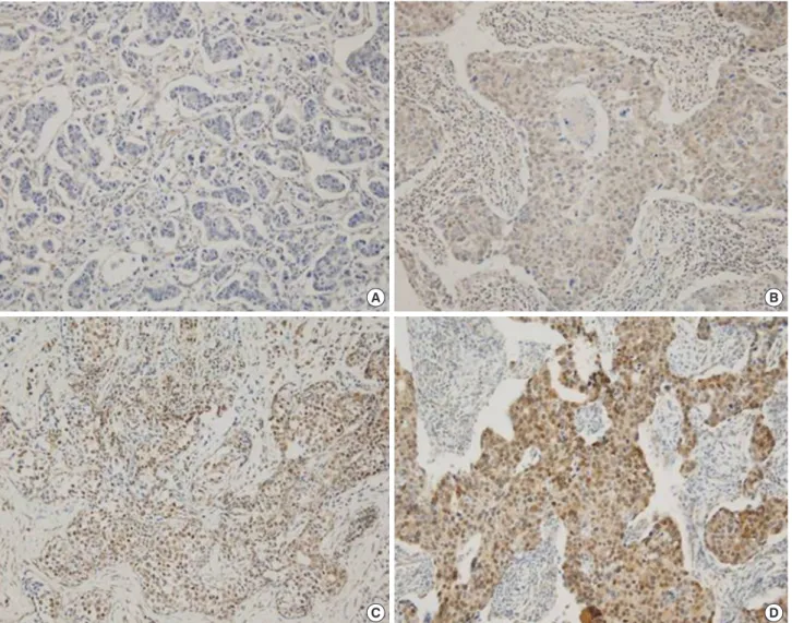
To analyze SMAD4 expression in breast cancer, we used the IHC assay with the application of immunoreactivity score (IRS) criteria [16]. The extensional standards were the “fraction of positive cells” (0: <5%, 1: 6%−25%, 2: 26%−50%, 3: 51%−75%, and 4: >75%) and the “staining intensity score” (0: colorless, 1: pallide-flavens, 2: yellow, and 3: brown). The IRS was calculated by multiplying the “staining inten-sity score” and the “fraction of positive cells” [17]. The staining score was stratified as absent (−, score 0), weak positive (+, score 1−4), mod-erate positive (++, score 5−8), and strong positive (+++, score 9−12) (Figure 1). To evaluate SMAD4 expression, specimens with absent and weak positive (0 ≤ IRS ≤4) scores were classified into the low SMAD4 expression (SMAD4 low) group. Specimens with moderate and strong positive scores (5 ≤ IRS ≤12) were classified into the high SMAD4 expression (SMAD4 high) group (Figure 1). The interpreta-tion of IHC staining in this study was performed by a single patholo-gist (SS Paik).
Statistical analysis
Fisher’s exact test, chi-squared test, and logistic regression analysis were used to evaluate the correlations between the clinicopathological features and SMAD4 expression. In the survival analyses, the plots were generated using the Kaplan-Meier curve and were compared us-ing the log-rank test. A multivariate analysis was performed to identi-fy the independent prognostic markers for disease-free survival (DFS) and overall survival (OS) using the Cox multistep regression model. Statistical analyses were performed using SAS version 9.4 (SAS Insti-tute Inc., Cary, USA) and R package version 3.5.1 (RStudio, Boston, USA).
RESULTS
The median follow-up period was 66.3 months (range 3.9 to 134.2 months) and the median age was 49 years (range 27 to 79 years). Among the 255 cases, 239 (93.7%) patients had primary tumor size of less than 2 cm and 16 (6.3%) patients had primary tumor size of more than 2 cm. Regional lymph node metastasis was present in 131 cases (51.4%). According to the AJCC classification scheme, 77 (30.2%) pa-tients had stage I cancer, while 178 (69.8%) had stage II and stage III cancer. ER expression was positive in 142 cases (55.7%) and PR expres-sion was positive in 134 cases (52.5%). Out of all the cases, 67 (26.3%) were positive for HER2 expression on IHC analysis and/or positive for HER2 gene amplification detected by the FISH analysis. One
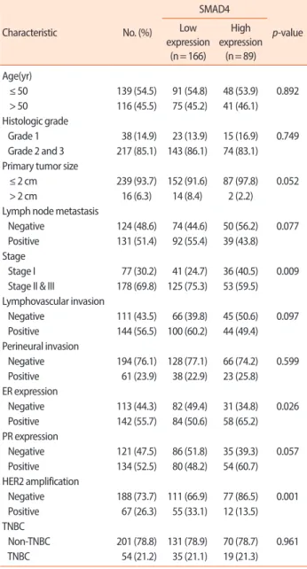
hun-dred sixty-six specimens (65.1%) were categorized into the SMAD4 low group and 89 specimens (34.9%) were categorized into the SMAD4 high group (Table 1, Figure 1).
Relationship between SMAD4 expression and clinicopathological parameters
High SMAD4 expression was positively associated with good clini-cal phenotypes of breast cancer such as low early AJCC stage (p = 0.009), ER positivity (p = 0.026), or HER2 negativity (p = 0.001). No significant correlation was detected between SMAD4 expression and the other clinicopathological parameters such as histological grade, age, lymph node metastasis, PR status, and triple-negative breast can-cer (TNBC) (Table 1).
Figure 1. Representative sections of the immunohistochemistry of SMAD4 in breast cancer tissue (magnification, × 200). (A) Negative SMAD4 staining. (B) Weak SMAD4 staining. (C) Intermediate SMAD4 staining. (D) Strong SMAD4 staining.
A
C
B
Comparison between survival outcome and SMAD4 expression
During the follow-up period, there were 64 (25.1%) recurrences among the 255 patients. Out of these, 47 cases were SMAD4 low and 17 were SMAD4 high (73.4% and 26.6%, respectively). In overall sur-vival, 55 events (21.6%) occurred. Out of these, 42 patients were SMAD4 low and 13 were SMAD4 high (76.4% and 23.6%, respective-ly). No difference was observed in DFS between the SMAD4 low group and the SMAD4 high group (p = 0.115), but the SMAD4 high
Table 1. Association between SMAD4 expression and clinicopathological
parameters in breast cancer patients (n=255)
Characteristic No. (%) SMAD4 p-value Low expression (n= 166) High expression (n= 89) Age(yr) ≤ 50 139 (54.5) 91 (54.8) 48 (53.9) 0.892 > 50 116 (45.5) 75 (45.2) 41 (46.1) Histologic grade Grade 1 38 (14.9) 23 (13.9) 15 (16.9) 0.749 Grade 2 and 3 217 (85.1) 143 (86.1) 74 (83.1) Primary tumor size
≤ 2 cm 239 (93.7) 152 (91.6) 87 (97.8) 0.052 > 2 cm 16 (6.3) 14 (8.4) 2 (2.2)
Lymph node metastasis
Negative 124 (48.6) 74 (44.6) 50 (56.2) 0.077 Positive 131 (51.4) 92 (55.4) 39 (43.8) Stage
Stage I 77 (30.2) 41 (24.7) 36 (40.5) 0.009 Stage II & III 178 (69.8) 125 (75.3) 53 (59.5) Lymphovascular invasion Negative 111 (43.5) 66 (39.8) 45 (50.6) 0.097 Positive 144 (56.5) 100 (60.2) 44 (49.4) Perineural invasion Negative 194 (76.1) 128 (77.1) 66 (74.2) 0.599 Positive 61 (23.9) 38 (22.9) 23 (25.8) ER expression Negative 113 (44.3) 82 (49.4) 31 (34.8) 0.026 Positive 142 (55.7) 84 (50.6) 58 (65.2) PR expression Negative 121 (47.5) 86 (51.8) 35 (39.3) 0.057 Positive 134 (52.5) 80 (48.2) 54 (60.7) HER2 amplification Negative 188 (73.7) 111 (66.9) 77 (86.5) 0.001 Positive 67 (26.3) 55 (33.1) 12 (13.5) TNBC Non-TNBC 201 (78.8) 131 (78.9) 70 (78.7) 0.961 TNBC 54 (21.2) 35 (21.1) 19 (21.3)
ER= estrogen receptor; PR= progesterone receptor; HER2= human epider-mal growth factor receptor 2; TNBC= triple negative breast cancer.
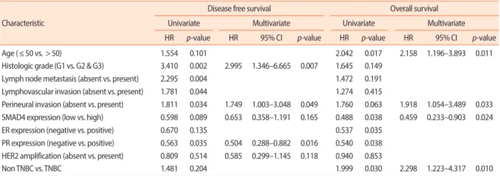
group showed significantly better OS (p= 0.045) than the SMAD4 low group (Figure 2A, 2B). In the subgroup analysis, high SMAD4 expres-sion was correlated with better OS in T1 stage tumor (size ≤2 cm) (p= 0.034). However, it did not show significant correlation with DFS (Figure 2C, 2D). In the multivariate analysis, a significant association was observed between OS and SMAD4. High SMAD4 expression was observed to be an independent prognostic factor for better OS among patients with T1 stage tumor in the multivariate analysis (haz-ard ratio: 0.459, p= 0.024) (Table 2).
DISCUSSION
SMAD4 is a key mediator of the TGF-β pathway. In the present study, the expression of SMAD4 was investigated using IHC analysis in 255 cases of IDC. The present data showed that high SMAD4 ex-pression was associated with favorable clinicopathological parameters (stage, ER positivity, and HER2 negativity) and OS. There was a sig-nificant association between SMAD4 expression and OS in patients with T1 stage tumors (tumor size ≤ 2 cm).
SMAD4 expression was found to be lower in breast tumor cells when compared with normal breast epithelium [11]. Two previous re-ports applied the IRS criteria with respect to the expression of SMAD4 in breast cancer. Lui et al. [13] reported that 43.1% of breast cancer cas-es were SMAD4 low (0 ≤ IRS ≤4) and 56.9% were SMAD4 high (5 ≤ IRS ≤ 2 ), whereas Stuelten et al. [11] reported the figures of 36.3% and 64%, respectively. Our results are closely analogous to those of these studies, with 34.9% SMAD4 low cases and 65.1% SMAD4 high cases. There are inconsistent reports on the correlation between SMAD4 expression and prognostic markers in breast cancer. In previous stud-ies, SMAD4 showed no significant correlation with tumor size, me-tastases, nodal status, histological grade, histological type, or estrogen receptor expression [11]. However, Lui et al. [13] reported that SMAD4 expression was negatively associated with histological grade.
The role of SMAD4 as a tumor suppressor is consistent with the observation that high expression of this protein is associated with a fa-vorable prognosis. Recent studies have indicated that inactivation of SMAD4 is related to progression of disease in various cancers [18-21]. In colon cancer, SMAD4 inactivation promotes malignancy and drug resistance [22]. In pancreatic cancer, low expression of SMAD4 is as-sociated with malignant progression [23]. Similarly, in non-small-cell
lung carcinoma, SMAD4 expression was higher in the normal bron-cho-tracheal epithelium, but lower in the tumor tissues and closely correlated with lymph node metastasis [24]. Thus, low expression of
SMAD4 shows an unfavorable outcome in other types of cancers. Our study demonstrated similar results in breast cancer.
Very few studies have reported the correlation between SMAD4
1.0 0.9 0.8 0.7 0.6 0.5 1.0 0.9 0.8 0.7 0.6 0.5 1.0 0.9 0.8 0.7 0.6 0.5 1.0 0.9 0.8 0.7 0.6 0.5 50 100 150 200 50 100 150 200 50 100 150 200 50 100 150 200 Disease -fr ee sur viv al Disease -fr ee sur viv al O ver all sur viv al O ver all sur viv al Time (mo) Time (mo) Time (mo) Time (mo) High High High High Low Low Low Low p = 0.115 p = 0.085 p = 0.045 p = 0.034
Figure 2. Kaplan-Meier curves for disease-free survival (DFS) and overall survival (OS) for breast cancer according to SMAD4 expression. (A) All patients –DFS. (B) All patient – OS. (C) T1 staged tumor (≤2 cm) – DFS. (D) T1 staged tumor (≤2 cm) – OS.
A
C
B
D
Table 2. Univariate and multivariate analysis of various prognostic parameters for survival in cancer patients with primary tumor size ≤ 2 cm Characteristic
Disease free survival Overall survival
Univariate Multivariate Univariate Multivariate
HR p-value HR 95% CI p-value HR p-value HR 95% CI p-value
Age (≤ 50 vs. > 50) 1.554 0.101 2.042 0.017 2.158 1.196–3.893 0.011
Histologic grade (G1 vs. G2 & G3) 3.410 0.002 2.995 1.346–6.665 0.007 1.645 0.149
Lymph node metastasis (absent vs. present) 2.295 0.004 1.472 0.191
Lymphovascular invasion (absent vs. present) 1.781 0.044 1.274 0.415
Perineural invasion (absent vs. present) 1.811 0.034 1.749 1.003–3.048 0.049 1.760 0.063 1.918 1.054–3.489 0.033 SMAD4 expression (low vs. high) 0.598 0.089 0.653 0.358–1.191 0.165 0.488 0.038 0.459 0.233–0.903 0.024
ER expression (negative vs. positive) 0.670 0.135 0.537 0.035
PR expression (negative vs. positive) 0.563 0.035 0.504 0.288–0.882 0.016 0.540 0.038 HER2 amplification (absent vs. present) 0.809 0.514 0.585 0.299–1.145 0.118 0.940 0.853
Non TNBC vs. TNBC 1.481 0.204 1.999 0.030 2.298 1.223–4.317 0.010
HR= hazard ratio; CI= confidence interval;ER= estrogen receptor; PR= progesterone receptor; HER2= human epidermal growth factor receptor 2; TNBC= triple negative breast cancer.
and prognosis in breast cancer. Lui et al. [13] reported that SMAD4 expression appeared to be decreased in breast cancer when compared with normal tissues and reduced SMAD4 expression levels tended to exhibit more poorly differentiated tumors, a higher risk of recurrence, and shorter OS. Kruijt et al. [12] also reported that low expression of SMAD4 had an unfavorable prognosis regarding progression-free survival. These results are in agreement with the present study. How-ever, one study suggested that a SMAD4-negative tumor showed marginally better overall 5-year survival than a SMAD4-positive tu-mor, though this difference was not statistically significant [11]. An-other study demonstrated that loss of SMAD4 was correlated with a decrease in axillary lymph node metastasis [25]. Since there are con-flicting reports in terms of prognosis, recent studies have attempted to determine whether SMAD4 as a prognostic factor combines with other genes or protein expressions [10,12].
TGF-β is considered to play a dual role in cancer development as it displays both tumorigenic and tumor-suppressive effects. TGF-β has been reported to act as a tumor suppressor by inhibiting the cell pro-liferation of breast cancer cell lines in early stage [26,27]. In contrast, in later stages of cancer, TGF-β has direct pro-tumorigenic effects through the stimulation of invasion, the migration of tumor cells, and the activation of the tumor stroma [28,29]. Positive association of high expression of SMAD4 in T1 stage early breast cancer with better sur-vival outcome in the present study could be explained by the fact that SMAD4 is a key mediator of TGF-β pathway and TGF-β acts as a tu-mor suppressor in the early stage.
The present study has several limitations. No stage IV patients were included in the TMA data. The IHC analysis using TMA may not re-flect the intratumoral heterogeneity. Moreover, the proportion of small-sized tumors was unintentionally higher than the large-sized tumors in our data. However, our results could provide data for SMAD4 expression and prognosis in the early stage breast cancer.
In conclusion, high SMAD4 expression was correlated with several favorable prognostic factors including tumor size, regional lymph node metastasis, AJCC stage, ER positivity, and HER2 negativity. Ad-ditionally, high SMAD4 expression was associated with favorable OS in T1 stage breast cancer. Therefore, SMAD4 in breast cancer has po-tential prognostic significance and further investigation and under-standing are needed to elucidate it.
CONFLICT OF INTEREST
The authors declare that they have no competing interests.
REFERENCES
1. Siegel RL, Miller KD, Fedewa SA, Ahnen DJ, Meester RGS, Barzi A, et al. Colorectal cancer statistics, 2017. CA Cancer J Clin 2017;67:177-93.
2. Guo F, Kuo YF, Shih YCT, Giordano SH, Berenson AB. Trends in breast cancer mortality by stage at diagnosis among young women in the United States. Cancer 2018;124:3500-9.
3. Fayanju OM, Park KU, Lucci A. Molecular genomic testing for breast cancer: utility for surgeons. Ann Surg Oncol 2018;25:512-9. 4. Heldin CH, Miyazono K, ten Dijke P. TGF-beta signalling from cell
membrane to nucleus through SMAD proteins. Nature 1997;390: 465-71.
5. Miyaki M, Iijima T, Konishi M, Sakai K, Ishii A, Yasuno M, et al. Higher frequency of Smad4 gene mutation in human colorectal cancer with distant metastasis. Oncogene 1999;18:3098-103. 6. Shi Y, Hata A, Lo RS, Massague J, Pavletich NP. A structural basis for
mutational inactivation of the tumour suppressor Smad4. Nature 1997;388:87-93.
7. Yan P, Klingbiel D, Saridaki Z, Ceppa P, Curto M, McKee TA, et al. Reduced expression of SMAD4 is associated with poor survival in colon cancer. Clin Cancer Res 2016;22:3037-47.
8. Singh P, Srinivasan R, Wig JD. SMAD4 genetic alterations predict a worse prognosis in patients with pancreatic ductal adenocarcino-ma. Pancreas 2012;41:541-6.
9. Singhi AD, Foxwell TJ, Nason K, Cressman KL, McGrath KM, Sun W, et al. Smad4 loss in esophageal adenocarcinoma is associated with an increased propensity for disease recurrence and poor sur-vival. Am J Surg Pathol 2015;39:487-95.
10. Min KW, Kim DH, Do SI, Chae SW, Kim K, Sohn JH, et al. Expres-sion pattern of Smad4/GATA3 as a predictor of survival in invasive ductal carcinoma of the breast. Pathobiology 2017;84:130-8. 11. Stuelten CH, Buck MB, Dippon J, Roberts AB, Fritz P, Knabbe C.
Smad4-expression is decreased in breast cancer tissues: a retrospec-tive study. BMC Cancer 2006;6:25.
JR, et al. The prognostic role of TGF-beta signaling pathway in breast cancer patients. Ann Oncol 2013;24:384-90.
13. Liu N, Yu C, Shi Y, Jiang J, Liu Y. SMAD4 expression in breast ductal carcinoma correlates with prognosis. Oncol Lett 2015;10:1709-15. 14. Harvey JM, Clark GM, Osborne CK, Allred DC. Estrogen receptor
status by immunohistochemistry is superior to the ligand-binding assay for predicting response to adjuvant endocrine therapy in breast cancer. J Clin Oncol 1999;17:1474-81.
15. Wolff AC, Hammond ME, Schwartz JN, Hagerty KL, Allred DC, Cote RJ, et al. American Society of Clinical Oncology/College of american pathologists guideline recommendations for human epi-dermal growth factor receptor 2 testing in breast cancer. Arch Pathol Lab Med 2007;131:18-43.
16. Brown RS, Wahl RL. Overexpression of Glut-1 glucose transporter in human breast cancer. An immunohistochemical study. Cancer 1993;72:2979-85.
17. Remmele W, Stegner HE. Recommendation for uniform definition of an immunoreactive score (IRS) for immunohistochemical estro-gen receptor detection (ER-ICA) in breast cancer tissue. Pathologe 1987;8:138-40.
18. Wilentz RE, Su GH, Dai JL, Sparks AB, Argani P, Sohn TA, et al. Im-munohistochemical labeling for dpc4 mirrors genetic status in pan-creatic adenocarcinomas : a new marker of DPC4 inactivation. Am J Pathol 2000;156:37-43.
19. Wilentz RE, Iacobuzio-Donahue CA, Argani P, McCarthy DM, Parsons JL, Yeo CJ, et al. Loss of expression of Dpc4 in pancreatic in-traepithelial neoplasia: evidence that DPC4 inactivation occurs late in neoplastic progression. Cancer Res 2000;60:2002-6.
20. Natsugoe S, Xiangming C, Matsumoto M, Okumura H, Nakashi-ma S, Sakita H, et al. SNakashi-mad4 and transforming growth factor beta1 expression in patients with squamous cell carcinoma of the esopha-gus. Clin Cancer Res 2002;8:1838-42.
21. Cardillo MR, Lazzereschi D, Gandini O, Di Silverio F, Colletta G. Transforming growth factor-beta pathway in human renal cell
car-cinoma and surrounding normal-appearing renal parenchyma. Anal Quant Cytol Histol 2001;23:109-17.
22. Papageorgis P, Cheng K, Ozturk S, Gong Y, Lambert AW, Abdol-maleky HM, et al. Smad4 inactivation promotes malignancy and drug resistance of colon cancer. Cancer Res 2011;71:998-1008. 23. Blackford A, Serrano OK, Wolfgang CL, Parmigiani G, Jones S,
Zhang X, et al. SMAD4 gene mutations are associated with poor prognosis in pancreatic cancer. Clin Cancer Res 2009;15:4674-9. 24. Ke Z, Zhang X, Ma L, Wang L. Deleted in pancreatic carcinoma
lo-cus 4/Smad4 participates in the regulation of apoptosis by affecting the Bcl-2/Bax balance in non-small cell lung cancer. Hum Pathol 2008;39:1438-45.
25. Xie W, Mertens JC, Reiss DJ, Rimm DL, Camp RL, Haffty BG, et al. Alterations of Smad signaling in human breast carcinoma are asso-ciated with poor outcome: a tissue microarray study. Cancer Res 2002;62:497-505.
26. Zugmaier G, Ennis BW, Deschauer B, Katz D, Knabbe C, Wilding G, et al. Transforming growth factors type beta 1 and beta 2 are equi-potent growth inhibitors of human breast cancer cell lines. J Cell Physiol 1989;141:353-61.
27. Gobbi H, Dupont WD, Simpson JF, Plummer WD Jr, Schuyler PA, Olson SJ, et al. Transforming growth factor-β and breast cancer risk in women with mammary epithelial hyperplasia. J Natl Cancer Inst 1999;91:2096-101.
28. Wiercinska E, Naber HP, Pardali E, van der Pluijm G, van Dam H, ten Dijke P. The TGF-beta/Smad pathway induces breast cancer cell invasion through the up-regulation of matrix metalloproteinase 2 and 9 in a spheroid invasion model system. Breast Cancer Res Treat 2011;128:657-66.
29. Ronnov-Jessen L, Petersen OW. Induction of alpha-smooth muscle actin by transforming growth factor-beta 1 in quiescent human breast gland fibroblasts. Implications for myofibroblast generation in breast neoplasia. Lab Invest 1993;68:696-707.