Tau Positron Emission
Tomography Imaging
in Degenerative
Parkinsonisms
Chul Hyoung Lyoo,1 Hanna Cho,1 Jae Yong Choi,2,3
Young Hoon Ryu,2 Myung Sik Lee1
1Departments of Neurology and 2Nuclear Medicine, Gangnam Severance Hospital,
Yonsei University College of Medicine, Seoul, Korea
3Division of RI-Convergence Research, Korea Institute Radiological and
Medical Sciences, Seoul, Korea
Received: October 16, 2017 Accepted: November 20, 2017
Corresponding author: Chul Hyoung Lyoo, MD, PhD, Department of Neurology, Gang-nam Severance Hospital, Yonsei University College of Medicine, Research Center for Future Medicine, 20 Eonju-ro 63-gil, Gangnam-gu, Seoul 06229, Korea
Tel: +82-2-2019-3326 Fax: +82-2-3462-5904 E-mail: [email protected]
ABSTRACT
In recent years, several radiotracers that selective-ly bind to pathological tau proteins have been de-veloped. Evidence is emerging that binding patterns of in vivo tau positron emission tomography (PET) studies in Alzheimer’s disease (AD) patients close-ly resemble the distribution patterns of known neu-rofibrillary tangle pathology, with the extent of tracer binding reflecting the clinical and pathologi-cal progression of AD. In Lewy body diseases (LBD), tau PET imaging has clearly revealed corti-cal tau burden with a distribution pattern distinct from AD and increased cortical binding within the LBD spectrum. In progressive supranuclear palsy, the globus pallidus and midbrain have shown in-creased binding most prominently. Tau PET pat-terns in patients with corticobasal syndrome are characterized by asymmetrical uptake in the motor cortex and underlying white matter, as well as in the basal ganglia. Even in the patients with multi-ple system atrophy, which is basically a synucle-inopathy, 18F-flortaucipir, a widely used tau PET
tracer, also binds to the atrophic posterior puta-men, possibly due to off-target binding. These dis-tinct patterns of tau-selective radiotracer binding in the various degenerative parkinsonisms suggest its utility as a potential imaging biomarker for the differential diagnosis of parkinsonisms.
Key Words
Tau; positron emission tomography; parkinsonism.
https://doi.org/10.14802/jmd.17071 / J Mov Disord 2018;11(1):1-12 pISSN 2005-940X / eISSN 2093-4939
cc This is an Open Access article distributed under the terms of the Creative Commons
Attri-bution Non-Commercial License (http://creativecommons.org/licenses/by-nc/4.0) which per-mits unrestricted non-commercial use, distribution, and reproduction in any medium, provided the original work is properly cited.
J Mov Disord 2018;11(1):1-12
JMD
INTRODUCTION
11C-Pittsburgh compound B (11C-PIB) is a
radio-tracer that selectively binds to amyloid-β (Aβ) in se-nile plaques, which are a pathological hallmark of Alzheimer’s disease (AD). This radiotracer has en-abled a new era of pathology-targeted molecular imaging of neurodegenerative diseases. The recent development of 18F-labelled radiotracers that are
se-lective for Aβ, including 18F-flutemetamol, 18
F-flor-betapir, 18F-florbetaben, and 18F-NAV4694
(former-ly 18F-AZD4694), has also facilitated the application
of Aβ-imaging for clinical use.1-4 Positron emission
tomography (PET) using these Aβ-selective radio-tracers clearly mirrors the extent of Aβ accumulation in the brain,5,6 thereby enabling an earlier diagnosis
of prodromal AD.7,8 However, because neocortical
Aβ pathology generally plateaus at an early stage of AD,9 Aβ-imaging is less effective in delineating the
progression of AD.10
Paired helical filaments (PHF) of hyperphosphor-ylated tau protein are a major constituent of neuro-fibrillary tangles (NFT), the second major patho-logical hallmark of AD.11 NFTs first appear in the
transentorhinal region, spreading hierarchically to the neighboring limbic areas and distant associa-tion neocortices before finally reaching the primary
cortices.11 Because the distant propagation of tau
pa-thology is preceded by an early and widespread dissemination of Aβ pathology in the neocortex,9,12
cortical tau burden is a better indicator of the clini-cal progression of AD.13,14 In addition, in contrast to
the limited number of Aβ-related diseases, the exis-tence of a wider clinical spectrum of tauopathies has necessitated the development of molecular imaging biomarkers for tau protein.15
The development of the first tau-selective radio-tracer, 18F-THK523, in 2011 was another major
break-through.16 Although this PET radiotracer is currently
no longer used for clinical research due to serious drawbacks that occurred in human studies,17 it
en-couraged the development of better tau PET radio-tracers that are now used in clinical research (Fig-ure 1).
Over recent years, clinical tau PET studies have primarily focused on the AD spectrum. Tau PET al-lows clear visualization of AD tau pathology with a high selectivity for PHF-tau18,19 and is now
general-ly accepted as a useful imaging biomarker for assess-ing the pathological and clinical progression of AD.20-22 In contrast to AD, postmortem
autoradiog-raphy and a smaller number of in vivo tau PET stud-ies in non-AD tauopathstud-ies have consistently report-ed weaker radiotracer binding to non-AD tau than
to PHF-tau in AD.18,19,23-32
In this review, we focus on recent progress in the knowledge of tau-selective tracers and clinical tau PET studies in degenerative parkinsonisms, such as Parkinson’s disease (PD), dementia with Lewy bod-ies (DLB), progressive supranuclear palsy (PSP), cor-ticobasal syndrome (CBS), and multiple system at-rophy (MSA).
CHARACTERISTICS OF TAU-
SELECTIVE RADIOTRACERS
18
F-THK series (
18F-THK-523,
18F-THK-5105,
18F-THK-5317, and
18F-THK-5351)
The first tau-selective radiotracer, 18F-THK523,
ex-hibited a 10-fold stronger binding affinity to patho-logical tau protein than to Aβ fibrils in vitro, selective binding to PHF-tau pathology in autoradiography studies with postmortem AD tissue, and stronger uptake in the brains of tau transgenic mice when compared to the wild-type or APP/PS1 transgenic mice.16 In contrast to these promising results,
subse-quent in vivo human PET studies with 18F-THK523
were quite disappointing due to high levels of white matter binding and low standardized uptake value ratio (SUVR) values, even in AD patients.17
Region-al differences in 18F-THK523 binding in AD were only
discernible with partial volume correction of the PET images. This regional difference was almost eliminated without the correction. Therefore, 18
F-THK523 has been deemed unsuitable for clinical tau PET imaging studies.17 To overcome these
is-sues, improvements to the 18F-THK series have
fo-cused on reducing white matter binding, and the second generation of the 18F-THK series, namely, 18F-THK-5117, 18F-THK-5317, and 18F-THK-5351,
have exhibited much lower white matter binding than their predecessor. The most recently devel-oped radiotracer in the 18F-THK series is 18F-THK-
5351, which has a higher affinity for PHF-tau and more rapid washout from white matter than the pre-vious version, 18F-THK-5117.33 For this reason, 18
F-THK-5351 PET achieves higher contrasts between true binding and background and a much lower de-gree of white matter binding than 18F-THK-5117
and is now considered a useful imaging biomarker for AD.
Even with these positive findings for 18F-THK-
5351, white matter binding is still a significant issue
in comparison to other tau-selective radiotracers.17
High white matter binding may mask small increas-es in 18F-THK-5351 binding in the gray matter due
to an overflow of PET signals from the adjacent white matter. Likewise, 18F-THK-5351 PET still
ex-hibits elevated binding in the pons. This may affect accurate determination of tau pathology in the brain-stem. Additionally, similar to 18F-flortaucipir,
off-target binding to the basal ganglia, even in healthy elderly individuals, is another common issue for the
18F-THK series.
A recent 18F-THK-5351 PET study reported
seri-ous problems relating to monoamine oxidase-B (MAO-B) binding.34 MAO-B is widely expressed in
the brain, most prominently in the basal ganglia, followed by the insular cortex.35 This topographical
pattern was replicated in several in vivo PET stud-ies.36-38 In healthy controls and patients with mild
cognitive impairment, AD, and PSP, one study con-ducted three 18F-THK-5351 PET scans acquired
be-fore and after 10 mg of the MAO-B inhibitor, selegi-line, was orally administered and again at 9–28 days after the selegiline treatment.34 Surprisingly, a single
oral dose of selegiline dramatically reduced 18F-THK-
5351 standardized uptake values by 37–52% across all regions, most prominently in the thalamus (52%) and basal ganglia (51%), and even in the cerebellar cortex (42%), which is generally used as a reference tissue. This suppressive effect was sustained until the third PET scan.34 Therefore, the MAO-B binding
characteristics of 18F-THK-5351 may limit its
applica-bility in tau imaging.
18
F-flortaucipir (formerly referred to as
18F-AV-1451 or
18F-T807)
18F-flortaucipir has exhibited a 25-fold greater
bind-ing affinity to PHF-tau than to Aβ, and very low white matter binding in several in vivo human PET studies.39 As a result, 18F-flortaucipir PET enables
high contrasts between binding and background, which are helpful for detecting small increases in cortical binding. Unlike the similar radiotracer, 18
F-T808, which shows a high skull uptake in some sub-jects due to serious defluorination,4018F-flortaucipir
does not exhibit defluorination issues in human.39,41
Due to these positive findings, 18F-flortaucipir has
been most widely used for clinical tau imaging studies.
J Mov Disord 2018;11(1):1-12
JMD
have consistently reported a stronger binding affin-ity of 18F-flortaucipir to PHF-tau in AD, in contrast
to its much weaker binding affinity to straight fila-ment tau in non-AD tauopathies.18,19 Therefore, 18
F-flortaucipir is better for tau imaging studies in AD rather than in various other non-AD tauopathies.
However, there are two significant issues with 18
F-flortaucipir. First, unlike the other types of tau-se-lective radiotracers, which show stable SUVR val-ues after a certain time point, 18F-flortaucipir has
unstable kinetics, causing the SUVR values to steadi-ly increase even after 60 mins.41 This characteristic
can limit quantification attempts, especially in lon-gitudinal studies,42 although data acquired 80–100
mins post-injection can provide reliable SUVR val-ues that correlate with the binding valval-ues deter-mined by compartmental modeling.43,44 A second
problem is the widely reported off-target binding.
18F-flortaucipir also exhibits a high affinity for
mel-anin-producing cells, including the substantia nig-ra, skin epithelium, retinal pigment epithelium, and melanomas. It, therefore, binds strongly to the sub-stantia nigra, in which a high concentration of neu-romelanin exists.18,4518F-flortaucipir also strongly
binds to the basal ganglia, even in healthy elderly in-dividuals with an absence of tau pathology.18,45 One
study showed a possible interaction with iron due to a correlation between age-related increases in bas-al ganglibas-al iron content and 18F-flortaucipir binding
in the basal ganglia.46 Nigral and basal ganglial
off-target binding is problematic for tau imaging, espe-cially in parkinsonisms. The choroid plexus is anoth-er off-target binding site. Although one study found tangle-like structures that were immunoreactive to phosphorylated tau antibody in the epithelial cells in the choroid plexus,47 the exact mechanism of this
off-target binding remains unknown. Off-target bind-ing in the choroid plexus also disturbs the precise quantitation of underlying hippocampal binding and can be an obstacle to early detection of hippocam-pal tau burden.
18F-flortaucipir binds to MAO-A with a high
affin-ity,48 but unlike 18F-THK-5351, there have been no
reports to date of 18F-flortaucipir binding to MAO-B.
18
F-MK6240
18F-MK6240 is the most recently developed
tau-selective radiotracer, and, thus, there is little infor-mation about it. One autoradiographical PET study
in monkeys reported a 5-fold higher binding poten-tial of 3H-MK6240, no off-target binding, and no
MAO-A binding when compared to 3
H-flortaucip-ir.48,49 In a small number of healthy elderly subjects
and AD patients, 18F-MK6240 exhibited fast
wash-out, high binding to the cortical regions vulnerable to AD pathology, and a good correlation with the severity of cognitive impairment in AD.50 Larger
clinical PET studies are needed to better character-ize the 18F-MK6240 radiotracer.
11
C-PBB3
Unlike 18F-labelled compounds with longer
half-lives (109 mins), the shorter (20 mins) half-life of
11C-labelled compound permits two PET scans in
the same day. Therefore, 11C-labelled compounds
are suitable for research-based PET, while 18F-labelled
compounds are suitable for clinical PET scans. 11
C-PBB3 is the only 11C-labelled tau-selective
radiotrac-er that has an approximate 50-fold highradiotrac-er affinity for PHF-tau than that for Aβ.51 An in vivo 11C-PBB3 PET
study also exhibited high tracer binding to the corti-cal regions of AD, similar to other types of tau-selec-tive radiotracers.51 More importantly, PBB3 also has
a higher affinity for 4-repeat (4R) or 3R tau than 18
F-flortaucipir and is considered to be a tau PET tracer specific for a broader range of tau.52 However, 11
C-PBB3 is rapidly metabolized in plasma, and radio-active metabolites that enter into the brain can con-taminate PET signals. This problem makes 11C-PBB3
unsuitable for quantification.53,54 In addition, high
tracer retention in the venous sinus in all human sub-jects may contaminate PET signals around the ve-nous sinus.51
LEWY BODY DISEASES
PD with normal cognition (PDNC), PD with mild cognitive impairment (PDMCI), PD with dementia (PDD), and DLB all share common clinical charac-teristics and neuropathology and are now consid-ered to be part of the Lewy body diseases (LBD) spec-trum.55,56 In addition to the well-known α-synuclein
pathologies presenting as Lewy bodies and Lewy neurites, AD-type pathologies containing Aβ and PHF-tau are also found in LBD.57,58 Although the
prevalence of Aβ-positivity seen in the 11C-PIB PET
studies of LBD, can be highly variable,59-64 a clearly
Aβ-positivity within the LBD spectrum (5% in PDM-CI, 34% in PDD, and 68% in DLB) has been ob-served.65 Therefore, a similar increasing trend of
cortical binding in tau PET studies can be expected. All 18F-flortaucipir PET studies in PD patients to
date have consistently shown no increased binding in the basal ganglia or in the cerebral cortex.25,66-69
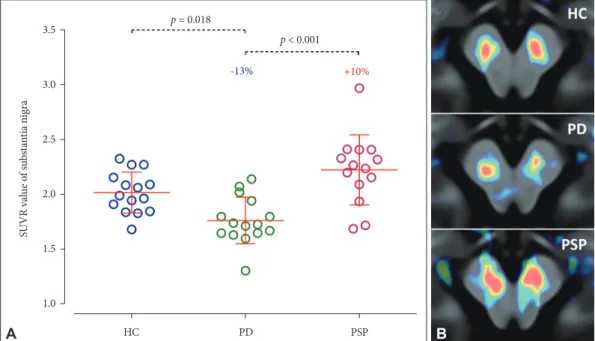
PD patients exhibited approximately 13% lower 18
F-flortaucipir binding in the substantia nigra com-pared to controls,25,66,67 due to off-target binding of 18F-flortaucipir to neuromelanin pigment, which
normally exists in the substantia nigra and is lost in PD (Figure 2).18,19 Reduced 18F-flortaucipir binding
was more prominent in the lateral part of the sub-stantia nigra than in the medial part. However, ni-gral 18F-flortaucipir binding did not correlate with
the motor severity of PD and did not reflect clinical asymmetry.25,66
DLB is positioned at the end of the LBD spectrum and can, therefore, be expected to exhibit the great-est cortical 18F-flortaucipir binding. The first 18
F-flor-taucipir PET study in a small number of patients within the LBD spectrum [7 DLB, 8 PD with cogni-tive impairment (PDCI), and 9 PDNC] showed an increasing trend of cortical 18F-flortaucipir binding.68 18F-flortaucipir binding was increased in the
inferi-or tempinferi-oral and precuneus cinferi-ortices in DLB and in
the same area in PDCI, with a lower level of statisti-cal significance. The binding in the inferior tempo-ral and precuneus cortices correlated with the sever-ity of cognitive impairment only in the composite group with DLB and PDCI.68
The second 18F-flortaucipir PET study involved
19 DLB and 19 AD patients.70 Compared to the
con-trols, the DLB patients showed greater binding in the posterior temporo-parietal and occipital corti-ces, in which 18F-flortaucipir binding correlated with
the global cortical 11C-PIB binding. Interestingly, the
medial temporal regions were relatively preserved in the DLB patients when compared to the AD pa-tients, and for this reason, medial temporal 18
F-flor-taucipir binding may be useful for differential diag-nosis between DLB and AD. However, they found no correlation between the 18F-flortaucipir binding
and the severities of cognitive impairment and par-kinsonian motor deficits.70
A recent 18F-flortaucipir PET study in a larger
num-ber of patients within the LBD spectrum (18 DLB, 22 PDCI, and 12 PDNC) showed a clearly increas-ing trend of cortical 18F-flortaucipir binding within
the LBD spectrum.69 In this report, 18F-flortaucipir
binding was dependent on Aβ-positivity, as deter-mined by 18F-florbetaben PET. Compared to the
controls, the Aβ-positive DLB group showed
signif-HC p = 0.018 p < 0.001 -13% +10% PD PSP SUVR va lue o f s ubs ta nt ia nig ra 3.5 3.0 2.5 2.0 1.5 1.0 A B
Figure 2. Different nigral 18F-flortaucipir binding in PD and PSP. A: Compared to the controls, 18F-flortaucipir SUVR
values in the substantia nigra were 13% lower in PD patients and 10% higher in PSP patients. B: A demonstration of different nigral 18F-flortaucipir binding in a control subject and patients with PD and PSP. HC: healthy controls,
J Mov Disord 2018;11(1):1-12
JMD
icantly increased binding in the sensorimotor, pri-mary visual, and parieto-temporal cortices, and the Aβ-positive PDCI group showed slightly increased binding in the middle and inferior temporal and parahippocampal cortices without surviving multi-ple comparisons. All Aβ-negative DLB, PDCI, and PDNC groups showed no increased binding in any of the cortical regions. In DLB, there was only a weak correlation between the severity of the cognitive im-pairment and binding in the prefrontal, sensorimo-tor, posterior cingulate, and occipital cortices.69
In summary, the cortical tau burden observed in the 18F-flortaucipir PET study increases within the
LBD spectrum (Figure 3). DLB patients exhibit the greatest tau burden, with distribution patterns dis-tinct from AD. Cortical Aβ accumulation may play a greater role in pathological tau accumulation than
α-synuclein does. The future development of radio-tracers targeting α-synuclein will be helpful in in-vestigating the interaction between the three patho-logical proteins, as well as for the differential diagnosis of LBD.
PROGRESSIVE SUPRANUCLEAR
PALSY
Unlike the 3R and 4R tau isoform found in AD pathology, the 4R tau isoform is associated with PSP.15 In PSP, central subcortical gray matter
struc-tures, such as the globus pallidus, subthalamic nu-cleus, and substantia nigra, are most vulnerable to the accumulation of pathological tau protein. In addition to these regions, the striatum, pontine nu-clei, dentate nucleus, and cerebellar white matter
Figure 3. Group-averaged 18F-flortaucipir PET images in various degenerative parkinsonisms. In LBD, 18F-flortaucipir PET shows an
in-creasing pattern of cortical binding with the advancement of the disease. In addition, different degenerative parkinsonisms show distinct patterns of 18F-flortaucipir binding; compared to the controls, lower binding has been observed in the substantia nigra in PD, in contrast to
higher binding in PSP, as well as higher binding in the globus pallidus and dentate nucleus in PSP, asymmetrically increased binding in the basal ganglia, substantia nigra and white matter underlying the motor cortex in CBS, and asymmetrically increased binding in the putamen in MSA. Color bars represent SUVR values. LBD: Lewy body diseases, HC: healthy controls, PDNC: Parkinson’s disease with normal cog-nition, PDMCI: Parkinson’s disease with mild cognitive impairment, PDD: Parkinson’s disease with dementia, DLB: dementia with Lewy bodies, AD: Alzheimer’s disease, PSP: progressive supranuclear palsy, CBS: corticobasal syndrome, MSA: multiple system atrophy, C/I: contralateral or ipsilateral to the clinically more affected side, SUVR: standardized uptake value ratio, PET: positron emission tomography.
are the second most vulnerable regions. Tau pathol-ogy is also frequently found in the frontal gray and white matter, predominantly in the posterior re-gion, while tau accumulation in the parietal cortex occurs in severely affected patients.71,72 Although the
pathological tau burden is most severe in the PSP-Richardson’s syndrome compared to the PSP-par-kinsonism and PSP-pure akinesia with gait freezing types, all PSP subtypes commonly feature the promi-nent involvement of the central subcortical gray mat-ter structures, and the clinical severity of PSP corre-lates with pathological tau burden.72
The first attempt at in vivo PET imaging of path-ological tau protein in PSP was performed with 2-(1-6-[(2-18F-fluoroethyl) (methyl)
amino]-2-naph-thylethylidene) malononitrile (18F-FDDNP) PET,
which non-selectively binds to tau, as well as to Aβ.73
In this study, PSP patients exhibited increased 18
F-FDDNP binding primarily in the subcortical regions, including the striatum, thalamus, subthalamic nu-cleus, midbrain, and cerebellar white matter. How-ever, PSP rating scale (PSPRS) scores correlated only with the binding in the frontal cortex.73
Following the development of the tau-selective radiotracers, six 18F-flortaucipir PET studies,
in-cluding one case report and one 18F-THK5351 PET
study, were published in 2017.25,26,30-32,67,74 All of
these studies commonly found highly increased ra-diotracer binding in the globus pallidus and mid-brain relative to controls. Five 18F-flortaucipir PET
studies additionally found increased binding in the striatum,25,30-32,67 and four studies additionally
ob-served increased binding in the cerebellar dentate nucleus.25,30,32,67 Only one study showed additionally
increased 18F-flortaucipir binding in the frontal
cortex.32 No correlation between disease severity
measured by the PSPRS scores and radiotracer binding in any regions was found in any of the three studies,25,30,67 while two studies found a weak
correlation between the PSPRS scores and binding in the globus pallidus,31 or that in the midbrain,
thalamus, dentate nucleus, precentral cortex, sup-plementary motor area, middle frontal cortex, and inferior frontal cortex (Figures 2 and 3).32 It is very
interesting to note that PSP patients can be dis-criminated by the high 18F-flortaucipir binding in
the globus pallidus with 93% sensitivity and 100% specificity.25 A recent large study including 33 PSP
patients and 26 PD patients replicated this finding
(85% sensitivity and 92% specificity).67
In contrast to the high in vivo 18F-flortaucipir
binding and a high amount of hyperphosphorylat-ed tau in the globus pallidus and midbrain, autora-diography studies of postmortem tissues of PSP brains have shown weak binding of 18F-flortaucipir,
as with other types of non-AD tauopathies.18,19,23,30
It is still questionable whether high in vivo 18
F-flor-taucipir binding in the globus pallidus and mid-brain in PSP is true specific binding with a weak af-finity or a result of unknown off-target binding.
Although there has been some variability seen in tau PET radiotracer binding, increased binding in the globus pallidus and midbrain, which are the most vulnerable to tau pathology in PSP, is consid-ered a characteristic tau PET finding in PSP (Fig-ures 2 and 3). Tau PET may, therefore, be helpful for the differential diagnosis of PD and PSP.
CORTICOBASAL SYNDROME
CBS is a pathologically heterogeneous clinical syndrome characterized by parkinsonism, dysto-nia, apraxia, alien hand phenomenon, and myoclo-nus.75-77 Corticobasal degeneration (CBD) is a
path-ological diagnosis accounting for almost half of CBS patients.78-82 Considering the prevalence of other
types of non-AD tauopathies in CBS, non-AD tau pathology can be found in over 70% of CBS pa-tients.78-82 Tau pathology featuring the 4R-isoform is
found most prominently in the superior frontal and parietal cortices, as well as the perirolandic areas and their underlying white matter, and subcortical gray matter structures.83,84
Excluding three CBS patients who showed asym-metrically increased 18F-flortaucipir binding in the
parietotemporal cortex due to AD,27,85 three 18
F-flor-taucipir PET studies including one pathologically confirmed CBD patient and one 18F-THK5351 study,
have been reported.24,27,81,86,87 One autoradiography
study with 3H-THK5351 showed strong 3H-THK5351
binding in the frontal subcortical white matter, es-pecially in the thread pathology.86 Moreover,
bind-ing intensity in the autoradiography results corre-lated with the extent of tissue tau pathology.86 In
contrast, another autoradiography study with 18
F-flortaucipir showed very weak binding in a small part of the basal ganglia in which the greatest tau pathology existed, but antemortem in vivo 18
F-flor-J Mov Disord 2018;11(1):1-12
JMD
taucipir PET binding correlated with tau burden, as measured by immunohistochemical stains of post-mortem tissue.81
The first tau PET study of 18F-THK5351 in five
CBS patients revealed highly increased binding to the perirolandic cortical gray matter and underly-ing white matter, as well as in the basal ganglia, that was predominant in the side contralateral to the clinically more-affected side.86 Likewise, two
subse-quent 18F-flortaucipir PET studies supported this
finding.24,27 Interestingly, these 18F-flortaucipir PET
studies commonly found a good correlation be-tween the severity of parkinsonian motor deficit and 18F-flortaucipir binding in the internal capsule27
or the precentral gray matter and underlying white matter.24
Tau PET distribution patterns in CBS patients are characterized by increased radiotracer binding predominantly in the motor cortex and the under-lying white matter, as well as in the basal ganglia (Fig-ure 3). Although 18F-flortaucipir binding is
general-ly much weaker in CBS compared to AD, cortical or parkinsonian motor deficits may be attributable to tau burden in motor-related cortical gray matter and white matter, and basal ganglia.
MULTIPLE SYSTEM ATROPHY
Glial cytoplasmic inclusion (GCI) containing α-synuclein is a pathological hallmark of MSA, and can, therefore, be considered a synucleinopathy.88
Although co-localization of tau pathology in GCIs has been reported in some patients with MSA,89-93
tau pathology was found to be very rare in a post-mortem study with a large number of MSA pa-tients.94 Therefore, it may be unlikely that there is
increased 18F-flortaucipir binding in the putamen,
where GCI pathology is most prominent. However, one 18F-flortaucipir PET study of four consecutive
parkinsonian-type MSA patients clearly showed asymmetrically increased 18F-flortaucipir binding
in the atrophic posterior putamen, which was more prominent in the side ipsilateral to the greater pu-taminal atrophy, together with lower uptake of do-pamine transporter PET contralateral to the clini-cally more affected side.95 Considering the very low
prevalence of tau pathology in MSA, it is unlikely that 18F-flortaucipir bound specifically to tau
pro-tein co-localized in the atrophic putamen. Instead,
the unexpected results could be attributable to un-known off-target binding.
In 18F-flortaucipir PET, basal ganglial off-target
binding is commonly observed even in healthy el-derly individuals.18,41,45 Interestingly, the topography
of subcortical nuclei showing 18F-flortaucipir
bind-ing is similar to that of iron in the brain, although an autoradiography study failed to find a spatial match within each region.19 Greater iron content was
dem-onstrated in the putamen of the MSA brains,96-98 an
effect that was replicated in quantitative MR imag-ing studies of brain iron.99,100 A recent iron-sensitive
quantitative magnetic resonance imaging and 18
F-flortaucipir PET study showed a direct correlation between age-related increases in basal ganglial iron content and 18F-flortaucipir binding.46 Therefore,
there may be an in vivo interaction between 18
F-flor-taucipir and iron. Another possible mechanism for this unexpected binding can be explained by off-target binding to the MAO-B expressed by reactive astrocytes, although 18F-flortaucipir binding to
MAO-B has not been proven.101 However, regardless of
the nature of the putaminal 18F-flortaucipir binding
in MSA, 18F-flortaucipir PET may be useful for the
differential diagnosis of parkinsonism due to its binding topography in the basal ganglia (Figure 3).
CONCLUSIONS
Although 18F-flortaucipir is the most promising
tau-selective radiotracer for imaging various tauop-athies among the tau-selective radiotracers already validated by clinical PET studies, it has drawbacks: off-target binding, unstable kinetics, weak affinity to non-AD tau, and possible MAO binding. Never-theless, 18F-flortaucipir binds in distinct patterns in
different degenerative parkinsonisms and is, there-fore, a potential imaging biomarker for the differ-ential diagnosis of parkinsonisms. Next generation tau-selective radiotracers without the problems that are common for the first generation radiotracers will be more helpful for the visualization of tau pa-thology in degenerative parkinsonisms, as well as in AD. Furthermore, tau PET will be a good imaging biomarker for monitoring the response to patholo-gy-targeted immunotherapy in these tauopathies.
Conflicts of Interest
Acknowledgments
This study was financially supported by the “Mirae Medical” Faculty Research Assistance Program of Yonsei University Col-lege of Medicine (grant number 6-2016-0162) and the Basic Sci-ence Research Program through the NRF funded by the Minis-try of Science, ICT & Future Planning (2017R1A2B2006694).
REFERENCES
1. Hatashita S, Yamasaki H, Suzuki Y, Tanaka K, Wakebe D, Hayakawa H. [18F]Flutemetamol amyloid-beta PET im-aging compared with [11C]PIB across the spectrum of Al-zheimer’s disease. Eur J Nucl Med Mol Imaging 2014;41: 290-300.
2. Landau SM, Breault C, Joshi AD, Pontecorvo M, Mathis CA, Jagust WJ, et al. Amyloid-β imaging with Pittsburgh compound B and florbetapir: comparing radiotracers and quantification methods. J Nucl Med 2013;54:70-77. 3. Rowe CC, Pejoska S, Mulligan RS, Jones G, Chan JG,
Svens-son S, et al. Head-to-head compariSvens-son of 11C-PiB and 18F-AZD4694 (NAV4694) for β-amyloid imaging in aging and dementia. J Nucl Med 2013;54:880-886.
4. Villemagne VL, Mulligan RS, Pejoska S, Ong K, Jones G, O’Keefe G, et al. Comparison of 11C-PiB and 18F-florbe-taben for Aβ imaging in ageing and Alzheimer’s disease. Eur J Nucl Med Mol Imaging 2012;39:983-989.
5. Driscoll I, Troncoso JC, Rudow G, Sojkova J, Pletnikova O, Zhou Y, et al. Correspondence between in vivo (11)C-PiB-PET amyloid imaging and postmortem, region-matched assessment of plaques. Acta Neuropathol 2012; 124:823-831.
6. Sojkova J, Driscoll I, Iacono D, Zhou Y, Codispoti KE, Kraut MA, et al. In vivo fibrillar β-amyloid detected using [11C]PiB positron emission tomography and neuropatho-logic assessment in older adults. Arch Neurol 2011;68: 232-240.
7. Kemppainen NM, Aalto S, Wilson IA, Någren K, Helin S, Brück A, et al. PET amyloid ligand [11C]PIB uptake is in-creased in mild cognitive impairment. Neurology 2007;68: 1603-1606.
8. Lowe VJ, Kemp BJ, Jack CR Jr, Senjem M, Weigand S, Shi-ung M, et al. Comparison of 18F-FDG and PiB PET in cognitive impairment. J Nucl Med 2009;50:878-886. 9. Jack CR Jr, Knopman DS, Jagust WJ, Petersen RC, Weiner
MW, Aisen PS, et al. Tracking pathophysiological pro-cesses in Alzheimer’s disease: an updated hypothetical model of dynamic biomarkers. Lancet Neurol 2013;12: 207-216.
10. Jack CR Jr, Lowe VJ, Weigand SD, Wiste HJ, Senjem ML, Knopman DS, et al. Serial PIB and MRI in normal, mild cognitive impairment and Alzheimer’s disease: implica-tions for sequence of pathological events in Alzheimer’s disease. Brain 2009;132:1355-1365.
11. Braak H, Braak E. Neuropathological stageing of Alzheim-er-related changes. Acta Neuropathol 1991;82:239-259. 12. Murray ME, Lowe VJ, Graff-Radford NR, Liesinger AM,
Cannon A, Przybelski SA, et al. Clinicopathologic and 11C-Pittsburgh compound B implications of Thal amyloid phase across the Alzheimer’s disease spectrum. Brain 2015; 138:1370-1381.
13. Arriagada PV, Growdon JH, Hedley-Whyte ET, Hyman BT. Neurofibrillary tangles but not senile plaques parallel duration and severity of Alzheimer’s disease. Neurology 1992;42:631-639.
14. Bierer LM, Hof PR, Purohit DP, Carlin L, Schmeidler J, Davis KL, et al. Neocortical neurofibrillary tangles corre-late with dementia severity in Alzheimer’s disease. Arch Neurol 1995;52:81-88.
15. Villemagne VL, Fodero-Tavoletti MT, Masters CL, Rowe CC. Tau imaging: early progress and future directions. Lan-cet Neurol 2015;14:114-124.
16. Fodero-Tavoletti MT, Okamura N, Furumoto S, Mulligan RS, Connor AR, McLean CA, et al. 18F-THK523: a novel in vivo tau imaging ligand for Alzheimer’s disease. Brain 2011;134:1089-1100.
17. Villemagne VL, Furumoto S, Fodero-Tavoletti MT, Mulli-gan RS, Hodges J, Harada R, et al. In vivo evaluation of a novel tau imaging tracer for Alzheimer’s disease. Eur J Nucl Med Mol Imaging 2014;41:816-826.
18. Marquié M, Normandin MD, Vanderburg CR, Costanti-no IM, Bien EA, Rycyna LG, et al. Validating Costanti-novel tau pos-itron emission tomography tracer [F-18]-AV-1451 (T807) on postmortem brain tissue. Ann Neurol 2015;78:787-800.
19. Lowe VJ, Curran G, Fang P, Liesinger AM, Josephs KA, Parisi JE, et al. An autoradiographic evaluation of AV-1451 Tau PET in dementia. Acta Neuropathol Commun 2016; 4:58.
20. Cho H, Choi JY, Hwang MS, Kim YJ, Lee HM, Lee HS, et al. In vivo cortical spreading pattern of tau and amyloid in the Alzheimer disease spectrum. Ann Neurol 2016;80:247-258.
21. Cho H, Choi JY, Hwang MS, Lee JH, Kim YJ, Lee HM, et al. Tau PET in Alzheimer disease and mild cognitive impair-ment. Neurology 2016;87:375-383.
22. Johnson KA, Schultz A, Betensky RA, Becker JA, Sepulcre J, Rentz D, et al. Tau positron emission tomographic imag-ing in agimag-ing and early Alzheimer disease. Ann Neurol 2016; 79:110-119.
23. Marquié M, Normandin MD, Meltzer AC, Siao Tick Chong M, Andrea NV, Antón-Fernández A, et al. Patho-logical correlations of [F-18]-AV-1451 imaging in non-al-zheimer tauopathies. Ann Neurol 2017;81:117-128. 24. Cho H, Baek MS, Choi JY, Lee SH, Kim JS, Ryu YH, et al.
18F-AV-1451 binds to motor-related subcortical gray and white matter in corticobasal syndrome. Neurology 2017;89: 1170-1178.
25. Cho H, Choi JY, Hwang MS, Lee SH, Ryu YH, Lee MS, et al. Subcortical 18 F-AV-1451 binding patterns in progres-sive supranuclear palsy. Mov Disord 2017;32:134-140. 26. Hammes J, Bischof GN, Giehl K, Faber J, Drzezga A,
Klock-gether T, et al. Elevated in vivo [18F]-AV-1451 uptake in a patient with progressive supranuclear palsy. Mov Disord 2017;32:170-171.
27. Smith R, Schöll M, Widner H, van Westen D, Svennings-son P, Hägerström D, et al. In vivo retention of 18F-AV-1451 in corticobasal syndrome. Neurology 2017;89:845-853.
28. Spina S, Schonhaut DR, Boeve BF, Seeley WW, Ossenkop-pele R, O’Neil JP, et al. Frontotemporal dementia with the V337M MAPT mutation: Tau-PET and pathology corre-lations. Neurology 2017;88:758-766.
29. Smith R, Puschmann A, Schöll M, Ohlsson T, van Swieten J, Honer M, et al. 18F-AV-1451 tau PET imaging correlates strongly with tau neuropathology in MAPT mutation car-riers. Brain 2016;139:2372-2379.
30. Passamonti L, Vázquez Rodríguez P, Hong YT, Allinson KS, Williamson D, Borchert RJ, et al. 18F-AV-1451 posi-tron emission tomography in Alzheimer’s disease and
J Mov Disord 2018;11(1):1-12
JMD
progressive supranuclear palsy. Brain 2017;140:781-791. 31. Smith R, Schain M, Nilsson C, Strandberg O, Olsson T,
Hägerström D, et al. Increased basal ganglia binding of 18 F-AV-1451 in patients with progressive supranuclear palsy. Mov Disord 2017;32:108-114.
32. Whitwell JL, Lowe VJ, Tosakulwong N, Weigand SD, Senjem ML, Schwarz CG, et al. [18F]AV-1451 tau positron emission tomography in progressive supranuclear palsy. Mov Dis-ord 2017;32:124-133.
33. Harada R, Okamura N, Furumoto S, Furukawa K, Ishiki A, Tomita N, et al. 18F-THK5351: a novel PET radiotracer for imaging neurofibrillary pathology in Alzheimer dis-ease. J Nucl Med 2016;57:208-214.
34. Ng KP, Pascoal TA, Mathotaarachchi S, Therriault J, Kang MS, Shin M, et al. Monoamine oxidase B inhibitor, selegi-line, reduces 18F-THK5351 uptake in the human brain. Alzheimers Res Ther 2017;9:25.
35. Jossan SS, Gillberg PG, d’Argy R, Aquilonius SM, Lång-ström B, Halldin C, et al. Quantitative localization of hu-man brain monoamine oxidase B by large section autora-diography using L-[3H]deprenyl. Brain Res 1991;547:69-76. 36. Fowler JS, MacGregor RR, Wolf AP, Arnett CD, Dewey
SL, Schlyer D, et al. Mapping human brain monoamine oxidase A and B with 11C-labeled suicide inactivators and PET. Science 1987;235:481-485.
37. Fowler JS, Volkow ND, Wang GJ, Logan J, Pappas N, Shea C, et al. Age-related increases in brain monoamine oxidase B in living healthy human subjects. Neurobiol Aging 1997; 18:431-435.
38. Ginovart N, Meyer JH, Boovariwala A, Hussey D, Rabiner EA, Houle S, et al. Positron emission tomography quanti-fication of [11C]-harmine binding to monoamine oxidase-a in the humoxidase-an broxidase-ain. J Cereb Blood Flow Metoxidase-ab 2006;26: 330-344.
39. Xia CF, Arteaga J, Chen G, Gangadharmath U, Gomez LF, Kasi D, et al. [(18)F]T807, a novel tau positron emission tomography imaging agent for Alzheimer’s disease. Al-zheimers Dement 2013;9:666-676.
40. Chien DT, Szardenings AK, Bahri S, Walsh JC, Mu F, Xia C, et al. Early clinical PET imaging results with the novel PHF-tau radioligand [F18]-T808. J Alzheimers Dis 2014;38: 171-184.
41. Chien DT, Bahri S, Szardenings AK, Walsh JC, Mu F, Su MY, et al. Early clinical PET imaging results with the nov-el PHF-tau radioligand [F-18]-T807. J Alzheimers Dis 2013; 34:457-468.
42. Baker SL, Lockhart SN, Price JC, He M, Huesman RH, Schonhaut D, et al. Reference tissue-based kinetic evalua-tion of 18F-AV-1451 for tau imaging. J Nucl Med 2017;58: 332-338.
43. Shcherbinin S, Schwarz AJ, Joshi A, Navitsky M, Flitter M, Shankle WR, et al. Kinetics of the tau PET tracer 18F-AV-1451 (T807) in subjects with normal cognitive function, mild cognitive impairment, and Alzheimer disease. J Nucl Med 2016;57:1535-1542.
44. Wooten DW, Guehl NJ, Verwer EE, Shoup TM, Yokell DL, Zubcevik N, et al. Pharmacokinetic evaluation of the tau PET radiotracer 18F-T807 (18F-AV-1451) in human subjects. J Nucl Med 2017;58:484-491.
45. Sander K, Lashley T, Gami P, Gendron T, Lythgoe MF, Rohrer JD, et al. Characterization of tau positron emission tomography tracer [18F]AV-1451 binding to postmortem tissue in Alzheimer’s disease, primary tauopathies, and other dementias. Alzheimers Dement 2016;12:1116-1124. 46. Choi JY, Cho H, Ahn SJ, Lee JH, Ryu YH, Lee MS, et al.
Off-target (18)F-AV-1451 binding in the basal ganglia cor-relates with age-related iron accumulation. J Nucl Med 2018;59:117-120.
47. Ikonomovic MD, Abrahamson EE, Price JC, Mathis CA, Klunk WE. [F-18]AV-1451 positron emission tomogra-phy retention in choroid plexus: more than “off-target” binding. Ann Neurol 2016;80:307-308.
48. Hostetler ED, Walji AM, Zeng Z, Miller P, Bennacef I, Sa-linas C, et al. Preclinical characterization of 18F-MK-6240, a promising PET tracer for in vivo quantification of human neurofibrillary tangles. J Nucl Med 2016;57:1599-1606.
49. Saint-Aubert L, Lemoine L, Chiotis K, Leuzy A, Rodriguez-Vieitez E, Nordberg A. Tau PET imaging: present and fu-ture directions. Mol Neurodegener 2017;12:19.
50. Lohith T, Bennacef I, Sur C, Declercq R, Serdons K, Bor-mans G, et al. Quantification of [18F]MK-6240, a new PET tracer targeting human neurofibrillary tangles (NFTs) in brain of healthy elderly and subjects with Alzheimer’s dis-ease. J Nucl Med 2017;58:277.
51. Maruyama M, Shimada H, Suhara T, Shinotoh H, Ji B, Maeda J, et al. Imaging of tau pathology in a tauopathy mouse model and in Alzheimer patients compared to nor-mal controls. Neuron 2013;79:1094-1108.
52. Ono M, Sahara N, Kumata K, Ji B, Ni R, Koga S, et al. Dis-tinct binding of PET ligands PBB3 and AV-1451 to tau fi-bril strains in neurodegenerative tauopathies. Brain 2017; 140:764-780.
53. Hashimoto H, Kawamura K, Igarashi N, Takei M, Fujishi-ro T, Aihara Y, et al. Radiosynthesis, photoisomerization, biodistribution, and metabolite analysis of 11C-PBB3 as a clinically useful PET probe for imaging of tau pathology. J Nucl Med 2014;55:1532-1538.
54. Kimura Y, Ichise M, Ito H, Shimada H, Ikoma Y, Seki C, et al. PET quantification of tau pathology in human brain with 11C-PBB3. J Nucl Med 2015;56:1359-1365. 55. Gomperts SN. Imaging the role of amyloid in PD
demen-tia and demendemen-tia with Lewy bodies. Curr Neurol Neurosci Rep 2014;14:472.
56. Lippa CF, Duda JE, Grossman M, Hurtig HI, Aarsland D, Boeve BF, et al. DLB and PDD boundary issues: diagnosis, treatment, molecular pathology, and biomarkers. Neurol-ogy 2007;68:812-819.
57. Ballard C, Ziabreva I, Perry R, Larsen JP, O’Brien J, McK-eith I, et al. Differences in neuropathologic characteristics across the Lewy body dementia spectrum. Neurology 2006; 67:1931-1934.
58. Jellinger KA, Attems J. Prevalence and impact of vascular and Alzheimer pathologies in Lewy body disease. Acta Neuropathol 2008;115:427-436.
59. Burack MA, Hartlein J, Flores HP, Taylor-Reinwald L, Perl-mutter JS, Cairns NJ. In vivo amyloid imaging in autopsy-confirmed Parkinson disease with dementia. Neurology 2010;74:77-84.
60. Foster ER, Campbell MC, Burack MA, Hartlein J, Flores HP, Cairns NJ, et al. Amyloid imaging of Lewy body-asso-ciated disorders. Mov Disord 2010;25:2516-2523. 61. Rowe CC, Ng S, Ackermann U, Gong SJ, Pike K, Savage G,
et al. Imaging beta-amyloid burden in aging and dementia. Neurology 2007;68:1718-1725.
62. Edison P, Rowe CC, Rinne JO, Ng S, Ahmed I, Kemppain-en N, et al. Amyloid load in Parkinson’s disease demKemppain-entia and Lewy body dementia measured with [11C]PIB posi-tron emission tomography. J Neurol Neurosurg Psychiatry 2008;79:1331-1338.
63. Maetzler W, Liepelt I, Reimold M, Reischl G, Solbach C, Becker C, et al. Cortical PIB binding in Lewy body disease is associated with Alzheimer-like characteristics. Neuro-biol Dis 2009;34:107-112.
64. Gomperts SN, Rentz DM, Moran E, Becker JA, Locascio JJ, Klunk WE, et al. Imaging amyloid deposition in Lewy body diseases. Neurology 2008;71:903-910.
65. Petrou M, Dwamena BA, Foerster BR, MacEachern MP, Bohnen NI, Müller ML, et al. Amyloid deposition in Par-kinson’s disease and cognitive impairment: a systematic review. Mov Disord 2015;30:928-935.
66. Hansen AK, Knudsen K, Lillethorup TP, Landau AM, Parbo P, Fedorova T, et al. In vivo imaging of neuromela-nin in Parkinson’s disease using 18F-AV-1451 PET. Brain 2016;139:2039-2049.
67. Schonhaut DR, McMillan CT, Spina S, Dickerson BC, Siderowf A, Devous MD, et al. 18F-flortaucipir tau posi-tron emission tomography distinguishes established pro-gressive supranuclear palsy from controls and Parkinson disease: a multicenter study. Ann Neurol 2017;82:622– 634.
68. Gomperts SN, Locascio JJ, Makaretz SJ, Schultz A, Caso C, Vasdev N, et al. Tau positron emission tomographic im-aging in the Lewy body diseases. JAMA Neurol 2016;73: 1334-1341.
69. Lee SH, Cho H, Choi JY, Lee JH, Ryu YH, Lee MS, et al. Distinct patterns of amyloid-dependent tau accumulation in Lewy body diseases. Mov Disord 2017 Nov 23 [Epub]. https://doi.org/10.1002/mds.27252.
70. Kantarci K, Lowe VJ, Boeve BF, Senjem ML, Tosakulwong N, Lesnick TG, et al. AV-1451 tau and β-amyloid positron emission tomography imaging in dementia with Lewy bodies. Ann Neurol 2017;81:58-67.
71. Williams DR, de Silva R, Paviour DC, Pittman A, Watt HC, Kilford L, et al. Characteristics of two distinct clinical phe-notypes in pathologically proven progressive supranuclear palsy: Richardson’s syndrome and PSP-parkinsonism. Brain 2005;128:1247-1258.
72. Williams DR, Holton JL, Strand C, Pittman A, de Silva R, Lees AJ, et al. Pathological tau burden and distribution distinguishes progressive supranuclear palsy-parkinsonism from Richardson’s syndrome. Brain 2007;130:1566-1576. 73. Kepe V, Bordelon Y, Boxer A, Huang SC, Liu J, Thiede FC,
et al. PET imaging of neuropathology in tauopathies: pro-gressive supranuclear palsy. J Alzheimers Dis 2013;36:145-153.
74. Ishiki A, Harada R, Okamura N, Tomita N, Rowe CC, Vil-lemagne VL, et al. Tau imaging with [18 F]THK-5351 in progressive supranuclear palsy. Eur J Neurol 2017;24:130-136.
75. Boeve BF, Maraganore DM, Parisi JE, Ahlskog JE, Graff-Radford N, Caselli RJ, et al. Pathologic heterogeneity in clinically diagnosed corticobasal degeneration. Neurology 1999;53:795-800.
76. Kouri N, Whitwell JL, Josephs KA, Rademakers R, Dick-son DW. Corticobasal degeneration: a pathologically dis-tinct 4R tauopathy. Nat Rev Neurol 2011;7:263-272. 77. Mathew R, Bak TH, Hodges JR. Diagnostic criteria for
corticobasal syndrome: a comparative study. J Neurol Neu-rosurg Psychiatry 2012;83:405-410.
78. McMonagle P, Blair M, Kertesz A. Corticobasal degenera-tion and progressive aphasia. Neurology 2006;67:1444-1451.
79. Boeve BF, Lang AE, Litvan I. Corticobasal degeneration and its relationship to progressive supranuclear palsy and
frontotemporal dementia. Ann Neurol 2003;54 Suppl 5:S15-S19.
80. Lee SE, Rabinovici GD, Mayo MC, Wilson SM, Seeley WW, DeArmond SJ, et al. Clinicopathological correlations in corticobasal degeneration. Ann Neurol 2011;70:327-340. 81. Josephs KA, Whitwell JL, Tacik P, Duffy JR, Senjem ML,
Tosakulwong N, et al. [18F]AV-1451 tau-PET uptake does correlate with quantitatively measured 4R-tau burden in autopsy-confirmed corticobasal degeneration. Acta Neu-ropathol 2016;132:931-933.
82. Ling H, O’Sullivan SS, Holton JL, Revesz T, Massey LA, Williams DR, et al. Does corticobasal degeneration exist? A clinicopathological re-evaluation. Brain 2010;133:2045-2057.
83. Dickson DW, Bergeron C, Chin SS, Duyckaerts C, Horou-pian D, Ikeda K, et al. Office of rare diseases neuropatho-logic criteria for corticobasal degeneration. J Neuropathol Exp Neurol 2002;61:935-946.
84. Ling H, Kovacs GG, Vonsattel JP, Davey K, Mok KY, Har-dy J, et al. Astrogliopathy predominates the earliest stage of corticobasal degeneration pathology. Brain 2016;139: 3237-3252.
85. Ossenkoppele R, Schonhaut DR, Schöll M, Lockhart SN, Ayakta N, Baker SL, et al. Tau PET patterns mirror clinical and neuroanatomical variability in Alzheimer’s disease. Brain 2016;139:1551-1567.
86. Kikuchi A, Okamura N, Hasegawa T, Harada R, Watanuki S, Funaki Y, et al. In vivo visualization of tau deposits in corticobasal syndrome by 18F-THK5351 PET. Neurology 2016;87:2309-2316.
87. Klunk WE, Engler H, Nordberg A, Wang Y, Blomqvist G, Holt DP, et al. Imaging brain amyloid in Alzheimer’s dis-ease with Pittsburgh Compound-B. Ann Neurol 2004;55: 306-319.
88. Jellinger KA. Neuropathology of multiple system atrophy: new thoughts about pathogenesis. Mov Disord 2014;29: 1720-1741.
89. Nagaishi M, Yokoo H, Nakazato Y. Tau-positive glial cyto-plasmic granules in multiple system atrophy. Neuropathol-ogy 2011;31:299-305.
90. Piao YS, Hayashi S, Hasegawa M, Wakabayashi K, Yama-da M, Yoshimoto M, et al. Co-localization of alpha-synu-clein and phosphorylated tau in neuronal and glial cyto-plasmic inclusions in a patient with multiple system atrophy of long duration. Acta Neuropathol 2001;101:285-293.
91. Takanashi M, Ohta S, Matsuoka S, Mori H, Mizuno Y. Mixed multiple system atrophy and progressive supranu-clear palsy: a clinical and pathological report of one case. Acta Neuropathol 2002;103:82-87.
92. Takeda A, Arai N, Komori T, Iseki E, Kato S, Oda M. Tau immunoreactivity in glial cytoplasmic inclusions in mul-tiple system atrophy. Neurosci Lett 1997;234:63-66. 93. Uchikado H, DelleDonne A, Uitti R, Dickson DW.
Coexis-tence of PSP and MSA: a case report and review of the lit-erature. Acta Neuropathol 2006;111:186-192.
94. Jellinger K. Unusual tau in MSA. Neuropathology 2012;32: 110-111.
95. Cho H, Choi JY, Lee SH, Ryu YH, Lee MS, Lyoo CH. 18 F-AV-1451 binds to putamen in multiple system atrophy. Mov Disord 2017;32:171-173.
96. Dexter DT, Carayon A, Javoy-Agid F, Agid Y, Wells FR, Daniel SE, et al. Alterations in the levels of iron, ferritin and other trace metals in Parkinson’s disease and other neurodegenerative diseases affecting the basal ganglia.
J Mov Disord 2018;11(1):1-12
JMD
Brain 1991;114:1953-1975.
97. Dickson DW, Lin W, Liu WK, Yen SH. Multiple system at-rophy: a sporadic synucleinopathy. Brain Pathol 1999;9: 721-732.
98. Kato S, Meshitsuka S, Ohama E, Tanaka J, Llena JF, Hirano A. Increased iron content in the putamen of patients with striatonigral degeneration. Acta Neuropathol 1992;84:328-330.
99. Lee JH, Han YH, Kang BM, Mun CW, Lee SJ, Baik SK. Quantitative assessment of subcortical atrophy and iron content in progressive supranuclear palsy and parkinso-nian variant of multiple system atrophy. J Neurol 2013;260:
2094-2101.
100. Lee JH, Kim TH, Mun CW, Kim TH, Han YH. Progression of subcortical atrophy and iron deposition in multiple system atrophy: a comparison between clinical subtypes. J Neurol 2015;262:1876-1882.
101. Bevan-Jones WR, Cope TE, Jones PS, Passamonti L, Hong YT, Fryer TD, et al. [18F]AV-1451 binding in vivo mirrors the expected distribution of TDP-43 pathology in the se-mantic variant of primary progressive aphasia. J Neurol Neurosurg Psychiatry 2017 Sep 14 [Epub]. https://doi.org/ 10.1136/jnnp-2017-316402.