Cavernous sinus cavernous hemangioma (CSCH) is a relative-ly rare benign lesion of the cavernous sinus. Despite its benign nature, it can manifest symptoms that can result from progres-sive mass growth and compression of cranial nerves. CSCHs could be misdiagnosed as meningioma or schwannomas.1-5 Magnetic resonance image (MRI) and red blood cell (RBC) scan are commonly used for differential diagnosis of these lesions.6
Surgical removal of CSCH can cause many morbidities or
sometimes mortality due to increased mass vascularization; the surgical mortality rate associated with CSCH is reported to be 38%.3 With advances in neurosurgical techniques, the risk is decreasing; however, surgical removal of CSCH is still challeng-ing. Since its discovery, Gamma Knife surgery (GKS) has been widely used to manage CSCH and avoid the surgery-related risks. The purpose of this study was to evaluate the efficacy and safety of GKS treatment of CSCH with a specific focus on their relationship with radiological results and clinical outcomes.
This study included all adult patients (age >18 years) who un-derwent stereotactic radiosurgery for a radiologically suspected CSCH between 2001 and 2017 at the Department of Neurosur-gery, Severance Hospital, Yonsei University. Patients with in-sufficient clinical data as well as patients with inin-sufficient fol-low-up time (less than 12 months) were excluded from the final analysis.
All CSCHs were diagnosed based on radiologic findings, ex-cept for one case of histopathologic diagnosis at the previous surgery. The diagnosis was made by experienced
neuroradiol-Temporal Volume Change of Cavernous Sinus
Cavernous Hemangiomas after Gamma Knife Surgery
Jin Mo Cho
1, Kyoung Su Sung
2, In-Ho Jung
3, Won Seok Chang
3,4, Hyun Ho Jung
3,4, and Jong Hee Chang
3,4,51Department of Neurosurgery, International St Mary’s Hospital, Catholic Kwandong University, Incheon; 2Department of Neurosurgery, Dong-A University Hospital, Dong-A University College of Medicine, Busan;
3Department of Neurosurgery, 4Gamma Knife Center, 5Brain Tumor Center, Yonsei University Health System, Seoul, Korea.
Cavernous hemangiomas occur very rarely in the cavernous sinus. This study aimed to evaluate the efficacy of Gamma Knife sur-gery (GKS) on cavernous sinus cavernous hemangioma (CSCH) and to analyze the temporal volume change. We retrospectively reviewed the clinical data of 26 CSCH patients who were treated with GKS between 2001 and 2017. Before GKS, 11 patients (42.3%) had cranial neuropathies and 5 patients (19.2%) complained of headache, whereas 10 patients (38.5%) were initially as-ymptomatic. The mean pre-GKS mass volume was 9.3 mL (range, 0.5–31.6 mL), and the margin dose ranged from 13 to 15 Gy ac-cording to the mass volume and the proximity to the optic pathway. All cranial neuropathy patients and half of headache patients showed clinical improvement. All 26 patients achieved mass control; remarkable responses (less than 1/3 of the initial mass vol-ume) were shown in 19 patients (73.1%) and moderate responses (more than 1/3 and less than 2/3) in 7 patients (26.9%). The mean final mass volume after GKS was 1.8 mL (range, 0–12.6 mL). The mean mass volume at 6 months after GKS was 45% (range, 5–80%) compared to the mass volume before GKS and 21% (range, 0–70%) at 12 months. The higher radiation dose tended to in-duce more rapid and greater volume reduction. No treatment-related complication was observed during the follow-up period. GKS could be an effective and safe therapeutic strategy for CSCH. GKS induced very rapid volume reduction compared to other benign brain tumors.
Key Words: Cavernous sinus, cavernous hemangiomas, cranial nerve palsy, radiosurgery
pISSN: 0513-5796 · eISSN: 1976-2437
Received: March 10, 2020 Revised: September 15, 2020 Accepted: September 25, 2020
Corresponding author: Jong Hee Chang, MD, PhD, Department of Neurosurgery,
Gamma Knife Center, Brain Tumor Center, Yonsei University Health System, 50-1 Yonsei-ro, Seodaemun-gu, Seoul 03722, Korea.
Tel: 82-2-2228-2162, Fax: 82-2-393-9979, E-mail: [email protected] •The authors have no potential conflicts of interest to disclose. © Copyright: Yonsei University College of Medicine 2020
This is an Open Access article distributed under the terms of the Creative Com-mons Attribution Non-Commercial License (https://creativecomCom-mons.org/licenses/ by-nc/4.0) which permits unrestricted non-commercial use, distribution, and repro-duction in any medium, provided the original work is properly cited.
Yonsei Med J 2020 Nov;61(11):976-980 https://doi.org/10.3349/ymj.2020.61.11.976
ogists based on typical MRI findings, including 1) high signal intensities on T2-weighted images, 2) strong delayed enhance-ment after contrast injection, and 3) a round shape without a “dural tail” sign to exclude the possibility of meningioma.7,8 In cases where the diagnosis was difficult to confirm using MRI scan, labeled red blood cell pool scintigraphy (RBC scan) was used for differential diagnosis.6,7 This study showed progressive and persistent tracer accumulation of labeled RBCs in the le-sion, establishing RBC scan as an effective method for the di-agnosis of CSCH.6,7
Single-session GKS was provided to all of the patients using the Leksell Gamma Knife (Elekta Instrument AB, Stockholm, Sweden), Model B, C, or Perfexion. Following the frame fixation, a T1-weighted, three-dimensional, multiplanar, rapid-acquisi-tion, gradient-echo MRI and a T2-weighted sequence were ob-tained before and after gadolinium enhancement. Those imag-es were exported to a computer workstation for dose planning using the Gamma Plan software (Elekta Instrument AB). The GKS dose was determined according to the proximity to the optic pathway and the mass volume calculated during the dose planning. Multiple isocenter planning method was applied to minimize the radiation exposure to the critical neuronal struc-tures, such as the optic nerve.
All GKS were outpatient-based, and performed with a rou-tine clinical follow-up schedule, as follows: clinical follow-up at 4 weeks after GKS, imaging follow-up at 6 and 12 months af-ter GKS, and then annual follow-up. Specifically, in case of sus-tained symptoms, imaging follow-up was performed, regard-less of the scheduled timeline.
We evaluated the changes in mass volume, symptom improve-ment, and adverse effects induced by GKS at regular follow-up. T1-weighted gadolinium-enhanced image and T2-weighted image were used for mass volume measurement. The mass ume change was defined as the ratio of the follow-up mass vol-ume to the initial mass volvol-ume. “Remarkable response” was defined as the follow-up mass volume less than 1/3 of the initial mass volume, “moderate response” as the follow-up mass vol-ume more than 1/3 and less than 2/3, and “minimal response” as the follow-up mass volume more than 2/3. As for the patient’s recovery, “complete recovery” was defined as the disappearance of symptoms before treatment, “partial recovery” as any im-provement in clinical feature, and “no change” as no clinical deterioration or improvement observed after treatment.
We used the SPSS version 21.0 (IBM Corp., Armonk, NY, USA) for statistical analyses and the Wilcoxon paired t test to analyze the volume change data. P-value less than 0.05 was considered statistically significant.
Among the 29 patients who were treated by GKS, three pa-tients missed a follow-up MRI due to general clinical improve-ment, as they personally declared. As a result, they were exclud-ed from this study, and a final total of 26 CSCH patients were included in this study. Among the 26 patients, GKS was per-formed for 24 patients as a primary treatment, and a
second-ary treatment for two patients (one residual mass after surgical resection and one recurred mass after conventional radiation therapy). The median age of the patients at the time of treat-ment was 54 years (range, 28–75 years), and the majority were female (n=20, 76.9%).
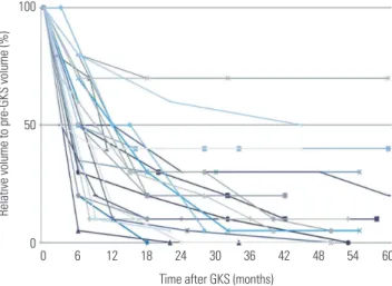
Patient demographics and outcomes are described in Table 1. Before treatment, 11 patients (42.3%) had cranial nerve dysfunc-tion characterized by ptosis, diplopia, facial sense change, or vision change; and 5 patients (19.2%) experienced headache. In contrast, the remaining 10 patients (38.5%) were incidentally diagnosed during their workup for assessing other conditions, such as head injury. The mean clinical follow-up period was 45.7 months (range, 12.1–131.1 months), and the mean mass volume before GKS was 9.3 mL (range, 0.5–31.6 mL). The mean marginal dose directed to the 50% isodose line was 13.7 Gy (range, 13–15 Gy). Clinical results showed good clinical out-comes in all patients; 10 patients had “complete recovery,” 6 pa-tients had “partial recovery,” and 10 papa-tients had “no change.” Moreover, there was no recurrence or aggravation of symptoms during the follow-up period. None of the patients showed mini-mal response, and all 26 patients achieved mass control; re-markable responses were observed in 19 patients (73.1%), and moderate responses in 7 patients (26.9%). The mean mass vol-ume at 6 months after GKS was 45% (range, 5–80%) of the mass volume before GKS and 21% (range, 0–70%) at 12 months. Post-GKS MRI in 26 patients revealed a mean post-treatment mass volume of 1.8 mL (range, 0–12.6 mL), which was significantly lower than the pre-treatment volumes (p<0.05). Fig. 1 shows Table 1. Patient Demographics and Outcomes
Variables Patients (n=26) Sex (%) Male 6 (23.1) Female 20 (76.9) Age (yr) 54.9±13.3 Side (%) Left 17 (65.4) Right 9 (34.6)
Follow-up duration (months) 45.7±27.7 Marginal dose (Gy) 13.7±0.6 Mass volume before GKS (mL) 9.3±7.3 Mass volume after GKS (mL) 1.8±2.9 Mass volume after GKS (%) 20.8±19.5 Radiologic results* (%) Remarkable response (<1/3) 19 (73.1) Moderate response (1/3–2/3) 7 (26.9) Minimal response (>2/3) 0 (0.0) Clinical results (%) Complete recovery 10 (38.5) Partial recovery 6 (23.1) No change 10 (38.5)
GKS, Gamma Knife surgery.
temporal volume change after GKS during follow-up. Then, we classified the 26 patients into two groups accord-ing to their radiologic results to identify the factors that influ-ence mass volume change after GKS; 7 patients were classified as moderate remission group and 19 patients as remarkable remission group. We checked to see whether there were any significant differences between the two groups using the chi-square or Student’s t-test (Table 2). Naturally, the mass volume after GKS showed a significant difference between the two groups (p=0.044, p<0.001). The marginal dose tended to be higher in the remarkable remission group than in the moderate remission group, but it did not reach statistical significance. We expect that if more sample sizes are secured, the results would have statistical significance. Other variables did not show statistical difference.
Cavernous hemangioma in cavernous sinus is a rarely
pre-sented lesion, which shares similar histologic, but different clini-cal features with intracerebral lesions. Many well-known neu-rosurgeons have reported their clinical experience of surgical removal of CSCHs, and specifically, suggested a total removal rate of 40–92.3%.2,8-11 Even though these surgeries were per-formed by very experienced neurosurgeons, the reported post-operative morbidity and mortality rates were as high as 80% and 20%, respectively.2,5,8,9,12 The reasons for the poor surgical outcomes were the high mass vascularization and deep location.
Before the era of GKS, radiotherapy was considered as an al-ternative treatment modality for avoiding the surgical risk. A number of studies have reported good clinical outcomes after radiotherapy, with a relatively high dose of radiation (>3 Gy) in fractions considered as an effective dose for CSCH manage-ment.3,13-15 However, radiation therapy can cause complica-tions in the central nervous system, especially after high-dose radiation.
According to Iwai, et al.,16 CSCH was firstly treated with GKS in 1999. The patient was surgically treated at first, and then, GKS was performed for the residual lesion, resulting in good clini-cal outcome. Following that case, many studies suggested that GKS could be an alternative to surgery or conventional radio-therapy.17-22
Wang, et al.22 published the meta-analysis results of GKS for 59 CSCH patients. Their study reported a remarkable mass shrinkage (more than 50%) in 40 patients (67.8%), partial shrink-age (25–50%) in 15 patients (25.4%), and no change (less than 25%) in 4 patients (6.8%). They also reported that there was no significant correlation between lesion volume and mass shrink-age. However, patients with remarkable mass shrinkage were associated with higher prescription radiation dose (14 Gy vs. 13.5 Gy, p=0.031).
Our study also demonstrated remarkable mass shrinkage during relatively early follow-up. Specifically, significant shrink-age was observed in 19 patients (73.1%), clinical symptoms were relieved within a short period after GKS for about half of the patients (53.8%), and no complication related to GKS was not-ed. These results were consistent with previous studies, and the rapid clinical improvement could be a result of rapid vol-ume reduction (Fig. 2).
The optimal radiation dose for mass control and symptom relief still remains debatable. The exposure to higher radiation doses facilitates higher rates of mass control, but it can nega-tively affect critical structures around the cavernous sinus, such as the optic apparatus or cranial nerves. Therefore, the possibil-ity of this complication usually limits the exposure to high ra-diation doses.22 Our results showed that a higher radiation dose tended to induce earlier and greater volume reduction.
Our study had several limitations. First, CSCHs were not diag-nosed according to pathologic findings, but imaging findings. Therefore, there was a small possibility of misdiagnosis, and we tried to minimize the risk of misdiagnosis by performing RBC scan in case of difficult differentiation from other pathology. Table 2. Comparisons between Moderate Remission Group and
Re-markable Remission Group Variables Radiologic results Moderate remission (n=7) Remarkable remission (n=19) p value Sex (%) 0.904 Male 1 (14.3) 5 (26.3) Female 6 (85.7) 14 (73.7) Age (yr) 60.5±10.7 52.8±13.8 0.201 Side (%) 1.000 Left 5 (71.4) 12 (63.2) Right 2 (28.6) 7 (36.8)
Marginal dose (Gy) 13.3±0.5 13.8±0.6 0.069 Mass volume before GKS (mL) 10.2±10.2 8.9±6.2 0.698 Mass volume after GKS (mL) (%) (48.6±9.0)4.7±4.0 (10.5±9.6)8.0±1.3
GKS, Gamma Knife surgery.
Fig. 1. Temporal volume changes after GKS in 26 patients. The graph shows rapid mass decrease (within 6–12 months after GKS). The mass volume gradually decreased over 2 years, and no volume re-expansion was observed. GKS, Gamma Knife surgery.
100
50
0
0 6 12 18 24 30 36 42 48 54 60
Time after GKS (months)
Second, as about half of the patients in our series were inciden-tally diagnosed or had no cranial nerve deficit, the rationality of upfront GKS for these lesions could be controversial. Nev-ertheless, the minimal invasiveness and higher safety of GKS could be a rationale for treating CSCHs that are incidentally found, as well.
Although surgical resection for CSCH is a curative treatment, it is not always easy and safe. Due to the highly radiosensitive nature of CSCH, GKS could be an effective and safe primary treatment modality for CSCH to prevent possible surgical com-plications. Further studies should be performed to define the natural history of and optimal treatment guidelines for CSCH.
AUTHOR CONTRIBUTIONS
Conceptualization: Jin Mo Cho, Won Seok Chang, Hyun Ho Jung, and
Jong Hee Chang. Data curation: Jin Mo Cho and Kyoung Su Sung.
Formal analysis: Jin Mo Cho and In-Ho Jung. Funding acquisition:
Jong Hee Chang. Investigation: Jin Mo Cho and In-Ho Jung.
Method-ology: Jin Mo Cho and Jong Hee Chang. Project administration: Jong
Hee Chang. Resources: Won Seok Chang, Hyun Ho Jung, and Jong
Hee Chang. Software: Jin Mo Cho and In-Ho Jung. Supervision: Jong
Hee Chang. Validation: Won Seok Chang, Hyun Ho Jung, and Jong
Hee Chang. Visualization: Jin Mo Cho, In-Ho Jung, and Jong Hee
Chang. Writing—original draft: Jin Mo Cho and Jong Hee Chang.
Writing—review & editing: Jin Mo Cho, In-Ho Jung, and Jong Hee Chang. Approval of final manuscript: all authors.
ORCID iDs
Jin Mo Cho https://orcid.org/0000-0002-1192-8993
Kyoung Su Sung https://orcid.org/0000-0003-3289-0143
In-Ho Jung https://orcid.org/0000-0002-4135-5743
Won Seok Chang https://orcid.org/0000-0003-3145-4016
Hyun Ho Jung https://orcid.org/0000-0002-8289-564X
Jong Hee Chang https://orcid.org/0000-0003-1509-9800
REFERENCES
1. Gonzalez LF, Lekovic GP, Eschbacher J, Coons S, Porter RW,
Spet-A
D
B C
E
Fig. 2. A 35-year-old female patient presented with incomplete right 6th cranial nerve palsy. T1-weighted (A) and T2-weighted images (B) showed a mass at the right cavernous sinus. Red blood cell scintigraphy (C) showed marked radioisotope uptake at the lesion, confirming the diagnosis of cavernous si-nus cavernous hemangioma. The measured volume was 5.3 mL. After GKS (14 Gy of marginal dose at the 50% isodose line), all symptoms were resolved within 1 week. The 6-month follow-up magnetic resonance image showed remarkable decrease of the lesion (D), while the lesion could be hardly seen at 35 months after GKS (E). GKS, Gamma Knife surgery.
zler RF. Are cavernous sinus hemangiomas and cavernous mal-formations different entities? Neurosurg Focus 2006;21:e6. 2. Zhou LF, Mao Y, Chen L. Diagnosis and surgical treatment of
cav-ernous sinus hemangiomas: an experience of 20 cases. Surg Neu-rol 2003;60:31-6; discussion 36-7.
3. Linskey ME, Sekhar LN. Cavernous sinus hemangiomas: a series, a review, and an hypothesis. Neurosurgery 1992;30:101-8. 4. Li P, Ren H, Zhang S, Wang W. Clinical results of Gamma Knife
surgery for cavernous sinus hemangiomas. J Neurosurg 2012;117 Suppl:89-95.
5. Dou Y, Meng Q, Yan Z, Xu J, Che S, Jiao Y, et al. Diagnosis and mi-crosurgical treatment of cavernous sinus hemangioma. Artif Cells Blood Substit Immobil Biotechnol 2010;38:109-12.
6. Salanitri GC, Stuckey SL, Murphy M. Extracerebral cavernous hemangioma of the cavernous sinus: diagnosis with MR imaging and labeled red cell blood pool scintigraphy. AJNR Am J Neurora-diol 2004;25:280-4.
7. Polito E, Burroni L, Pichierri P, Loffredo A, Vattimo AG. Techne-tium tc 99m-labeled red blood cells in the preoperative diagnosis of cavernous hemangioma and other vascular orbital tumors. Arch Ophthalmol 2005;123:1678-83.
8. Shi J, Hang C, Pan Y, Liu C, Zhang Z. Cavernous hemangiomas in the cavernous sinus. Neurosurgery 1999;45:1308-13; discussion 1313-4.
9. Ohata K, El-Naggar A, Takami T, Morino M, El-Adawy Y, El-Sheik K, et al. Efficacy of induced hypotension in the surgical treatment of large cavernous sinus cavernomas. J Neurosurg 1999;90:702-8. 10. Bansal S, Suri A, Singh M, Kale SS, Agarwal D, Sharma MS, et al.
Cavernous sinus hemangioma: a fourteen year single institution experience. J Clin Neurosci 2014;21:968-74.
11. Goel A. The extradural approach to lesions involving the cavern-ous sinus. Br J Neurosurg 1997;11:134-8.
12. Suri A, Ahmad FU, Mahapatra AK. Extradural transcavernous
ap-proach to cavernous sinus hemangiomas. Neurosurgery 2007;60: 483-8; discussion 488-9.
13. Shibata S, Mori K. Effect of radiation therapy on extracerebral cavernous hemangioma in the middle fossa. Report of three cas-es. J Neurosurg 1987;67:919-22.
14. Rigamonti D, Pappas CT, Spetzler RF, Johnson PC. Extracerebral cavernous angiomas of the middle fossa. Neurosurgery 1990;27: 306-10.
15. Maruishi M, Shima T, Okada Y, Nishida M, Yamane K, Okita S. Cavernous sinus cavernoma treated with radiation therapy--case report. Neurol Med Chir (Tokyo) 1994;34:773-7.
16. Iwai Y, Yamanaka K, Nakajima H, Yasui T. Stereotactic radiosur-gery for cavernous sinus cavernous hemangioma. Neurol Med Chir (Tokyo) 1999;39:288-90.
17. Chou CW, Wu HM, Huang CI, Chung WY, Guo WY, Shih YH, et al. Gamma knife surgery for cavernous hemangiomas in the cavern-ous sinus. Neurosurgery 2010;67:611-6; discussion 616.
18. Yamamoto M, Kida Y, Fukuoka S, Iwai Y, Jokura H, Akabane A, et al. Gamma Knife radiosurgery for hemangiomas of the cavernous sinus: a seven-institute study in Japan. J Neurosurg 2010;112:772-9. 19. Thompson TP, Lunsford LD, Flickinger JC. Radiosurgery for hem-angiomas of the cavernous sinus and orbit: technical case report. Neurosurgery 2000;47:778-83.
20. Peker S, Kiliç T, Sengöz M, Pamir MN. Radiosurgical treatment of cavernous sinus cavernous haemangiomas. Acta Neurochir (Wien) 2004;146:337-41; discussion 340.
21. Khan AA, Niranjan A, Kano H, Kondziolka D, Flickinger JC, Lun-sford LD. Stereotactic radiosurgery for cavernous sinus or orbital hemangiomas. Neurosurgery 2009;65:914-8; discussion 918. 22. Wang X, Mei G, Liu X, Dai J, Pan L, Wang E. The role of
stereotac-tic radiosurgery in cavernous sinus hemangiomas: a systemastereotac-tic review and meta-analysis. J Neurooncol 2012;107:239-45.