84
책임저자 : 김은경, 서울시 서대문구 신촌동 134 120-752, 연세대학교 의과대학 영상의학교실 Tel: 02-2228-7400, Fax: 02-393-3035
E-mail: ekkim@yuhs.ac
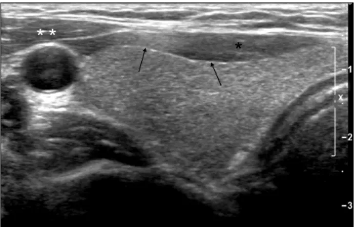
Fig. 1. Normal thyroid gland. Right thyroid gland shows
homoge-neous hyperechoic parenchyma than adjacent muscle (*: strap muscle, **: sternocleidomastoid muscle). Hypere-choic linear thyroid capsule is node (arrows).
갑상선 결절의 초음파
연세대학교 의과대학 영상의학교실
김 은 경
Sonographic Evaluation of Thyroid Nodules
Eun-Kyung Kim, M.D.
With the improvements in the technology, ultrasonography
of the thyroid has been applied to characterize the
appear-ance and distinct featuresof thyroid nodules. In this review,
we discuss the sonographic findings of thyroid nodules and
we confirm that sonography has a definite role for
diagnos-ing and evaluatdiagnos-ing thyroid nodules. (Korean J Endocrine
Surg 2008;8:84-88)
Key Words: Thyroid nodule, Ultrasonography, Diagnosis
중심 단어:
갑상선 결절, 초음파, 진단
Department of Radiology, Yonsei University College of
Medicine, Seoul, Korea
초음파로 갑상선의 혹은 일반적으로 40∼50%에서 발견되 나 이중 악성인 경우는 약 5∼10%이고 대부분은 양성 혹이 다. 최근 초음파 기기의 발달로 결절의 성상을 잘 파악하게 함으로써 결절의 양성, 악성을 감별하고자 하는 노력이 있 다. 본 종설에서는 갑상선 초음파의 방법, 갑상선 결절의 감 별에서의 초음파의 역할에 대해 정리하고자 한다.
갑상선 초음파의 방법
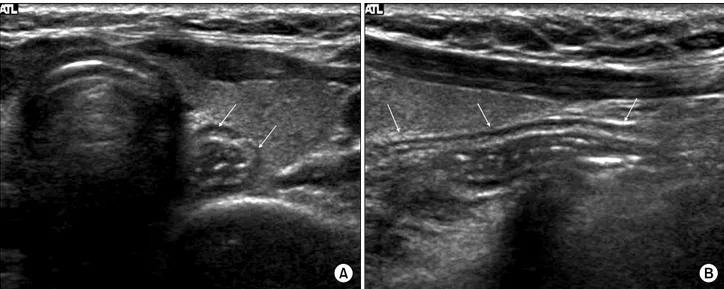
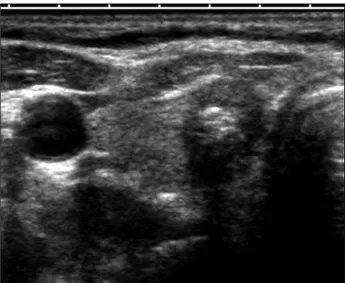
갑상선은 표재성 기관이므로 7∼10 MHz의 고주파 탐촉 자(high-frequency transducer)를 이용해야 좋은 영상을 얻을 수 있다. 둥근 모양의 탐촉자보다 직선형의 탐촉자를 이용 해야 더 좋은 근거리 영상을 얻을 수 있다.(1,2) 환자는 앙와위(supine position)에서 고개 뒤에 베게를 받 쳐 목이 신전된 자세를 취한다. 초음파 검사는 갑상선의 횡 단 및 종단을 모두 관찰해야 하며 경동맥, 경정맥 주위를 포함해 임파선 종대 등의 소견도 검사해야 한다. 초음파에서 정상 갑상선은 중간에서 약간 높은 정도의 에코이며 고에코의 선이 갑상선을 둘러싸고 있는데 이는 갑상선의 피막이다(Fig. 1). 흉골설골근(sternohyoid), 흉골갑 상근(sternothyroid)으로 이루어진 띠근육이 갑상선의 앞쪽 에 저에코로 관찰된다. 흉쇄유돌근(sternocleidomastoid mus-cle)은 갑상선의 양 옆에 위치한다. 식도는 보통 기관의 왼 쪽에 위치하며 간혹 갑상선 결절로 오인될 수 있으나 과녁 모양과 연동운동, 시상면에서 관상의 구조를 확인함으로써 구별할 수 있다(Fig. 2). 또한 경정맥(jugular vein)이 간혹 비 대칭적으로 보일 수 있는데 이는 압박을 하면 쉽게 압박되 므로 정상구조임을 확인할 수 있다(Fig. 3). 그러므로 초음 파를 할 경우, 여러 각도에서 탐촉자를 움직여가면서, 적당 한 압박을 하면서 혹은 풀면서 정상구조인지 병변이지를 확인하여야 한다.갑상선 결절의 감별
갑상선 결절의 감별에 있어서 흔히 초음파유도하 세침흡 인생검(US-FNAB)이 시행되지만, 갑상선 초음파가 결절의 발견과 감별에 매우 중요한 역할을 한다. 많은 검사에서 악 성과 양성을 감별하는 여러가지 초음파 소견들에 대해 논Fig. 2. Normal esophagus in posterior aspect of left thyroid gland. Transverse US image (A) shows target appearance mass like lesion
(arrow) in posterior aspect of left thyroid gland. At longitudinal US (B), it changes to be elongated shaped tubular structure (arrows) suggesting normal esophagus.
Fig. 3. Normal internal jugular vein. Transverse US image (A) shows round shaped anechoic lesion in lateral aspect of the carotid artery.
It is easily collapsed with compression (B).
해왔고(3-8) 각각의 소견들에 대해 정리하면 다음과 같다.
1) 내부 성분
초음파에서 갑상선 결절이 발견된 경우 크게 무에코 (anechoic, cystic) 결절, 혼합성(mixed echoic) 결절, 고형 (solid) 결절로 나눌 수 있다. 무에코 결절은 내부 에코가 없 는 경계가 좋은 결절을 의미하며, 내부에 혜성꼬리허상 (comet tail artifact)가 동반된 고에코 점이 보일 수 있다(Fig. 4). 이러한 무에코 결절은 갑상선 낭종을 의미하며, 단순 낭 종은 거의 악성의 가능성이 없다. 이런 낭종은 갑상선 국소 결절의 1% 정도를 차지한다. 혜성꼬리허상은 콜로이드 낭 종내의 미세크리스탈과 관련이 있으며 이 허상은 악성 결 절에서 보이는 미세석회화와 구분하여야 하고, 낭종내에 떠있다는 점, 고에코 뒤에 허상이 보인다는 점들이 구별점 이나 감별이 힘든 경우도 있으므로 이 둘을 구분하기 위해 서 초음파 검사를 신중하게 하여야 한다. 혼합성 결절은 고형 성분과 낭종 성분이 섞여 있는 결절 을 의미하며(Fig. 5) 이는 이미 있던 고형 종괴가 변성되면 서 이차적으로 생긴다. 이러한 혼합성 낭종은 단순 낭종과 구별되어야하며 약 10∼15%가 악성과 연관이 있다. Frates 등(9)은 혼합성 결절의 낭성 성분에 따라 25% 이하의 낭성 성분이 있으며 석회질이 있는 경우 31.6%에서 악성이, 낭성 성분이 75% 이상이면서 석회질이 없는 경우는 1%에서 악 성이 있었다고 보고하였다. 저자는 혼합성 결절의 초음파
Fig. 4. Typical cystic lesions in thyroid gland. Complete anechoic lesion (A) and anechoic lesion with internal hyperechoic dot showing
comet-tail artifact (B) are typical cysts in thyroid gland.
Fig. 5. Mixed echoic nodule. US shows mass containing both
cyst-ic and solid portion, called mixed echocyst-ic mass. It was diag-nosed as adenomatous hyperplasia by US-FNAB.
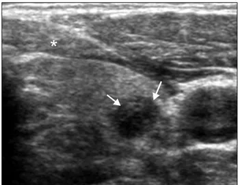
Fig. 6. Marked hypoechogenicity of the nodule. The echogenicity
of the thyroid mass (arrow) is lower than the one of strap muscle. It is called as marked hypoechogenicity, one of suspicious sonographic criteria.
소견 중 악성의 가능성이 있는 소견을 알아보았으며 양성 에 비해 악성에서 낭성 성분이 50% 이하인 경우가 흔하였 고, 석회질을 동반한 경우가 많았다.(10) 고형 결절인 경우 혼합형에 비해 악성의 가능성이 높으며, 고형 결절 자체가 갑상선암의 매우 민감한 소견이나 대부분의 양성 결절도 고형으로 보이므로 악성과 양성 결절을 감별하는 데는 도 움이 되지 않는다. 2) 에코정도 고형 결절이나 혼합형 결절의 고형성분을 기준으로 주변 갑상선 조직의 에코와 비교하여 저에코(hypoechoic), 동일에 코(isoechoic), 고에코(hyperechoic)로 나눌 수 있다. 저에코의 고형 결절은 일반적으로 악성 종양에서 유의하게 많이 나 타나는 것으로 보고되어있지만(5,11) 양성 결절과 많은 부 분에서 겹치는 소견이며, 띠근육보다 더 낮은 에코를 보이 는 경우를 현저한 저에코(Fig. 6)로 분류하였을 때 악성에서 유의하게 차이를 보였다.(3,12) 3) 모양 결절의 모양은 타원형, 원형 혹은 길쭉한 모양(taller than wide)으로 나눌 때 길쭉한 모양이(Fig. 7) 좀 더 악성에서 많 이 보이는 소견이다(3,8,12,13).
Fig. 7. Taller than wide shape suggesting malignancy. US shows
hypoechoic nodule with taller than wide shape (anterior posterior dimension is greater than transverse dimension).
Fig. 8. Microcalcification in thyroid mass. US shows numerous
mi-crocalcifications within the thyroid mass.
Fig. 9. Malignant thyroid mass containing benign looking dense
calcification. Thyroid nodule contains large dense calcifica-tion but it has microlobulated margin with taller than wide shape. So the sonographic interpretation should be suspi-cious malignancy.
Table 1. Diagnostic index of malignant US characteristics and classification (3)
Characteristics Sensitivity (%) Specificity (%) PPV (%) NPV (%) Accuracy (%) Microcalcification
Irregular or microlobulated margin Marked hypoechogenicity
Taller than wide shape
29/49 (59.1) 27/49 (55.1) 13/49 (26.5) 16/49 (32.7) 91/106 (85.8) 88/106 (83) 100/106 (94.3) 98/106 (92.5) 29/41 (70.7) 27/45 (60) 13/19 (68.4) 16/24 (66.7) 91/114 (79.8) 88/110 (80) 100/136 (73.5) 98/131 (74.8) 120/155 (77.4) 115/155 (74.1) 113/155 (72.9) 114/155 (73.5) 4) 경계 경계가 선명한 경우보다는 미세소엽을 보이거나 불규칙 한 경우 악성의 가능성이 높다. 5) 석회화 주변부, 계란껍질 석회화는 양성의 가능성이 높은 반변 결전 내에 소금을 뿌려놓은 듯한 미세석회화(Fig. 8)는 악성 의 가능성이 높다.(3,14) 그러나 양성 가능성이 높은 석회화 가 있더라도 다른 악성의 소견을 가지고 있는 경우(Fig. 9) 악성의 가능성이 있는 종양으로 간주해야 한다.(15) 6) 혈류 갑상선 결절의 감별에 있어서 혈류가 도움이 되는지에 대한 논란은 많다.(16-18) 결절내부의 혈류가 있는 경우 민 감도는 66∼91%로 보고되어 있나 특이도와(34∼80%) 양성 예측도(23∼34%)가 낮아 감별에 이용되는 데는 어려움이 있다. 7) 여러 초음파 소견의 조합 초음파에서 발견한 갑상선 결절의 진단을 위해 US-FNAB 가 시행된다. 그렇지만 초음파에서 보이는 모든 결절을 US- FNAB를 하는 것은 현실적으로 불가능하며, 너무나 많은 불필요한 조직검사로 인한 의료비의 증가와 환자의 불편함 을 초래할 수 있다. 그러므로 갑상선 결절이 초음파에서 발 견될 경우 초음파에서 양성의 소견을 가지는 결절은 보존 적인 처치를 하고, 악성이 의심되는 결절의 경우 조직검사
를 시행한다면 악성을 놓치지 않으면서 양성질환을 가진 환자에서는 불필요한 검사를 줄일 수 있다. 이런 방법이 아 주 실질적이고 의료비를 고려 시 효과적이지만, 초음파로 양성과 악성의 감별은 아주 신중하게 그리고 전문가에 의 해서 시행되어야 한다. 각각의 초음파 소견은 양성과 악성 갑상선 결절에 겹치 는 소견이 많다. 그러나 몇 개의 의심스러운 초음파 소견이 있는 경우 이를 조합하였을 때 우수한 진단적 가치가 있었 다.(3) 초음파 소견 중 미세석회화, 미세소엽 또는 불규칙한 경계, 현저한 저에코, 가로보다 세로가 긴 모양 이러한 4가 지 기준 중 현격한 저에코, 불규칙하거나 미세소엽 가장자 리, 점상 석회화, 길쭉한 모양을 악성 초음파 소견으로 간주 하고, 이중 한가지 소견이라도 포함하는 결절을 악성의 가 능성이 있는 병변으로 간주할 때, 민감도 93.8%, 특이도 66%, 양성예측도 56%, 음성예측도 95.9%, 정확도 74.8%의 좋은 결과를 얻었다(Table 1).(3) 그렇지만 초음파 검사는 검사자 에 따라 상당히 주관적인 판단이 이루어지는 검사이므로, 같은 결절을 보면서도 다른 결론에 도달할 수 있다. 따라서 검사자는 자신의 판독과 결과를 꾸준히 비교하는 훈련이 반드시 필요하다.
결 론
갑상선 초음파 검사는 결절의 발견, 감별에 가장 예민한 검사이며 초음파 소견에 따라 향후 FNAB를 할 것이지 결 정하는 중요한 정보를 제공한다. 검사자는 악성을 시사하 는 초음파 소견을 숙지하여야 하며 한가지라도 의심스러운 초음파 소견이 있는 경우 FNAB를 시행하는 것이 바람직하다.REFERENCES
1) Solbiati L. The thyroid gland. In: Rumack CM, editor. Diagnostic Ultrasound. 3rd ed. St. Louis: Elsevier Mosby; 2005. p.735-70.
2) 김은경, 곽진영: 갑상선 초음파학. 1st ed. 서울: 가본의학; 2006. p.7-38.
3) Kim EK, Park CS, Chung WY. New sonographic criteria for recommending fine-needle aspiration biopsy of nonpalpable solid nodules of the thyroid. AJR Am J Roentgenol 2002;178: 687-91.
4) Cappelli C, Castellano M, Pirola I, Cumetti D, Agosti B, Gandossi E, et al. The predictive value of ultrasound findings in the management of thyroid nodules. QJM 2007;100:29-35. 5) Papini E, Guglielmi R, Bianchini A. Risk of malignancy in
nonpalpable thyroid nodules: predictive value of ultrasound and color-Doppler features. J Clin Endocrinol Metab 2002;87:
1941-6.
6) Nam-Goong IS, Kim HY, Gong G. Ultrasonography-guided fine-needle aspiration of thyroid incidentaloma: correlation with pathological findings. Clin Endocrinol (Oxf) 2004;60: 21-8.
7) Kovacevic O, Skurla MA, Gong G. Sonographic diagnosis of thyroid nodules: correlation with the results of sonographically guided fine-needle aspiration biopsy. J Clin Ultrasound 2007; 35:63-7.
8) Cappelli C, Pirola I, Cumetti D, Micheletti L, Tironi A, Gandossi E, et al. Is the anteroposterior and transverse dia-meter ratio of nonpalpable thyroid nodules a sonographic criteria for recommending fine-needle aspiration cytology? Clin Endocrinol (Oxf) 2005;63:689-93.
9) Frates MC, Benson CB, Doubilet PM, Kunreuther E, Con-treras M, Cinas ES, et al. Prevalence and distribution of carci-noma in patients with solitary and multiple thyroid nodules on sonography. J Clin Endocrinol Metab 2006;91:3411-7. 10) Lee MJ, Kim EK, Kwak JY, Kim MJ. Mixed echoic thyroid
nodules on ultrasound: Probability of malignancy and predi-ctive value of sonographic findings. Thyroid 2008 (accepted). 11) Frates MC, Benson CB, Charboneau JW, Cibas ES, Clark OH,
Coleman BG, et al. Management of thyroid nodules detected at US: Society of Radiologists in Ultrasound consensus con-ference statement. Radiology 2005;237:794-800.
12) Moon WJ, Jung SL, Lee JH, Na DG, Baek JH, Lee YH, et al. Benign and malignant thyroid nodules: US differentiation- multicenter retrospective study. Radiology 2008;247:762-70. 13) Castellano M, Pirola I, Gandossi E, De Martino E, Cumetti
D, Agosti B, et al. Thyroid nodule shape suggests malignancy. Eur J Endocrinol 2006;155:27-31.
14) Kwak JY, Kim EK, Son EJ, Kim MJ, Oh KK, Kim JY, et al. Papillary thyroid carcinoma manifested solely as micro-calcifications on sonography. AJR Am J Roentgenol 2007;189: 227-31.
15) Kim MJ, Kim EK, Kwak JY, Park CS, Chung WY, Nam KH, et al. Differentiation of thyroid nodules with macrocalci-fications: role of suspicious sonographic findings. J Ultrasound Med 2008;27:1179-84.
16) Appetecchia M, Solivetti FM. The association of color flow Doppler sonography and conventional ultrasonography im-proves the diagnosis of thyroid carcinoma. Hormone Research 2006;66:249-56.
17) Frates MC, Benson CB, Doubilet PM, Cibas ES, Marqusee E. Can color Doppler sonography aid in the prediction of mali-gnancy of thyroid nodules? J Ultrasound Med 2004;22:127-31. 18) Iannuccilli JD, Cronan JJ, Monchik JM. Risk for malignancy of thyroid nodules as assessed by sonographic criteria: the need for biopsy. J Ultrasound Med 2004;23:1455-64.


