© 2012 The Korean Academy of Medical Sciences.
This is an Open Access article distributed under the terms of the Creative Commons Attribution Non-Commercial License (http://creativecommons.org/licenses/by-nc/3.0) which permits unrestricted non-commercial use, distribution, and reproduction in any medium, provided the original work is properly cited.
pISSN 1011-8934 eISSN 1598-6357
Meningitis by Toxocara canis after Ingestion of Raw Ostrich Liver
Recently reports on toxocariasis are increasing by serodiagnosis in Korea. A previously healthy 17-yr-old boy complained of headache, fever, dyspnea, and anorexia. He showed symptoms and signs of eosinophilic meningitis with involvement of the lungs and liver. Specific IgG antibody to Toxocara canis larval antigen was positive in serum and cerebrospinal fluid by ELISA. He took raw ostrich liver with his parents 4 weeks before the symptom onset. His parents were seropositive for T. canis antigen but had no symptoms or signs suggesting toxocariasis. This is the first report of toxocariasis in a family due to ingestion of raw ostrich liver in Korea.
Key Words: Toxocara canis; Visceral Larva Migrans; Meningitis; Raw Liver; Ostrich
Young Noh1, Sung-Tae Hong2,
Ji Young Yun3, Hong-Kyun Park4,
Jung-Hwan Oh4, Young Eun Kim4,
and Beom S. Jeon4
1Department of Neurology, Sungkyunkwan
University School of Medicine, Samsung Medical Center, Seoul; 2Department of Parasitology and
Tropical Medicine, Institute of Endemic Diseases, Seoul National University College of Medicine, Seoul; 3Department of Neurology, Ewha Womans
University Medical Center, Seoul; 4Department of
Neurology and Movement Disorder Center, Seoul National University Hospital, Seoul National University College of Medicine, Seoul, Korea Received: 5 March 2012
Accepted: 25 May 2012 Address for Correspondence: Beom S. Jeon, MD
Department of Neurology and Movement Disorder Center, Seoul National University Hospital, 101 Daehak-ro, Jongno-gu, Seoul 110-744, Korea
Tel: +82.2-2072-2876, Fax: +82.2-744-1785 E-mail: [email protected]
http://dx.doi.org/10.3346/jkms.2012.27.9.1105 • J Korean Med Sci 2012; 27: 1105-1108
CASE REPORT
Infectious Diseases, Microbiology & Parasitology
INTRODUCTION
Toxocara canis is a nematode of the Ascaridae family, which lives
in the lumen of the small intestine of dogs (1, 2). It produces eggs, which are passed in feces and embryonate in the soil. T. canis is known to infect various unusual paratenic hosts such as cats, foxes, cows, monkeys, pigs, lambs, mice, rats, chickens and pi-geons, by means of eggs or larvae being ingested with their prey or soil (1-3). Humans are infected by ingestion of contaminated embryonated eggs from soil, through direct contact with dogs or cats, eating raw vegetables, or ingestion of larvae from raw or undercooked giblets and muscles (4, 5). In infected humans, the eggs hatch in the intestine and the developing larvae cross the intestinal wall to enter the blood stream, and then migrate to the liver, lungs, eyes, and the central nervous system (CNS), myocardium or musculature (6, 7).
Recently reports on toxocariasis are increasing by serodiag-nosis in Korea (8). A Korean survey reported a seropositive rate for toxocariasis was 5% in rural area (9). One of the major causes of high incidence of toxocariasis may result from eating raw an-imal liver (3, 4, 10). A study revealed 66.7% subjects were
Toxo-cara seropositive among the patients with unexplained
pulmo-nary patchy infiltrate on chest computed tomography (CT) scans.
Eating raw cow liver is known to increase incidence of the sero-positivity by 7.8 times among them (10). We describe a patient’s clinical record with meningitis by Toxocara canis and his family with asymptomatic toxocariasis after ingestion of raw ostrich liver.
CASE DESCRIPTION
In February 2010, a previously healthy 17-yr-old boy was trans-ferred to Seoul National University Hospital with frontal head-ache, fever, dyspnea, and anorexia, which developed 8 days be-fore the transfer. Physical and neurologic examination recog-nized no remarkable findings, such as wheezing, hepatomega-ly, and nuchal rigidity. Fundoscopic examination was normal. Initial laboratory results showed slightly elevated white blood cell (WBC) count of 10,050 cells/μL with eosinophilia (2,440 cells/ μL) and serum IgG elevated to 529.4 (normal range, 1.0-183 IU/ mL). There was marked lymphocytic pleocytosis in the cere-brospinal fluid (CSF) (1,420 cell/μL), in which 75% of WBCs were eosinophils. CSF protein, glucose, and IgG index were all within normal ranges. Gram stain, culture, and latex agglutinin tests for bacteria were negative, and polymerase chain reaction (PCR) for Mycobacterium tuberculosis and viruses as well as
cul-Noh Y, et al. • Toxocara Meningitis by Raw Ostrich Liver
1106 http://jkms.org http://dx.doi.org/10.3346/jkms.2012.27.9.1105
tures for fungi showed also negative results. Magnetic resonance (MR) scans of the brain showed no abnormal findings. Parana-sal sinus radiography showed no specific findings. On CT of the chest revealed multifocal slight, patch ground glass opacities in both lung fields, which was consistent with eosinophilic pneu-monia (Fig. 1). Abdominal CT showed a small (12 mm) hypo- dense nodule in the liver, which was most likely eosinophilic granuloma. The results of tests for Legionella spp., (L. longbeachae sg., L. micdadei), Leptospira, Brucella, Coxiella burnetii, tsutsu-gamushi, and ameba were all negative. Anti-neutrophil cyto-plasmic antibodies (ANCAs), double stranded DNA anti-body, antinuclear antianti-body, and rheumatoid factor were also negative. Suspecting of eosinophilic meningitis with the lung and liver infiltration caused by helminth infection, specific IgG antibodies to various parasite antigens were measured by en-zyme-linked immunosorbent assay (ELISA). The specific IgG antibody to Toxocara canis larval antigen was positive by ELISA with absorbances of 0.375 and 0.439 (cut off value > 0.250) in both serum and CSF respectively. The results of ELISA for
Echi-nococcus sp., Anisakis simplex, Fasciola hepatica, Tricinella
spi-ralis, Schistosoma spp., Angiostrongylus cantonensis, Clonorchis sinensis, Paragnoimus sp., cysticercus cellulosae, and
sparga-num were all negative.
He had no allergy, autoimmune disease and malignancy. He denied regular consumption of medication. He had no history of travel abroad or recent contact to pets. He lived in a dormito-ry located in an urban area and occasionally visited his parents’ in the urban area during weekends. His parents owned a stock farm away from their dwelling house and raised ostriches, cows and dogs. He ate raw ostrich liver with his parents about 4 weeks before the symptom onset. Although his parents had no specific symptoms or signs, the results of ELISA using their serum showed positive absorbance of 0.391 for his 54-yr-old father and 0.316 for 50-yr-old mother to T. canis antigen. His 21-yr-old brother, who did not eat the raw ostrich liver, showed no reactivity to T. canis antigen with absorbance of 0.098. Complete blood cell (CBC) tests on his mother and brother showed eosinophilia only in his mother (WBC 7,100, eosinophil 15.5%).
Albendazole was administered to the patient and his parents for 2 weeks. This led to significant symptomatic improvement.
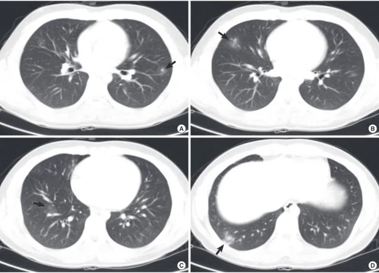
Fig. 1. Computed tomography (CT) scan findings of the chest. Multiple nodules with ground glass opacity halo in the left upper lung field (A) and ill defined patches of nodular ground glass opacity are shown in the right middle (B) and the right lower lung field (C, D), which involve mainly the peripheral regions of the both lungs (arrows).
A B
Noh Y, et al. • Toxocara Meningitis by Raw Ostrich Liver
http://jkms.org 1107 http://dx.doi.org/10.3346/jkms.2012.27.9.1105
Follow-up CBC of the patient showed reduced eosinophil count (WBC 7,500, eosinophil 10.2%) 1 month later and showed nor-malization (WBC 7,400, eosinophil 2.3%) 1 yr later.
DISCUSSION
Toxocariasis is classified into 4 types (1). Type 1 is asymptomat-ic toxocariasis whasymptomat-ich remains inapparent and composes of ma-jority. Type 2, covert or common toxocariasis, is usually mani-fested as mild and focal impairment such as weakness, pruritus, rash, and so on (11). Type 3, classical visceral larva migrans usu-ally seen in children, presents with general illness with fever, skin rash, pharyngitis, cervical lymphadenitis, lethargy, respira-tory symptoms such as wheeze, cough, and pneumonia due to lung involvement. Gastrointestinal symptoms such as anorexia, vomiting, and abdominal pain due to hepatomegaly or spleno-megaly, myalgia, arthralgias, and hypereosinophilia or hyper-gammaglobulinemia are also common (12, 13). The last one, type 4, is neurological toxocariasis including ocular involvement. In experiments, T. canis larvae are neurotropic in infected pri-mates (14) as well as rodents (15, 16). Reported CNS involve-ment of T. canis is presented as meningitis, encephalitis, myeli-tis, or some combinations. Cerebral vasculimyeli-tis, optic neurimyeli-tis, extramedullary tumors, seizure and isolated behavior disorder are rarer clinical manifestation of CNS infection (1, 17, 18). The present boy complained headache, fever, dyspnea, and anorexia. Most of them were nonspecific but the symptom com-bination assumed a certain systemic infection. Since his CSF included numerous WBCs with dominant eosinophils, the symp-toms must have been induced by eosinophilic meningitis. In addition to this, his chest CT visualized multifocal slight, patch ground glass opacities in both lung fields. The findings in the lungs were compatible with those of eosinophilic pneumonia. The abdominal CT recognized a focal dense nodule of 12 mm diameter in the liver. The liver nodule may be an image of the liver granuloma. The clinical manifestations and CT images of the patient strongly suggested that any helminthiasis had in-volved the meninges, lungs, and liver simultaneously. ELISA using multi-antigens could differentiate the causative agents among several tissue invading helminthiases. The serum and CSF of the patient were strongly positive for specific antibodies to the antigen of Toxocara canis larvae but negative for 10 other antigens of helminthes. Any other bacterial or viral infections were excluded by intensive laboratory works. The present case was diagnosed as the type 3 and 4 toxocariasis, showing menin-gitis, pneumonitis, and liver granuloma.
The present patient had a history of eating raw liver of ostrich-es 4 weeks before the onset. The period of 4-weeks might be an enough duration of worm invasion and inflammatory response in the host. During the period, he must have been involved of his liver and lungs before the CNS but missed it. He could note
subjective symptoms due to meningitis and pneumonitis to-gether. The source of his infection must be the raw liver of os-triches because eating the liver was the only related history. Tox-ocariasis by ingestion of raw liver of cows, chickens, pigs or lamb has been well-known (3-5) and the impacts of the habitual eat-ing of raw cow liver on toxocariasis have been evaluated (3, 10). Ostrich (Struchio camelus domesticus) is an imported animal for commercial farming to produce feather and meat. Since the eggs of T. canis remain viable in soil for months or years, the in-fection of farmed ostriches was presumed to be from contami-nating feces of nearby dogs.
The severity of host damage in toxocariasis depends on the host’s age and immunocompetence, affected tissues, the amount of ingested larvae and whether previous exposure had occurred (13, 19). The reason, why the boy alone manifested overt symp-toms although his parents were exposed to the pathogen togeth-er and stogeth-eropositive, may be related with the numbtogeth-er of migrat-ing larvae or host’s age.
We report a human case of neurotoxocariasis and visceral larva migrans caused by ingestion of raw ostrich liver. In addi-tion, this is the first case of toxocariasis in family. Misbelief that raw liver of animals is beneficial to health can lead to familial infection. Eating raw liver of animals should be actively discour-aged. Physicians should pay more attention to toxocariasis for patients with eosinophilia and organ involvement.
REFERENCES
1. Finsterer J, Auer H. Neurotoxocariasis. Rev Inst Med Trop Sao Paulo 2007; 49: 279-87.
2. Graeff-Teixeira C, da Silva AD, Yoshimura K. Update on eosinophilicc meningoencephalitis and its clinical relevance. Clin Microbiol Rev 2009; 22: 322-48.
3. Choi D, Lim JH, Choi DC, Paik SW, Kim SH, Huh S. Toxocariasis and ingestion of raw cow liver in patients with eosinophilia. Korean J Parasi-tol 2008; 46: 139-43.
4. Nagakura K, Tachibana H, Kaneda Y, Keto Y. Toxocariasis possibly caused by ingesting raw chicken. J Infect Dis 1989; 160: 735-6.
5. Sturchler D, Weiss N, Gassner M. Transmission of toxocariasis. J Infect Dis 1990; 162: 571.
6. Overgaauw PA. Aspects of Toxocara epidemiology: human toxocariosis. Crit Rev Microbiol 1997; 23: 215-31.
7. Haralambidou S, Vlacheaki E, Ioannidou E, Milioni V, Haralambidis S, Klonizakis I. Pulmonary and myocardial manifestations due to Toxo-cara canis infection. Eur J Intern Med 2005; 16: 601-2.
8. Lim JH. Foodborne eosinophilia due to visceral larva migrans: a disease abandoned. J Korean Med Sci 2012; 27: 1-2.
9. Park HY, Lee SU, Huh S, Kong Y, Magnaval JF. A seroepidemiological survey for toxocariasis in apparently healthy residents in Gangwon-do, Korea. Korean J Parasitol 2002; 40: 113-7.
10. Yoon YS, Lee CH, Kang YA, Kwon SY, Yoon HI, Lee JH, Lee CH. Impact of toxocariasis in patients with unexplained patchy pulmonary infiltrate in Korea. J Korean Med Sci 2009; 24: 40-5.
Noh Y, et al. • Toxocara Meningitis by Raw Ostrich Liver
1108 http://jkms.org http://dx.doi.org/10.3346/jkms.2012.27.9.1105
11. Taylor MR, Keane CT, O’Connor P, Girdwood RW, Smith H. Clinical features of covert toxocariasis. Scand J Infect Dis 1987; 19: 693-6. 12. Magnaval JF, Glickman, LT, Dorchies P, Morassin B. Highlights of
hu-man toxocariasis. Korean J Parasitol 2001; 39: 1-11.
13. Xinou E, Lefkopoulos A, Gelagoti M, Drevelegas A, Diaakou A, Milonas I, Dimitriadis AS. CT and MR imaging findings in cerebral toxocaral disease. AJNR Am J Neuroradiol 2003; 24: 714-8.
14. Glickman L, Summers BA. Experimental Toxocara canis infection in Cynomolgus macaques (Macaca fascicularis). Am J Vet Res 1983; 44: 2347-54.
15. Good B, Holland CV, Stafford P. The influence of inoculum size and time post-infection on the number and position of Toxocara canis larvae re-covered from the brains of outbred CD1 mice. J Helminthol 2001; 75:
175-81.
16. Fan CK, Lin YH, Du WY, Su KE. Infectivity and pathogenicity of 14-month-cultured embryonated eggs of Toxocara canis in mice. Vet Parasitol 2003; 113: 145-55.
17. Vidal JE, Sztajnbok J, Seguro AC. Eosinophilic meningoencephalitis due to Toxocara canis: case report and review of the literature. Am J Trop Med Hyg 2003; 69: 341-3.
18. Eberhardt O, Bialek R, Nagele R, Dichgans J. Eosinophilic meningomy-elitis in toxocariasis: case report and review of the literature. Clin Neurol Neurosurg 2005; 107: 432-8.
19. Despommier D. Toxocariasis: clinical aspects, epidemiology, medical ecology, and molecular aspects. Clin Microbiol Rev 2003; 16: 265-72.