Gastrointestinal Hemorrhage after Concurrent Chemoradiotherapy in
Locally Advanced Pancreatic Cancer
Kyong Joo Lee*, Hee Man Kim*, Joo Won Jung*, Moon Jae Chung*, Jeong Youp Park*, Seungmin Bang*, Seung Woo Park*, Woo Jung Lee†, Jin Sil Seong‡, and Si Young Song*,§,Ⅱ
*Division of Gastroenterology, Department of Internal Medicine, Departments of †Surgery, ‡Radiation Oncology, §Brain Korea 21 Project for
Medical Science, and ⅡSeverance Biomedical Science Institute, Yonsei University College of Medicine, Seoul, Korea
Background/Aims: While chemoradiotherapy (CRT) is con-sidered to be a reasonable treatment for locally advanced pancreatic cancer (LAPC), there is little information about the associated risk of gastrointestinal (GI) hemorrhage. We investigated the clinical features of GI toxicity after CRT in patients with LAPC and examined the effect of GI hemor-rhage on survival. Methods: Patients enrolled in this study had received CRT for pathologically proven LAPC. Their medi-cal records were retrospectively reviewed. Results: A total of 156 patients with LAPC (median age, 65 years; range, 39 to 90 years) who received treatment between August 2005 and March 2009 were included in this study. The most common GI toxicities were ulcer formation (25.6%) and hemorrhage (25.6%), and the most common grade 3 to grade 5 GI toxic-ity was hemorrhage (65%). The origins of GI hemorrhage were gastric ulcer (37.5%), duodenal ulcer (37.5%), and ra-diation gastritis (15.0%). The independent risk factor for GI hemorrhage was tumor location in the pancreatic body. The median overall survival of the patients with a GI hemorrhage was 13.8 months (range, 2.8 to 50.8 months) and was not significantly different from that of patients without GI hemor-rhage. Conclusions: GI hemorrhage was common in patients with LAPC after CRT. Although GI hemorrhage was controlled with endoscopic hemostasis, preventive measures should be investigated to reduce needless suffering. (Gut Liver 2013;7:106-111)
Key Words: Chemoradiotherapy; Gastrointestinal hemor-rhage; Toxicities; Pancreatic neoplasms
Correspondence to: Si Young Song
Department of Internal Medicine, Yonsei University College of Medicine, 50 Yonsei-ro, Seodaemun-gu, Seoul 120-752, Korea Tel: +82-2-2228-1957, Fax: +82-2-393-6884, E-mail: [email protected]
Received on February 16, 2012. Revised on March 12, 2012. Accepted on March 12, 2012. Published online on December 5, 2012. pISSN 1976-2283 eISSN 2005-1212 http://dx.doi.org/10.5009/gnl.2013.7.1.106
This is an Open Access article distributed under the terms of the Creative Commons Attribution Non-Commercial License (http://creativecommons.org/licenses/by-nc/3.0) which permits unrestricted non-commercial use, distribution, and reproduction in any medium, provided the original work is properly cited.
INTRODUCTION
Pancreatic cancer is the fourth leading cause of cancer-related deaths in the United States.1
Surgical resection is the only cura-tive treatment for pancreatic cancer.2 However, only 5% to 25% of patients with pancreatic cancer are candidates for curative pancreatectomy.
Locally advanced pancreatic cancer (LAPC) is surgically un-resectable nonmetastatic disease, which includes the cases of extensive peripancreatic lymph node involvement and major vasculatures involvement.3 The median survival time for pa-tients with LAPC is only 9 to 10 months.4
Chemoradiotherapy (CRT) is a reasonable treatment modality for LAPC because it is known to increase survival in patients with LAPC.5-7 CRT has been frequently used instead of chemo-therapy alone or radiochemo-therapy alone. However, overall toxic effects of CRT are greater than those of chemotherapy alone.8 Such adverse effects of CRT limit the maximum dose of chemo-therapy and radiochemo-therapy, and lead to unfavorable treatment results.9
CRT for treatment of LAPC produces unique gastrointestinal (GI) toxicities, including ulcer and hemorrhage in the stomach and duodenum that are included in the radiation field. However, to our knowledge, there are a few data focusing on GI hemor-rhage of CRT for treatment of LAPC. In addition, the effect of GI hemorrhage on survival of patients with LAPC has not been evaluated. GI hemorrhage of CRT needs to be examined to prevent adverse events and to develop methods to reduce the severity of such events. Indeed, in clinical practice, we experi-enced many patients with CRT-induced GI hemorrhage, which led us to perform this study. Thus, we examined clinical features of GI hemorrhage of CRT in patients with LAPC, and the effect of GI hemorrhage on survival.
MATERIALS AND METHODS
1. PatientsPatients who received concurrent CRT for treatment of LAPC at Severance Hospital in Seoul, Korea, between August 2005 and March 2009 were selected for this study. Inclusion criteria included pathologically-proven pancreatic adenocarcinoma, age of over 20 years, and the Eastern Cooperative Oncology Group performance status of 0 to 2. Exclusion criteria included patients who had received chemotherapy or surgery before CRT, and pa-tients with scheduled radiotherapy less than 4,000 cGy. Papa-tients who had not completed their scheduled radiation therapy were also excluded for per protocol analysis.
2. Treatment for LAPC
For regression analysis, regimens of chemotherapy and radio-therapy were classified into several groups. All chemoradio-therapy regimens of CRT performed for LAPC were classified into three groups: gemcitabine, 5-fluorouracil (5-FU), and 5-FU plus gem-citabine. Gemcitabine group was given 1,000 mg/m2
of gem-citabine on days 1, 8, and 15 of a 4-week regimen or gemcitabi-ne (same as above) along with 70 mg/m2
of cisplatin on day 1 of the regimen. 5-FU group received either 5-FU (1,000 mg/m2 on days 1 to 3 of a 4-week regimen) or; TS-1 (60 to 80 mg for 2 weeks); or a combination of 5-FU (1,000 mg/m2 on days 1 to 3), etoposide (100 mg/m2
on days 1 to 3), and cisplatin (70 mg/ m2 on day 1). For 5-FU plus gemcitabine group, 1,000 mg/m2 of 5-FU was given on days 1 to 3, and 1,000 mg/m2
gemcitabine on days 1, 8, and 15 of a 4-week regimen.
All radiotherapy regimens of CRT performed for LAPC were either three-dimensional (3D) conformal radiotherapy (total dose, 4,000 to 5,400 cGy; one dose, 180 to 250 cGy; fraction, 28) or intensity modulated radiotherapy (total dose, 4,200 to 6,000 cGy; one dose, 200 to 293 cGy; fraction, 25).
3. GI toxicities
GI toxicities were classified according to the National Cancer Institute Common Terminology Criteria for Adverse Events ver-sion 4.0. In our study, radiation-induced injuries observed with endoscopy were defined as telangiectasia, diffuse erythema of mucosa, ulcers, and scar formation.10,11
4. Statistical analysis
To investigate the risk factors of GI toxicities by CRT, Pear-son’s chi-square test for univariate analysis and logistic regres-sion for multivariate analysis were used. To evaluate survival effect of GI hemorrhage, Cox regression test was used. The Kaplan-Meier method and the log-rank test were used to com-pare survival between patients with GI hemorrhage and patients without GI hemorrhage.
All analyses were performed using statistical software SPSS version 11 (SPSS Inc., Chicago, IL, USA). A p-values lower than
0.05 indicated significance.
RESULTS
1. Patient characteristics
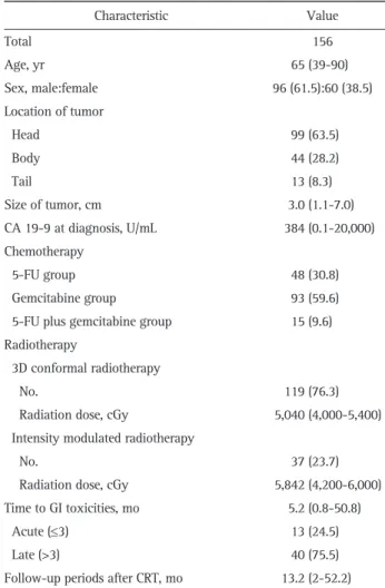
One hundred fifty-six patients with LAPC were eligible for analysis (Table 1). The median age at the time of the diagnosis of pancreatic cancer was 65 years, ranging from 35 to 90 years. Male patients accounted for 61.5% of the population. The tu-mors were mostly located at the pancreatic head (63.5%). The median size of the tumor was 2.9 cm, ranging from 1.1 to 7.0 cm. The median level of CA 19-9 was 384 U/mL (range, 0.1 to 20,000 U/mL). The 3D conformal radiotherapy was delivered to 119 patients (76.3%), and the median delivered dose was 5,040 cGy (range, 4,000 to 5,400 cGy). Intensity modulated radio-therapy was delivered to 37 patients (23.7%), and the median delivered dose was 5,842 cGy (range, 4,200 to 6,000 cGy). The median follow-up period was 13.2 months (range, 2 to 52.2 months).
Table 1. Baseline Patient Characteristics
Characteristic Value Total 156 Age, yr 65 (39-90) Sex, male:female 96 (61.5):60 (38.5) Location of tumor Head 99 (63.5) Body 44 (28.2) Tail 13 (8.3) Size of tumor, cm 3.0 (1.1-7.0) CA 19-9 at diagnosis, U/mL 384 (0.1-20,000) Chemotherapy 5-FU group 48 (30.8) Gemcitabine group 93 (59.6)
5-FU plus gemcitabine group 15 (9.6) Radiotherapy
3D conformal radiotherapy
No. 119 (76.3)
Radiation dose, cGy 5,040 (4,000-5,400) Intensity modulated radiotherapy
No. 37 (23.7)
Radiation dose, cGy 5,842 (4,200-6,000)
Time to GI toxicities, mo 5.2 (0.8-50.8)
Acute (≤3) 13 (24.5)
Late (>3) 40 (75.5)
Follow-up periods after CRT, mo 13.2 (2-52.2) Data are presented as median (range) or number (%).
5-FU, 5-fluorouracil; GI, gastrointestinal; 3D, three-dimensional; CRT, chemoradiotherapy.
2. GI toxicities
The prevalence of GI toxicities was 57.7% (Table 2). There were 30 patients with grade 1 or 2 abdominal pain or dyspep-sia. Two patients had grade 3 anorexia. Nausea and vomiting developed in four patients, and these were well controlled with appropriate medications. Forty patients (25.6%) had GI hem-orrhage: nine patients with hematemesis (22.5%), 21 patients with melena (52.5%), and 10 patients with hematochezia (25%). Eighteen patients (11.5%) had grade 3 or 4 GI hemorrhage, and eight patients (5.1%) had grade 5 (death) hemorrhage.
Seventy-eight patients (50%) underwent upper GI endoscopy after CRT. There were mucositis in 27.2% of patients, ulcer in 45%, and GI hemorrhage with above grade 3 toxicity in 65%. The median time of development of GI toxicities was 5.2 months.
3. GI hemorrhage
Forty patients (25.6%) suffered from GI hemorrhage after CRT (Table 3). The median initial hemoglobin was 10.1 g/dL (range, 7.1 to 15.3 g/dL), which decreased to 7.1 g/dL (range, 3.5 to 10.8 g/dL) when bleeding. Thirty-five patients underwent upper GI endoscopy. The results showed the cause of bleeding to be a gastric ulcer in 15 patients (37.5%), duodenal ulcer in 15 (37.5%), and radiation gastritis in five (15%). Upper GI endoscopy was not performed in five patients upon their guardians’ rejection. As the patients were in terminal stages, the guardians did not want them to undergo any more examinations. The remaining 35 patients received endoscopic treatment. Hemorrhage was successfully stopped by endoscopic treatment in 31 patients
(77.5%). The methods of endoscopic hemostasis were hypertonic saline-epinephrine injection, human plasmin thrombin injec-tion, argon plasma coagulainjec-tion, or hemoclipping. Embolization
Table 2. Gastrointestinal Toxicities after Chemoradiotherapy According to the National Cancer Institute Common Terminology Criteria Version 4.0
Variable Total G3-G5 G1 G2 G3 G4 G5
Abdominal pain or dyspepsia 30 (19.2) 0 16 14 0 0 0
Anorexia 5 (3.2) 2 (40) 2 1 2 0 0 Nausea 3 (1.9) 0 0 3 0 0 0 Vomiting 1 (0.6) 0 0 1 0 0 0 Mucositis 22 (14.1) 6 (27.2) 15 1 6 0 0 Stomach 17 (10.8) 5 (29.4) 11 1 5 0 0 Duodenum 5 (3.2) 1 (20) 4 0 1 0 0 Ulcer 40 (25.6) 18 (45) 11 11 14 1 3 Stomach 22 (14.1) 7 (31.8) 7 8 7 0 0 Duodenum 18 (11.5) 11 (61.1) 4 3 7 1 3 Other GI hemorrhage 40 (25.6) 26 (65) 0 14 17 1 8 Stomach 20 (12.8) 9 (45) 0 11 9 0 0 Duodenum 15 (9.6) 12 (80) 0 3 8 1 3 Not confirmed* 5 (3.2) 5 (100) 0 0 0 0 5
Data are presented as number (%). GI, gastrointestinal.
*Endoscopy was not performed, and the focus of bleeding was not confirmed.
Table 3. Gastrointestinal Hemorrhage after CRT (n=40)
Variable Value Hemoglobin, g/dL Initial 10.1 (7.1-15.3) At bleeding 7.1 (3.5-10.8) Origin Gastric ulcer 15 (37.5) Duodenal ulcer 15 (37.5) Radiation gastritis 5 (15.0) Not confirmed* 5 (15.0) Severity Mild (G1) 0 Moderate (G2, G3) 31 (77.5) Severe (G4, G5) 9 (22.5) Treatment Endoscopic hemostasis 35 (87.5)
Angiography and embolization 1 (2.5)
Conservative care 5 (12.5)
Time to GI hemorrhage from CRT, mo 5.4 (0.8-50.8) Survival time from CRT, mo 13.8 (2.8-50.8) Data are presented as median (range) or number (%).
CRT, chemoradiotherapy; GI, gastrointestinal.
*Endoscopy was not performed, and the focus of bleeding was not confirmed.
was performed in one of the four other patients, and hemor-rhage was finally stopped. However, three others died due to GI hemorrhage. The mortality of GI hemorrhage was eight patients
in total (Fig. 1).
The median time from CRT to GI hemorrhage was 5.4 months (range, 0.8 to 50.8 months), and the median overall survival was
Fig. 2. Comparison of survival between gastrointestinal (GI) hemor-rhage patients and non-GI hemorhemor-rhage patients. The overall median survival time was 13.1 months in the non-GI hemorrhage group and 13.5 months in the GI hemorrhage group.
Fig. 1. Treatment flow chart. Treatment of gastrointestinal hemor-rhage after chemoradiotherapy.
Table 4. Risk Factors for GI Hemorrhage in All Patients (n=156)
Variable GI hemorrhage GI hemorrhage
Presence (%) p-value* OR 95% CI p-value†
Age, yr 0.144 ≤65 25 (30.5) 1 >65 15 (20.3) 0.61 0.27-1.49 0.304 Sex 0.602 Female 14 (23.3) 1 Male 26 (27.1) 1.56 0.67-3.64 0.304 Location of tumor 0.007 Head 19 (19.2) 1 Body 19 (43.2) 2.99 1.27-7.03 0.033 Tail 2 (15.4) 0.42 0.08-2.26 0.314 Size of tumor, cm 0.042 ≤3 16 (19.0) 1 >3 24 (33.3) 1.94 0.87-4.31 0.102 Chemotherapy 0.311 5-FU group 10 (20.4) 1 Gemcitabine group 24 (26.1) 1.58 0.64-3.88 0.319
5-FU plus gemcitabine group 6 (40.0) 2.70 0.65-11.17 0.168
Radiation modality 0.834
3D conformal radiotherapy 31 (26.1) 1
Intensity modulated radiotherapy 9 (24.3) 0.97 0.38-2.42 0.945
CA 19-9, U/mL 0.554
≤1,200 27 (24.3) 1
>1,200 13 (28.9) 1.27 0.54-2.97 0.322
GI, gastrointestinal; OR, odds ratio; CI, confidence interval; 5-FU, 5-fluorouracil; 3D, three-dimensional. *Chi-square test was used; †Logistic regression was used.
13.8 months (range, 2.8 to 50.8 months). 4. Risk factors for GI hemorrhage
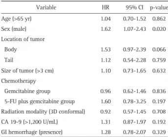
The association between clinical parameters and the risk of GI hemorrhage were analyzed (Table 4). In univariate analysis, location of tumor (p=0.007) and size of tumor (p=0.042) were risk factors for GI hemorrhage. In multivariate analysis, only tumors located on the pancreatic body (odds ratio [OR], 2.99; 95% confidence interval [CI], 1.27 to 7.03) was a significant risk factor for GI hemorrhage. The hazard ratio (HR) was 1.28 (95% CI, 0.78 to 2.07) for the effect of GI hemorrhage on survival, but it was not significant (Table 5). Male patients had signifi-cantly higher HR than females (HR, 1.62; 95% CI, 1.07 to 2.43; p=0.020). However, there were no difference in the number of patients with hypertension or diabetes mellitus between males and females (OR, 1.26; 95% CI, 0.66 to 2.40; p=0.492).
5. Survival
At the time of final analysis, 117 patients (75%) were dead. The median overall survival after the start of CRT was 13.1 months (range, 11.3 to 14.9 months). The median overall sur-vival was 13.1 months (range, 9.9 to 16.3 months) in patients without GI hemorrhage and 13.8 months (range, 2.8 to 50.8 months) in patients with GI hemorrhage (Fig. 2). Although over-all survival was longer in patients with GI hemorrhage, this dif-ference was not significant.
DISCUSSION
Our analysis showed that the prevalence of CRT-induced GI hemorrhage is frequent and serious if not treated properly. The body of the pancreas was the risk factor of GI hemorrhage after CRT. The median overall survival was similar with other stud-ies.5,8,12
This study also showed that GI hemorrhage after CRT did not reduce survival of patients with LAPC.
CRT was first introduced in the Gastrointestinal Tumor Study Group trial.13 Many studies reported the benefit of CRT, and CRT became one of the treatment options for pancreatic cancer.6,7 Based on the results of several studies, the RT dose of 50 to 60 Gy (182 cGy/day) is generally used.14,15
A study reported that the toxicity was higher in the LAPC group where the radiation dose increased up to 55 Gy than in the LAPC group with the dose up to 50 Gy; however, patient compliance was similar between the groups, and the treatment performance in the former was better than that in the latter.16 In studies comparing CRT and chemotherapy, however, more cases of toxicity were found in the CRT group; thus, care must be taken with regard to the use of CRT.17,18
Due to low awareness of GI hemorrhage, the frequency of en-doscopic examination was quite low. Of 156 patients, 20 were examined with endoscopy before CRT and 78 after CRT. Very few patients who had GI hemorrhage underwent endoscopic
study before the onset of bleeding. Had endoscopy also been performed in other patients, the chances of finding complica-tions such as radiation gastritis would have been higher.
The location of the tumor was related with GI hemorrhage. As the body of the pancreas is located close to both the stomach and duodenum, radiation on the pancreas affects the two organs as well. There is no consensus over the best time to perform en-doscopy after radiotherapy. After CRT, however, GI toxicites are likely to develop at anytime. Therefore, it is recommended that endoscopy be performed as was done in this study. If abnormal findings are found in endoscopy before CRT, preemptive treat-ment is necessary. Moreover, endoscopy as a baseline study is recommended for the comparison with post-CRT endoscopic results. Usually CRT is followed by chemotherapy or surgery about one month later. Endoscopy is recommended before such therapies as GI ulcer or hemorrhage can occur even within 90 days after CRT. As ulcerative bleeding is highly responsive to proton pump inhibitor, its early detection and treatment may prevent adverse events. Although the best frequency of endos-copy may be debatable, yearly or more frequent endosendos-copy, particularly in patients with a history of GI hemorrhage, is recommended considering the possibility of delayed ulcer and hemorrhage.
This study has several limitations. First, the results were ob-tained by retrospectively reviewing the medical charts. Second, regarding CRT, the chemotherapy-induced adverse effects could not be excluded. In this study, a greater number of patients received gemcitabine treatment than those who received 5-FU. Several studies reported that gemcitabine was more toxic than 5-FU.19,20 In addition, it was difficult to identify the cause of GI hemorrhage after surgery as well as after beginning a chemo-therapy only regimen following CRT. Third, the low number of
Table 5. Cox Regression Analysis of the Effect of GI Hemorrhage on Survival Variable HR 95% CI p-value Age (>65 yr) 1.04 0.70-1.52 0.862 Sex (male) 1.62 1.07-2.43 0.020 Location of tumor Body 1.53 0.97-2.39 0.066 Tail 1.12 0.54-2.28 0.759 Size of tumor (>3 cm) 1.10 0.73-1.65 0.632 Chemotherapy Gemcitabine group 0.96 0.62-1.46 0.836
5-FU plus gemcitabine group 1.60 0.78-3.25 0.197 Radiation modality (3D conformal) 0.92 0.57-1.45 0.708 CA 19-9 (>1,200 U/mL) 1.31 0.87-1.97 0.192 GI hemorrhage (presence) 1.28 0.78-2.07 0.329 GI, gastrointestinal; HR, hazard ratio; CI, confidence interval; 5-FU, 5-fluorouracil.
patients who received endoscopy before CRT made it impossible to determine if patients developed an ulcer before CRT. Fourth, patients who received 3D conformal radiotherapy and inten-sity modulated radiotherapy were analyzed together, and 37 of them received intensity-modulated radiotherapy. To the best of our knowledge, there has been no study that compared 3D conformal radiotherapy and intensity-modulated radiotherapy. Thus, a study is required to investigate if there is any difference between the two modalities.
In conclusion, the present results show that GI hemorrhage is common in LAPC after CRT. Although the median survival was similar regardless of GI hemorrhage, the clinicians need to con-sider the GI hemorrhage of CRT in LAPC. Extensive studies are required to compare the benefits and risks in terms of survival and complications between CRT and chemotherapy. In addition, studies are required to identify tests or treatments that can re-duce CRT-inre-duced GI hemorrahge.
CONFLICTS OF INTEREST
No potential conflict of interest relevant to this article was reported.
REFERENCES
1. Jemal A, Siegel R, Ward E, Hao Y, Xu J, Thun MJ. Cancer statis-tics, 2009. CA Cancer J Clin 2009;59:225-249.
2. Winek T, Hamre D, Mozell E, Vetto RM. Prognostic factors for sur-vival after pancreaticoduodenectomy for malignant disease. Am J Surg 1990;159:454-456.
3. Willett CG, Czito BG, Bendell JC, Ryan DP. Locally advanced pan-creatic cancer. J Clin Oncol 2005;23:4538-4544.
4. Hidalgo M. Pancreatic cancer. N Engl J Med 2010;362:1605-1617. 5. Shinchi H, Takao S, Noma H, et al. Length and quality of survival
after external-beam radiotherapy with concurrent continuous 5-fluorouracil infusion for locally unresectable pancreatic cancer. Int J Radiat Oncol Biol Phys 2002;53:146-150.
6. Huguet F, André T, Hammel P, et al. Impact of chemoradiotherapy after disease control with chemotherapy in locally advanced pan-creatic adenocarcinoma in GERCOR phase II and III studies. J Clin Oncol 2007;25:326-331.
7. Nakachi K, Furuse J, Kinoshita T, et al. A phase II study of in-duction chemotherapy with gemcitabine plus S-1 followed by chemoradiotherapy for locally advanced pancreatic cancer. Cancer Chemother Pharmacol 2010;66:527-534.
8. Chauffert B, Mornex F, Bonnetain F, et al. Phase III trial compar-ing intensive induction chemoradiotherapy (60 Gy, infusional 5-FU and intermittent cisplatin) followed by maintenance
gem-citabine with gemgem-citabine alone for locally advanced unresectable pancreatic cancer. Definitive results of the 2000-01 FFCD/SFRO study. Ann Oncol 2008;19:1592-1599.
9. Sawaki A, Hoki N, Ito S, et al. Clinical impact of radiotherapy for locally advanced pancreatic cancer. J Gastroenterol 2009;44:1209-1214.
10. DeCosse JJ, Rhodes RS, Wentz WB, Reagan JW, Dworken HJ, Holden WD. The natural history and management of radiation in-duced injury of the gastrointestinal tract. Ann Surg 1969;170:369-384.
11. Sell A, Jensen TS. Acute gastric ulcers induced by radiation. Acta Radiol Ther Phys Biol 1966;4:289-297.
12. Li CP, Chao Y, Chi KH, et al. Concurrent chemoradiotherapy treat-ment of locally advanced pancreatic cancer: gemcitabine versus 5-fluorouracil, a randomized controlled study. Int J Radiat Oncol Biol Phys 2003;57:98-104.
13. Moertel CG, Frytak S, Hahn RG, et al. Therapy of locally unresect-able pancreatic carcinoma: a randomized comparison of high dose (6000 rads) radiation alone, moderate dose radiation (4000 rads + 5-fluorouracil), and high dose radiation + 5-fluorouracil: The Gas-trointestinal Tumor Study Group. Cancer 1981;48:1705-1710. 14. Boz G, De Paoli A, Innocente R, et al. Radiotherapy and
continu-ous infusion 5-fluorouracil in patients with nonresectable pancre-atic carcinoma. Int J Radiat Oncol Biol Phys 2001;51:736-740. 15. Mehta VK, Poen JC, Ford JM, et al. Protracted venous infusion
5-fluorouracil with concomitant radiotherapy compared with bolus 5-fluorouracil for unresectable pancreatic cancer. Am J Clin Oncol 2001;24:155-159.
16. Henry AM, Ryder WD, Moore C, et al. Chemoradiotherapy for lo-cally advanced pancreatic cancer: a radiotherapy dose escalation and organ motion study. Clin Oncol (R Coll Radiol) 2008;20:541-547.
17. Gastrointestinal Tumor Study Group. Treatment of locally unre-sectable carcinoma of the pancreas: comparison of combined-mo-dality therapy (chemotherapy plus radiotherapy) to chemotherapy alone. J Natl Cancer Inst 1988;80:751-755.
18. Krishnan S, Rana V, Janjan NA, et al. Induction chemotherapy se-lects patients with locally advanced, unresectable pancreatic can-cer for optimal benefit from consolidative chemoradiation therapy. Cancer 2007;110:47-55.
19. Crane CH, Abbruzzese JL, Evans DB, et al. Is the therapeutic index better with gemcitabine-based chemoradiation than with 5-flu-orouracil-based chemoradiation in locally advanced pancreatic cancer? Int J Radiat Oncol Biol Phys 2002;52:1293-1302. 20. Huguet F, Girard N, Guerche CS, Hennequin C, Mornex F, Azria D.
Chemoradiotherapy in the management of locally advanced pan-creatic carcinoma: a qualitative systematic review. J Clin Oncol 2009;27:2269-2277.