Tuberc Respir Dis 2013;74:226-230
CopyrightⒸ2013. The Korean Academy of Tuberculosis and Respiratory Diseases. All rights reserved.
A Case of Locally Advanced Well-Differentiated Fetal Adenocarcinoma
of the Lung Treated with Concurrent Chemoradiation Therapy
Chanhee Kyung, M.D.1, Sang Young Kim, M.D.1, Beom Jin Lim, M.D., Ph.D.2, Jung-Joon Cha, M.D.1, Hyung Jung Kim, M.D, Ph.D.1, Chul Min Ahn, M.D., Ph.D.1, Heejin Park, M.D.1, Eun Na Cho, M.D.1, Yoon Soo Chang, M.D., Ph.D.1
Departments of 1Internal Medicine and 2Pathology, Yonsei University College of Medicine, Seoul, Korea
Fetal adenocarcinoma is a rare adenocarcinoma subtype of pulmonary blastoma. A 48-year-old male patient is being referred to our hospital due to progressive dyspnea. A chest X-ray showed a lung mass of unknown origin that was obstructing the right main bronchus. After relieving the airway obstruction with stent insertion via bronchoscopy, a diagnosis of fetal adenocarcinoma is being confirmed through thoracoscopic biopsy. Due to the locally advanced state of the lung cancer, it seemed to be inoperable, and concurrent chemo-radiation therapy was being administered with docetaxel. The stent was removed after improvements in the airway obstruction followed by a lung mass shrinkage. Comparing to other contexts which describe fetal adenocarcinoma as lower grade malignancy with low-associated mortality, herein, we describe a case of locally-advanced fetal adeno-carcinoma (T4N3M0). This is the first documented case being treated with concurrent chemoradiation therapy. The followed-up image studies represent a partial response and the patient is currently under further observations. Key Words: Adenocarcinoma; Drug Therapy; Radiotherapy
Address for correspondence: Yoon Soo Chang, M.D., Ph.D. Department of Internal Medicine, Yonsei University College of Medicine, 211 Eonju-ro, Gangnam-gu, Seoul 135-720, Korea Phone: 82-2-2019-3317, Fax: 82-2-3463-3882 E-mail: [email protected] Received: Aug. 20, 2012 Revised: Sep. 3, 2012 Accepted: Sep. 28, 2012
CCIt is identical to the Creative Commons Attribution Non-Commercial
License (http://creativecommons.org/licenses/by-nc/3.0/).
Introduction
Fetal adenocarcinoma is a very rare subtype of malig-nant lung cancer. It was first described by Kradin et al.1 as one subtype of pulmonary blastoma resembling the epithelial component of the fetal lung without a sarcom-atous component. The updated World Health Organiza-tion (WHO) classificaOrganiza-tion lists four rare variants of in-vasive adenocarcinoma of the lung: inin-vasive mucinous adenocarcinoma (formerly known as mucinous bron-chioloalveolar carcinoma), colloid adenocarcinoma, fe-tal adenocarcinoma, and enteric adenocarcinoma2.
Al-though an estimate of the incidence of fetal ad-enocarcinoma in Korea is unavailable, it is reported to be the cause of approximately 0.1% of all primary lung cancers2. Moreover, most of the reported cases in Korea were diagnosed in early stages, which enabled com-plete resection of the tumor3,4.
Histologically, fetal adenocarcinoma is generally well- differentiated (low grade) and consists of columnar, gly-cogen-rich cells growing in squamoid morules2. It is a low-grade malignancy and mortality is only 15%5. Although most fetal adenocarcinomas reported to date were operable2,6,7, we describe a case of inoperable ad-vanced fetal adenocarcinoma extending to the proximal airway causing dyspnea and multiple lymph node meta-stasis. We treated this case with endobronchial stent in-sertion via an endoscopic procedure followed by con-current chemoradiation therapy, which was successful in improving the patient's symptoms.
Figure 1. (A) Chest X-ray showed a large mass in the right paratracheal area, causing tracheal deviations to the left, with mild bron-chopneumonia in the right lower lobe. (B) Three-mon-th followed-up chest X-ray scans indicated that the airway obstruction and tra-cheal deviation had been improved.
Figure 2. (A) Computed tomography (CT) scans revealed an approximately 7.7×6.4 cm lobulated, heterogeneous, en-hancing mass abutting the trachea, carina, and both main bronchi. (B) One-month followed-up CT showed that the size of the mass in the right paratracheal area had decreased (5.8 cm decreased to 4.6 cm), indicating a stable disease.
Case Report
A 48-year-old man was referred to our hospital due to a lung mass of unknown origin. The patient had a 60 pack-year smoking history. Prior to admission, the patient visited an outside hospital with a one-month his-tory of dyspnea, cough and sputum. Chest imaging studies demonstrated a lung mass (initial size, 7.7×6.4 cm) at the right mediastinal border with deviation of the trachea. After placing an endobronchial stent in the left main bronchus from the carina, the patient was trans-ferred to our hospital.
Chest X-ray (Figure 1A) showed a large mass in the right paratracheal area, causing tracheal deviation to the left with mild bronchopneumonia in the right lower
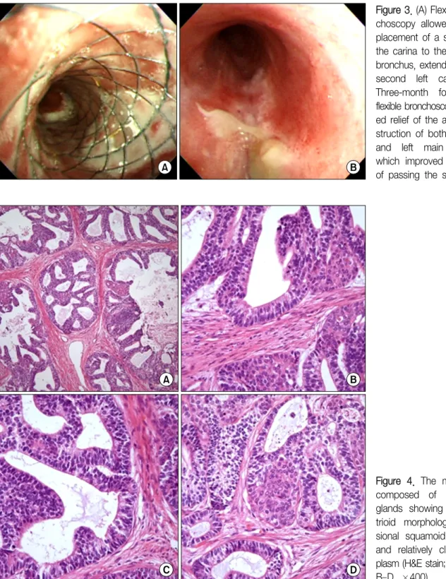
lobe. The stent in the left main bronchus remained patent. Computed tomography (CT) revealed a 7.7×6.4 cm lobulated, heterogeneous enhancing mass abutting the trachea, carina, and both main bronchi, as well as multiple enlarged lymph nodes in the subclavian, right hilar, subcarinal, upper and lower paratracheal areas (Figure 2A). A metallic stent was placed in the left main bronchus and irregular narrowing of the right main bronchial wall was also observed. Flexible broncho-scopy (FBS) showed the placement of a stent from the carina to the left main bronchus, extending to the left second carina (Figure 3A). The left upper and lower bronchi appeared normal. A white fungating mass ob-structing the right main bronchus and a pin- point open-ing due to a mass-like lesion were detected.
Figure 4. The mass was composed of numerous glands showing endome-trioid morphology, occa-sional squamoid morules, and relatively clear cyto-plasm (H&E stain; A, ×100; B–D, ×400).
Figure 3. (A) Flexible bron-choscopy allowed for the placement of a stent from the carina to the left main bronchus, extending to the second left carina. (B) Three-month followed-up flexible bronchoscopy show-ed relief of the airway ob-struction of both the right and left main bronchi, which improved the ease of passing the scope.
Thoracoscopic biopsy of the mediastinal lymph node was performed. Gross examination revealed a single, 2 cm, white, well-defined mass that bled easily. Histo-pathologically, the mass was composed of numerous glands showing an endometrioid morphology,
occa-sional squamoid morules (×400), and relatively clear cytoplasm (Figure 4). Based on these histopathological findings, we finalized the diagnosis as fetal adeno-carcinoma.
in this case, concurrent chemoradiation therapy (Tomo-Helical; 5,500 cGy) was administered. Treatment was comprised of 25 mg/m2 docetaxel weekly for six weeks with concomitant radiation therapy; the patient com-pleted the planned treatment regimen. Repeat CT per-formed at a 1-month follow-up visit (Figure 2B) showed the mass in the right paratracheal area had decreased in size (5.8 cm decreased to 4.6 cm), indicating a stable disease. Additional follow-up chest CT and FBS were scheduled at 3-month intervals. On follow-up studies at 4 months, positron emission tomography-CT showed significantly decreased size and activity (5.5×4.6 cm to 4.5×3.4 cm) of the mediastinal mass, which was con-sidered a favorable response to the treatment, and no other newly developed hypermetabolic foci to suggest distant metastasis were seen. Bronchoscopy (Figure 3B) showed relief of the airway obstruction of the right and left main bronchi, with increased ease of passing the scope. Compared with the previous chest X-ray (Figure 1A), the airway obstruction and tracheal deviation were improved (Figure 1B). We were able to remove the stent from the left main bronchus. Previous symptoms of dyspnea and cough were also improved. A further partial response was noted on the 7-month follow-up studies, and another CT follow-up is planned in 3 months.
Discussion
Fetal adenocarcinoma was formerly classified as a subtype of pulmonary blastoma termed well-differen-tiated fetal adenocarcinoma (WDFA)6. Because WDFA is characterized by a monophasic pattern consisting of an epithelial component alone, distinct from other pul-monary blastoma subtypes, the WHO in 1999 removed WDFA tumors from the pulmonary blastoma category and classified them as a variant of adenocarcinoma7. Our understanding of fetal adenocarcinoma is derived from case reports or small case series. It is estimated that approximately 0.1% of lung neoplasms are fetal adenocarcinomas. It is generally found in young pa-tients with a unimodal age peak in the third decade,
has a slight female predominance, and is most com-monly early stage disease at diagnosis with no lymph node involvement. It is generally treated and frequently cured by surgical resection, and has better survival com-pared with similarly staged and treated adenocarcinoma of the lung2,7. According to a study of 25 cases of fetal adenocarcinoma by Sato et al.7, cases in which the tu-mor was detected on a chest radiograph as part of rou-tine health maintenance accounted for 76% of the total. The tumor size at resection was comparatively small, with a median size of 3.5 cm (range, 1.4–12.0 cm). The tumors tended to be localized in the right lung, espe-cially in the upper lobe. Among the 24 cases in which the pathological stage was reported, there were two cas-es with N1 lymph node metastasis at the time of re-section, and only one had a distant metastasis to the eye at the time of diagnosis. There were no N2-positive cases, and the vast majority of cases were N0 (22 cases, 88%)7,8.
Contrasting with other cases, we described a 48- year-old male patient with locally advanced lung cancer in this paper. The lung mass was found by chest imag-ing performed due to the patient's symptoms rather than through routine health maintenance screening. Operative intervention was impossible because of the degree of local advancement and the presence of multi-ple lymph node involvement and tracheal deviation. The standard treatment for fetal adenocarcinoma is surgical resection. However, there have been no estab-lished treatment guidelines for inoperable cases up to this point. Van Loo et al.6 previously reported that among 63 cases of pulmonary blastoma, 9 were classi-fied as WDFA. Of these nine WDFA cases, three were beyond T3 stage and underwent postoperative chemo-therapy or radiation chemo-therapy. However, the benefit of this therapy was unclear6. In our case, after ameliorating the airway obstruction with stent insertion via broncho-scopy, concurrent chemoradiation therapy (CCRT) was performed with docetaxel for inoperable fetal adeno-carcinoma. A partial response was observed on fol-low-up chest CT at 1 month and 4 months after treat-ment, and follow-up chest CT and FBS were scheduled
at 3-month intervals.
Patients with fetal adenocarcinoma tend to have a better prognosis than those with other types of adeno-carcinoma. Surgical resection is the treatment of choice for operable cases; however, no treatment guidelines have been established for inoperable cases. We encoun-tered an inoperable case of fetal adenocarcinoma of the lung, and a favorable response was achieved with doce-taxel-based CCRT. Therefore, surgical resection follow-ing tumor down-stagfollow-ing with neo-adjuvant chemo-therapy or radiation chemo-therapy is one option for effective treatment.
References
1. Kradin RL, Young RH, Dickersin GR, Kirkham SE, Mark EJ. Pulmonary blastoma with argyrophil cells and lack-ing sarcomatous features (pulmonary endodermal tu-mor resembling fetal lung). Am J Surg Pathol 1982;6: 165-72.
2. Travis WD, Brambilla E, Noguchi M, Nicholson AG, Geisinger KR, Yatabe Y, et al. International Association for the Study of Lung Cancer/American Thoracic Society/European Respiratory Society international
mul-tidisciplinary classification of lung adenocarcinoma. J Thorac Oncol 2011;6:244-85.
3. Kang CU, Cho DG, Jo MS, Cho KD, Moon YK, Park JK. Well-differentiated fetal adenocarcinoma of the lung: 3 cases report. Korean J Thorac Cardiovasc Surg 2009;42:388-91.
4. Song DS, Chung WS, Kim H, Kim YH, Kang JH, Lee CB, et al. Surgical treatment of well-differentiated fetal adenocarcinoma: a case report. Korean J Thorac Cardiovasc Surg 2001;34:566-8.
5. Nakatani Y, Kitamura H, Inayama Y, Kamijo S, Nagashima Y, Shimoyama K, et al. Pulmonary ad-enocarcinomas of the fetal lung type: a clinicopatho-logic study indicating differences in histology, epidemi-ology, and natural history of low-grade and high-grade forms. Am J Surg Pathol 1998;22:399-411.
6. Van Loo S, Boeykens E, Stappaerts I, Rutsaert R. Classic biphasic pulmonary blastoma: a case report and review of the literature. Lung Cancer 2011;73:127-32. 7. Sato S, Koike T, Yamato Y, Yoshiya K, Honma K,
Tsukada H. Resected well-differentiated fetal pulmo-nary adenocarcinoma and summary of 25 cases re-ported in Japan. Jpn J Thorac Cardiovasc Surg 2006; 54:539-42.
8. Koss MN, Hochholzer L, O'Leary T. Pulmonary blastomas. Cancer 1991;67:2368-81.