의학
의학
의학
의학 석사학위
석사학위
석사학위
석사학위 논문
논문
논문
논문
Albumin Therapy in Acute Stroke
Patients
아
아
아
아 주
주
주
주 대
대
대
대 학
학
학 교
학
교 대
교
교
대
대 학
대
학
학
학 원
원
원
원
의
의
의
의 학
학
학 과
학
과
과
과
신
신
신
신 동
동
동 훈
동
훈
훈
훈
Albumin Therapy in Acute Stroke
Patients
지도교수
지도교수
지도교수
지도교수 방
방
방
방 오
오
오 영
오
영
영
영
이
이
이
이 논문을
논문을
논문을
논문을 의학
의학
의학 석사학위
의학
석사학위
석사학위
석사학위 논문으로
논문으로
논문으로
논문으로 제출함
제출함
제출함.
제출함
2006 년
년
년 8 월
년
월
월
월
아
아
아
아 주
주
주
주 대
대
대
대 학
학
학 교
학
교 대
교
교
대
대 학
대
학
학
학 원
원
원
원
의
의
의
의 학
학
학 과
학
과
과
과
신
신
신
신 동
동
동 훈
동
훈
훈
훈
신동훈의
신동훈의
신동훈의
신동훈의 의학
의학
의학 석사학위
의학
석사학위
석사학위
석사학위 논문을
논문을
논문을
논문을 인준함
인준함
인준함
인준함.
심사위
심사위
심사위
심사위원장
원장
원장 허
원장
허
허
허 균
균
균
균 인
인
인
인
심사위원
심사위원
심사위원
심사위원 방
방
방
방 오
오
오
오 영
영
영 인
영
인
인
인
심사위원
심사위원
심사위원
심사위원 김
김
김
김 선
선
선
선 용
용
용
용 인
인
인
인
아
아
아
아 주
주
주
주 대
대
대
대 학
학
학 교
학
교 대
교
교
대
대 학
대
학
학
학 원
원
원
원
2006 년
년
년
년 6 월
월
월
월 22 일
일
일
일
Albumin Therapy in Acute Stroke Patient
by
Dong Hoon Shin
A Dissertation Submitted to The Graduate School of Ajou University
in Partial Fulfillment of the Requirements for the Degree of
MASTER OF MEDICAL SCIENCES
Supervised by
Oh Young Bang, M.D., Ph.D.
Department of Medical Sciences
The Graduate School, Ajou University
i
- ABSTRCTS -
Albumin Therapy in Acute Stroke Patient
Preclinical studies have recently shown that albumin has neuroprotective effects for stroke in animal models. Thus, we sought to evaluate the effects of albumin therapy in patients with acute cerebral infarcts.
We prospectively studied 49 patients with moderate-to-severe cerebral infarcts within the middle cerebral arterial territory into one of two groups: the control group (N=18) received saline, whereas the albumin group (N=31) received either 40 g or 80 g of albumin within 24 h from symptom onset. The National Institutes of Health Stroke Scale (NIHSS) and diffusion-weighted imaging (DWI) were serially checked.
There was no adverse effect related to albumin therapy. Although there was no significant difference in both baseline NIHSS score and DWI lesion volume on admission, the NIHSS scores at the 14th day after treatment and the increase in DWI lesion volume 72–96 h after treatment were significantly reduced in patients of the albumin group (P=0.001 and 0.012, respectively); these effects were dose- and time-related. The outcome on the 90th day after stroke onset was more favorable in the albumin group than in the control group. However, among the albumin group, significant improvement was nearly exclusively observed in patients who had patent or recanalized vessels (P=0.004).
Our results indicate that albumin therapy is a safe and effective modality in patients with acute cerebral infarction. This study also suggests that the effects of albumin therapy may vary depending on vessel status of the patient.
Key words : Albumin, Acute stroke, Human
ii
TABLE OF CONTENTS
ABSTRCT ··· i
TABLE OF CONTENTS ···ii
LIST OF FIGURES ···iii
LIST OF TABLES ··· iv
I. INTRODUCTION ···1
II. MATERIALS AND METHODS ···3
A. PATIENTS AND GROUPING···3
B. MEASUREMENTS ···4 C. STATISTICAL ANALYSIS ···6 III. RESULTS ···7 IV. DISCUSSION ···17 V. CONCLUSION ···23 REFERENCES ···24 국문요약···31
iii
LIST OF FIGURES
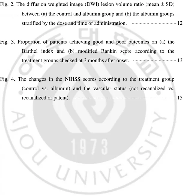
Fig. 1. The changes in the National Institutes of Health Stroke Scale (NIHSS) score (mean ± SD) between (a) the control and albumin group and (b) the albumin groups stratified by the dose and time of administration ····11 Fig. 2. The diffusion weighted image (DWI) lesion volume ratio (mean ± SD)
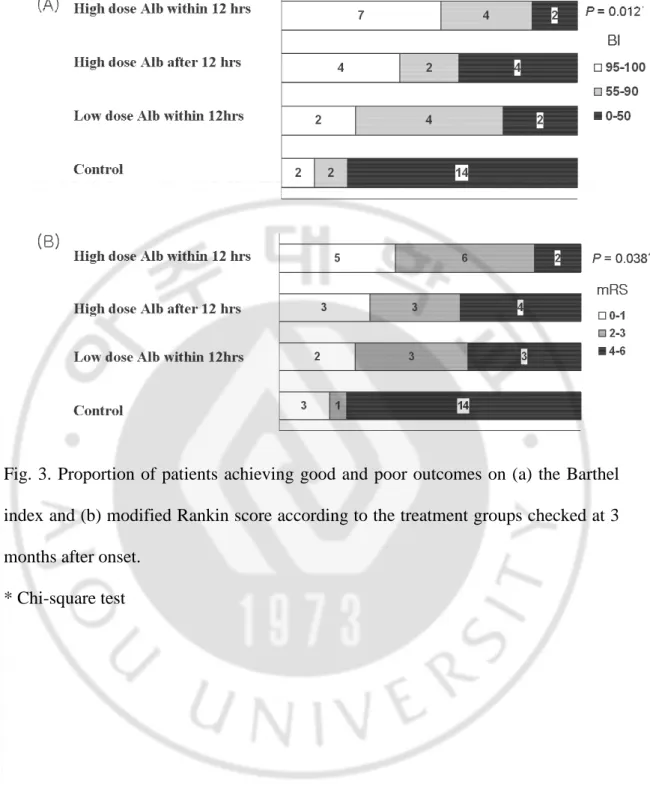
between (a) the control and albumin group and (b) the albumin groups stratified by the dose and time of administration. ···12 Fig. 3. Proportion of patients achieving good and poor outcomes on (a) the
Barthel index and (b) modified Rankin score according to the treatment groups checked at 3 months after onset. ···13 Fig. 4. The changes in the NIHSS scores according to the treatment group
(control vs. albumin) and the vascular status (not recanalized vs. recanalized or patent). ···15
iv
LIST OF TABLES
Table 1. Characteristics of patients ···9 Table 2. Factors affecting the effects of albumin ···16
1
I. INTRODUCTION
Ischemic stroke is a major cause of death and long-term disability in the elderly. Due to the limited potential for renewal of neuronal cells, severe neurologic deficits hardly improve once neurons are severely damaged even after long periods of time. Therefore, treatment during the acute stage of damage is important in the management of cerebral ischemic stroke. Unfortunately, an effective method for the management of acute stroke that can diminish the extent of neuronal damage and improve the functional outcome other than recombinant tissue-type plasminogen activator (tPA) has not been available. Although tPA has been shown to improve the functional outcome in ischemic stroke if administered within 3 h of the stroke onset in a randomized North American clinical trial and other prospective multicenter surveys of tPA use, the narrow therapeutic window and possibility of intracerebral hemorrhage limit its clinical use (NINDS rt-PA Stroke Study Group, 1995; Katzan et al, 2000).
Due to the laboratory investigations of cerebral ischemia over the past decades, key biochemical and molecular mechanisms that contribute to the death of brain tissue, such as excitotoxicity, tissue calcium overload, oxygen radicals, and inflammatory mediators, have been identified (Choi, 1992; Tymianski, 1998; Chan, 1996; Feuerstein et al, 1998). Because an antagonist against the mechanisms of ischemic injury could theoretically prevent damage and protect brain tissue, huge
2
efforts have been made to develop a pharmaceutical antagonist. The development of a neuroprotective agent has been hindered by the many side effects and inadequate efficacy experienced in randomized clinical trials involving ischemic stroke patients (Koroshetz and Moskowitz, 1996; Muir and Teal, 2005). Thus, a neuroprotective agent that effectively stops the vicious process triggering and propagating neuronal injury before severe damage occurs with an acceptable level of side effects, and can be administered easily without the need for time-consuming laboratory tests and specific delivery system must be developed.
Supplementation with human serum albumin has been used for patients whose level of albumin is low. In several recent experimental studies involving a rat model of ischemic stroke, albumin was found to be a multifunctional protein with potent neuroprotective effects. High-dose albumin therapy in preclinical studies has been found to provide protective effects by diminishing the volume of brain infarction and swelling, ameliorating behavioral and neurological functions, and improving the local perfusion to zones of critical blood flow reduction (Belayev et al, 1997; Belayev et al, 1998; Belayev et al, 1999; Belayev et al, 2001; Liu et al, 2001). In this study, we prospectively evaluated the efficacy and safety of high-dose albumin therapy in non-lacunar stroke patients with acute cerebral infarcts within the territory of the middle cerebral artery (MCA).
3
II. MATERIALS AND METHODS
A. Patients and Grouping
This study was a randomized, controlled, phase I/II clinical trial. The clinical trial protocol and the consent form were approved by the Institutional Review Board for Human Investigation of Ajou University Hospital, South Korea.
Between October 2004 and November 2005, we prospectively studied consecutive patients who were admitted with ischemic stroke within 12 h after the onset of symptoms and had relevant lesions within the MCA territory on diffusion-weighted imaging (DWI), non-lacunar infarcts clinically (Fisher, 1991), and moderate neurological deficits, i.e., patients with ≥5 and <20 points at admission according to the National Institutes of Health Stroke Scale (NIHSS) (Meyer et al, 2002). The exclusion criteria was as follows: severe hypertension above 180/100 mmHg; heart failure, defined as previously diagnosed or compatible with Framingham criteria (Eriksson et al, 1987); severe anemia, defined as hemoglobin below 8 g/dL; severe dehydration, defined as decreased skin turgor, dry oral mucous membrane, tachycardia (>100/min), and oliguria; or a candidate for thrombolytics or thrombolytics had already been used because thrombolytics can improve clinical outcome by itself. Patients were randomly allocated to one of two groups, the albumin group or the control group, based on the day of admission; patients who were admitted from Monday to Thursday received human serum albumin (20%
4
100cc albumin/bottle, Green Cross Corp, South Korea) (the albumin group), while those who were admitted from Friday to Sunday received saline as much as albumin volume (the control group). The albumin group was divided into three subgroups based on the time of albumin administration after the onset of symptoms, i.e., early (within 12 h of symptom onset) or delayed (12–24 h), and the dosage of albumin, i.e., either the high (1.25 g/kg over 4 h) or low dose (0.63 g/kg over 2 h), using random cards as follows: Alb low dose, early (specified the administration of the low albumin
dose within 12 h of the onset of acute stroke); Alb high dose, early (specified the
administration of the high albumin dose within 12 h of onset); and Alb high dose, delayed
(specified the administration of the high albumin dose between 12 and 24 h of onset).
B. Measurements
Patients were evaluated according to the protocol that included demographic data, medical history, vascular risk factors, stroke scales, and laboratory tests, as in our previous study (Bang et al, 2003). The laboratory tests included brain magnetic resonance imaging (MRI, 1.5T), vascular studies (including digital subtraction, magnetic resonance, or computed tomographic angiography), echocardiograms, electrocardiograms, and routine blood tests. Based on their clinical syndrome, infarct size on DWI, and the results of the vascular and cardiological studies, the mechanisms of stroke were documented in 47 (96%) patients (Adams et al, 1993).
5
All the patients underwent MRI studies including DWI and apparent diffusion coefficiency (ADC). Routine hematological and serological studies including inflammatory markers (CRP and fibrinogen) and albumin level at both admission and 72–96 h after albumin or saline infusion were performed. A follow-up vascular study (computed tomographic angiography) was performed 72–96 h after admission in all patients who showed major stenosis on the initial vascular studies.
To evaluate the effect of albumin, changes in the NIHSS score, were serially checked at admission, 1st day, 3rd day, 7th day, 14th day, and Barthel Index (BI), and modified Rankin score (mRS) were checked at admission, 7th day, 14th day, 1st month, and 3rd month after the onset of symptoms. Either one or two indices among the three (the NIHSS, BI, and mRS) are usually used to evaluate the therapeutic efficacy of new drug or treatment methods; in this study, however, we performed a comprehensive approach using the NIHSS to evaluate the neurologic deficit and BI and mRS to gauge the functional recovery. We also performed volumetric analysis of the DWI lesion volume using the DWI volume ratio, which was defined as the following: [(the volume of the DWI lesion at 72–96 h after onset – the volume of the DWI lesion at admission) / (the volume of the DWI lesion volume at admission)] × 100. Volumes were computed by multiplying the measured area per slice by the section thickness (slice thickness, 4 mm; gap, 1 mm). We assessed the safety of intravenous albumin infusion by checking whether immediate allergic reactions occurred, checking follow up Hb/Hct for aggravation of anemia, and checking chest X-ray after one day for cardiovascular overload.
6
C. Statistical Analysis
Differences between the groups with respect to the clinical and radiological features and the prognoses were examined using the Chi-square test, Fisher’s exact test, and Student’s t-test or one-way analysis of variance with post hoc comparisons. Statistical significance was established at P < 0.05.
7
III. RESULTS
A. General data
From the 131 patients with acute ischemic stroke within the MCA territory who were admitted during the study period, 49 were included in the study; 26 patients presented with lacunar syndrome, 49 patients showed mild deficits or met on exclusion criteria at the time of randomization, and 7 patients refused to participate in the study. The 49 patients included in the study were divided as follows: 18 were placed in the control group, and 31 were assigned to the albumin group, with 8 patients in the Alb low dose, early-treated subgroup, 13 in the Alb high dose, early-treated
subgroup, and 10 in the Alb high dose, delayed-treated subgroup.
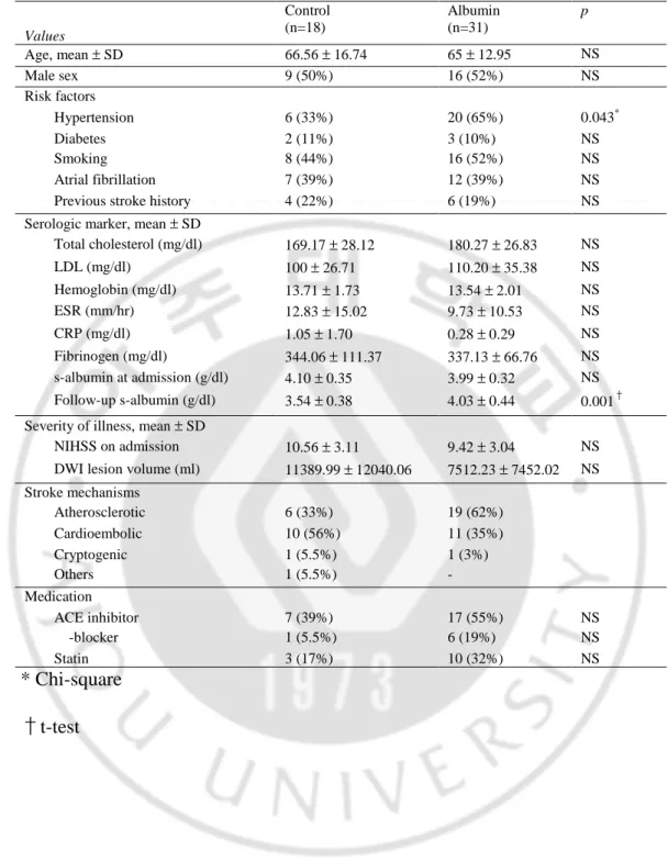
The characteristics of the patients are shown in Table 1. The clinical and radiological characteristics of the albumin group were not significantly different from those of the control group. The age and sex, risk factors for stroke, and stroke mechanisms were similar in both groups except hypertension was more frequent in albumin group (p=0.043) (Table 1). There was no significant difference in the baseline values of hematological and serological tests between the groups. The stroke severity, NIHSS score on admission, and initial DWI lesion volume among the patients were not different between the groups (P>0.05).
Although the serum albumin levels on admission were not different between the groups, the follow-up serum albumin levels in patients treated with albumin were
8
higher than in patients of the control groups (P=0.001) (Table 1). Compared to the initial serum albumin levels, the follow-up levels decreased in most of the control group (–0.57 ± 0.38 g/dl), decreased to lesser degree in patients treated with low dose albumin (–0.23 ± 0.33 g/dl), and increased in patients treated with high dose albumin (0.17 ± 0.48 g/dl).
9
Table 1. Characteristics of patients.
Values Control (n=18) Albumin (n=31) p Age, mean ± SD 66.56 ± 16.74 65 ± 12.95 NS Male sex 9 (50%) 16 (52%) NS Risk factors Hypertension 6 (33%) 20 (65%) 0.043* Diabetes 2 (11%) 3 (10%) NS Smoking 8 (44%) 16 (52%) NS Atrial fibrillation 7 (39%) 12 (39%) NS
Previous stroke history 4 (22%) 6 (19%) NS
Serologic marker, mean ± SD
Total cholesterol (mg/dl) 169.17 ± 28.12 180.27 ± 26.83 NS LDL (mg/dl) 100 ± 26.71 110.20 ± 35.38 NS Hemoglobin (mg/dl) 13.71 ± 1.73 13.54 ± 2.01 NS ESR (mm/hr) 12.83 ± 15.02 9.73 ± 10.53 NS CRP (mg/dl) 1.05 ± 1.70 0.28 ± 0.29 NS Fibrinogen (mg/dl) 344.06 ± 111.37 337.13 ± 66.76 NS s-albumin at admission (g/dl) 4.10 ± 0.35 3.99 ± 0.32 NS Follow-up s-albumin (g/dl) 3.54 ± 0.38 4.03 ± 0.44 0.001† Severity of illness, mean ± SD
NIHSS on admission 10.56 ± 3.11 9.42 ± 3.04 NS
DWI lesion volume (ml) 11389.99 ± 12040.06 7512.23 ± 7452.02 NS Stroke mechanisms Atherosclerotic 6 (33%) 19 (62%) Cardioembolic 10 (56%) 11 (35%) Cryptogenic 1 (5.5%) 1 (3%) Others 1 (5.5%) - Medication ACE inhibitor 7 (39%) 17 (55%) NS -blocker 1 (5.5%) 6 (19%) NS Statin 3 (17%) 10 (32%) NS * Chi-square †t-test
10
B. Improvements in Clinical and Neuroimaging Parameters
Several of the parameters tested in this study showed significant differences between the albumin and control groups. Despite similar initial values, the serial NIHSS scores at the 3rd, 7th, and 14th day were significantly lower in the albumin group than in the control patients (P=0.039, 0.009, and 0.001 at the 3rd, 7th, and 14th day, respectively) (Fig. 1A). This trend was also found in volumetric analysis (Fig. 2A). Although the infarct volume increased in both groups, the increase in the DWI lesion volume (as measured by the DWI volume ratio) was significantly higher in the control group than in the albumin group (67.2% vs. 26.0%, P=0.012). When patients in the albumin group were stratified based on the dose and time of administration, patients who were treated earlier with the high dose of albumin showed much more improvement than the other patients in both clinical (Figure 1B) and neuroimaging (Fig. 2B) features.
The effects of albumin therapy were consistently observed until the follow-up period (Figure 3). The results of the mRS and BI at the 90th day after symptom onset showed that patients who were treated with albumin maintained a better functional status than those of the control groups (P=0.004 for mRS and 0.002 for BI, not shown). In addition, the dose-dependent and infusion time-related differential effects of albumin were also observed in the long-term prognosis as in the early course after symptom onset; the prevalence of functional independence was highest in patients
11
who were treated earlier with the high dose (P=0.038 for mRS and 0.012 for BI) (Fig. 3).
C. Subgroup Analysis in the albumin group
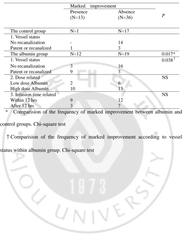
Despite of overall difference between the control and albumin group, a dramatic response after albumin therapy was observed in several patients. When significant improvement was defined as
Fig. 1. The changes in the National Institutes of Health Stroke Scale (NIHSS) score (mean ± SD) between (a) the control and albumin group and (b) the albumin groups stratified by the dose and time of administration.
12
Fig. 2. The diffusion weighted image (DWI) lesion volume ratio (mean ± SD) between (a) the control and albumin group and (b) the albumin groups stratified by the dose and time of administration.
* The increase in DWI lesion volume ratio
= (DWI volume 72–96 h after onset – initial DWI volume) × 100 Initial DWI volume
13
Fig. 3. Proportion of patients achieving good and poor outcomes on (a) the Barthel index and (b) modified Rankin score according to the treatment groups checked at 3 months after onset.
14
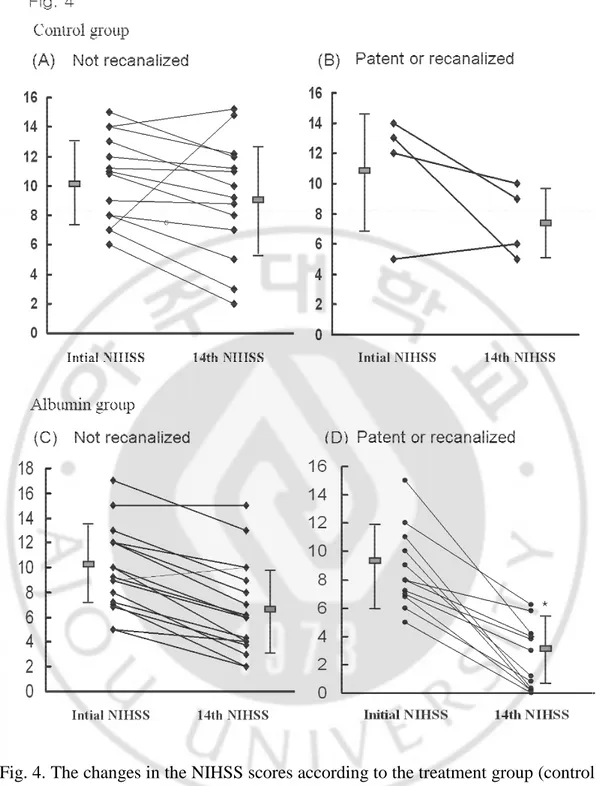
lowered by above 5 points NIHSS scores to less than 6 points within 14th days after the onset of symptoms, there were 12 patients in the albumin group but only one in the control group who showed significant improvement (P=0.017) (Table 2). Recanalized vessel was defined as initially occluded but visualized distant vessel at follow up angio-computerized tomography imaging. Compared within albumin group, significant improvement was more frequent in patients with a patent or recanalized cerebral vessel (P=0.038) (Table 2). Although not significant, more patients improved when high dose albumin was infused within 12 h as previously noted. The reduction in the NIHSS at the 14th day was significantly higher in the albumin group with patent or recanalized vessels than in the albumin group with occluded vessel and control group (P=0.004) (Fig. 4).
D. Safety
We did not encounter any immediate or delayed complications related to the albumin infusion, such as adverse allergic reactions, aggravated cardiac symptoms (dyspnea, chest pain), pulmonary edema documented on chest X-ray at 1st, 3rd, 7th day after albumin infusion, or worsening anemia.
15
Fig. 4. The changes in the NIHSS scores according to the treatment group (control vs. albumin) and the vascular status (not recanalized vs. recanalized or patent).
16
Table 2. Factors affecting the effects of albumin. Marked improvement Presence
(N=13)
Absence
(N=36) P
The control group N=1 N=17 1. Vessel status
No recanalization - 14 Patent or recanalized 1 3
The albumin group N=12 N=19 0.017* 1. Vessel status 0.038† No recanalization 3 16
Patent or recanalized 9 3
2. Dose related NS
Low dose Albumin 2 6 High dose Albumin 10 13
3. Infusion time related NS Within 12 hrs 9 12
After 12 hrs 3 7
* Comparision of the frequency of marked improvement between albumin and control groups. Chi-square test
†Comparision of the frequency of marked improvement according to vessel
17
IV. DISCUSSION
Patients who were so severe not to respond to any treatment (NIHSS <20) or so mild to improve spontaneously without treatment (NIHSS <4) were excluded. We targeted subject who was occluded in MCA and infused equivalent dose according to result of preclinical trial. The goal of this study was to determine the neuroprotective effect of albumin in humans as has been shown in many animal studies. Belayev et al. reported many cases on the neuroprotective effects of albumin. In rats with a temporary MCA occlusion, high-dose albumin infusion significantly improved behavioral function and reduced the total infarct volume by 34% and the cortical infarct volume by 57% compared to vehicle-treated rats (Belayev et al, 1997; Belayev et al, 1998). Furthermore, albumin therapy histologically salvaged some brain tissue in the ischemic zone by fostering the partial preservation of glial and endothelial elements (Belayev et al, 1998). Researchers have recently reported that a lower albumin dose was also strongly neuroprotective, with a broad therapeutic window extending to 4 h after the onset of stroke (Belayev et al, 2001). Treatment of albumin led to 48% increases in cortical perfusion (Liu et al, 2001) and improves microcirculatory dynamics in ischemia (Belayev et al, 2002). Other researchers reported albumin improves fatty acid delivery to the ischemic brain (Rodriguez et al, 2002). In addition, one recent clinical study reported that a relatively high serum
18
albumin level in acute stroke patients decreased the risk of poor outcome (Dziedzic et al, 2004).
Our present study showed dose-dependent effects of albumin in patients with acute ischemic stroke. These findings are compatible with the recent results of the ALIAS study that showed that albumin therapy appears to be well tolerated with few adverse events and at higher dose group may improve neurological outcome (Ginsberg et al, 2004; Ginsberg et al, 2005). Our results also showed that human albumin therapy significantly reduces the volume of cerebral infarction and significantly improves long-term neurological functions. In this study, we followed the recommendations for clinical trial evaluation of acute stroke therapy as follows (See Supplementary table below) (Stroke Therapy Academic Industry Roundtable II, 2001; Muir and Teal, 2005). First, we used an equivalent dose that had been shown to be effective in animal studies (Belayev, 2001). Second, the time window for albumin administration was decided to be 3–24 h to avoid possible confounding effects of tPA. Third, patients with ischemic stroke within the MCA territory were included, and patients with lacunar infarcts that mostly have good prognosis and predominantly involve white matter were excluded in this study because most neuroprotective substances have been tested in animal models of MCA occlusion. The data on the actions of neuroprotective substances in white matter are limited and conflicting. Lastly, patients with moderately severe disability were chosen as the target population. Many failed neuroprotectant trials that enrolled patients with either mild or very
19
severe strokes, which may have reduced the chances of demonstrating efficacy (Stroke Therapy Academic Industry Roundtable II, 2001; Muir and Teal, 2005).
A. Mechanisms of Neuroprotection
Albumin’s neuroprotective effect may be explained as follows. First, the albumin molecule has a prolonged circulating half-life (~20 days), and its high molecular weight prevents it from easily leaking from the intravascular space, which substantially increases the plasma oncotic pressure but does not affect the plasma osmolality (Halliwell and Gutteridge, 1990). These effects cause the expansion of the intravascular blood volume, improving the microcirculation and facilitating hemodilution. However, the neuroprotective effect of human albumin therapy may not be mediated solely by hemodilution because the results of experimental and clinical studies using other hemodiluents have generally been disappointing (Scandinavian Stroke Study Group, 1988; Italian Acute Stroke Study Group, 1988; Hemodilution in Stroke Study Group, 1989; Aichner et al, 1998).
The second and most important explanation is that albumin is a potent antioxidant, being 10- to 20-fold more powerful than vitamin E (Wayner et al, 1985). Albumin can access the zone of reduced blood flow through the dysfunction of the blood– brain barrier that results from ischemia and may then protect neurons and vascular elements from ischemic injury. Human serum albumin binds to free fatty acids (Halliwell, 1988) and exists in relatively high concentrations in both plasma and
20
interstitial fluid, so it can play a role as a scavenger of endogenously and exogenously derived oxygen free radicals. By avidly binding to copper ions, albumin also inhibits copper-ion-dependent lipid peroxidation and retards the formation of highly reactive hydroxyl radical species (Halliwell and Gutteridge, 1990). Our present study showed that the beneficial effect of albumin was observed in patients who had recanalized vessels but not in patients for whom vessel reopening did not occur. Thus, it is conceivable that the antioxidant effects of albumin against reperfusion injury may be one of the major pleiotropic effects of albumin beyond hemodilution. Further studies using markers for oxidative stress are needed. Other possible mechanisms of neuroprotection are albumin’s vasodilating and protective effects on endothelium (Keaney et al, 1993; He and Curry, 1993; Zoellner et al, 1996) and metabolic role in astrocytes and neurons (Bogert et al, 1994; Tabernero et al, 1999).
B. Clinical Applications
Albumin is a strong neuroprotectant as shown in this study, so considerable brain tissue in the ischemic zone could be salvaged if albumin is given at the doses examined within 24 h of onset of an acute stroke. A more favorable functional recovery and greater chance of maintaining the ability to independently perform activities of daily living may then be expected. In the aspect of safety profile (such as hemorrhagic complication), albumin has a wider therapeutic window than
21
thrombolytics and can therefore be used in patients admitted more than 3 h past the onset of stroke.
Recent clinical trials of prehospital, paramedic administered intravenous magnesium sulfate, a neuroprotective agent, in focal stroke patients showed that field initiation is feasible, safe, and time-saving compared with the in-hospital initiation of neuroprotective therapy (Saver et al, 2004). Our results showed that the effect of albumin therapy was definitely present at a dose of 0.63 to 1.25 g/kg, which is a feasible clinical dose. Adverse effects did not occur at any of the doses tested in this study. Albumin has been widely used in practice for various aims and is easily available, relatively cheap compared to new drugs such as thrombolytics, and infusable by paramedics without the use of sophisticated devices in the hyperacute stage of cerebral infarct. Moreover, it may be possible to use albumin in a stroke patient without neuroimaging data because albumin is well-known to reduce swelling in patients with spontaneous or traumatic hemorrhagic strokes. Several studies have reported that the use of albumin in patients with cerebral hemorrhage or subarachnoid hemorrhage had beneficial effects. For example, putaminal hemorrhage treated with 12.5 to 25 g per day of albumin for 2 weeks showed reductions in cerebral edema and improved the outcome (Tomita et al, 1994), and the administration of human albumin after subarachnoid hemorrhage may improve the clinical outcome and reduce hospital costs (Suarez et al, 2004). On the basis of these results, albumin infusion will be an amenable, effective initial treatment for any kind of cerebrovascular accident during the hyperacute stage.
22
The role of albumin might be strengthened by the conjunctive use of thrombolytics. Our present study showed that albumin therapy may not have beneficial effects in all patients with ischemic stroke; the effects of albumin was minimal, if present at all, in the absence of recanalization, which may have been caused for two reasons. First, albumin will exert its neuroprotective effects and reduce the neurologic deficit only after reaching the lesion site. Thus, one could expect to obtain a better response in a case of stroke with a patent vessel and cortical infarct. Second, as previously mentioned above, our results suggest that among the numerous mechanisms of cell death after ischemic injury, reperfusion injury might be a target of albumin. Therefore, to augment the effects of albumin, its combination with thrombolysis should be considered.
23
V. CONCLUSIONS
This study has several limitations. The chief limitation is that our study included a relatively small number of patients. Further studies with more patients such as ALIAS trial will exclude the sampling error and overcome this limitation. And the study was not blinded. This is a design flaw as bias could be introduced. Although we had checked clinical neurologic outcome during early admission period non-blindly, nurse practioner who didn’t know about albumin study have checked long term clinical neurologic outcome during outpatient follow up period. So this study is not entire blind study, but partial blind study. We think the possible bias has been minimized. In addition, even though we tried to perform DWI and perfusion-weigthed image in all the patients, we could not perform in many incooperated patients due to severe deficits and could not analyze the effect of albumin to perfusion.
In summary, human albumin therapy is a safe and effective modality with a relatively broad therapeutic window in acute cerebral infarction. Because the effect of albumin was different among the patients, further studies about the mechanisms of its action and a refinement of the candidate characteristics for this therapeutic modality are warranted.
24
REFERENCES
1. Adams HP Jr, Bendixen BH, Kappelle LJ, Biller J, Love BB, Gordon DL, Marsh EE: 3rd. Classification of subtype of acute ischemic stroke: definitions for use in a multicenter clinical trial. TOAST. Trial of Org 10172 in Acute Stroke Treatment. Stroke 24: 35-41, 1993
2. Aichner FT, Fazekas F, Brainin M, Polz W, Mamoli B, Zeiler K: Hypervolemic hemodilution in acute ischemic stroke: the Multicenter Austrian Hemodilution Stroke Trial (MAHST). Stroke 29: 743-749, 1998
3. Bang OY, Lee PH, Joo SY, Lee JS, Joo IS, Huh K: Frequency and mechanisms of stroke recurrence after cryptogenic stroke. Ann Neurol 54: 227-234, 2003
4., Belayev L, Busto R, Zhao W, Clemens JA, Ginsberg MD: Effect of delayed albumin hemodilution on infarction volume and brain edema after transient middle cerebral artery occlusion in rats. J Neurosurg 87: 595-601, 1997
5. Belayev L, Liu Y, Zhao W, Busto R, Ginsberg MD: Human albumin therapy of acute ischemic stroke: marked neuroprotective efficacy at moderate doses and with a broad therapeutic window. Stroke 32: 553-560, 2001
25
6. Belayev L, Pinard E, Nallet H, Seylaz J, Liu Y, Riyamongkol P, Zhao W, Busto R, Ginsberg MD: Albumin therapy of transient focal cerebral ischemia: in vivo analysis of dynamic microvascular responses. Stroke 33: 1077-1084, 2002
7. Belayev L, Saul I, Huh PW, Finotti N, Zhao W, Busto R, Ginsberg MD: Neuroprotective effect of high-dose albumin therapy against global ischemic brain injury in rats. Brain Res 845: 107-111, 1999
8. Belayev L, Zhao W, Pattany PM, Weaver RG, Huh PW, Lin B, Busto R, Ginsberg MD: Diffusion-weighted magnetic resonance imaging confirms marked neuroprotective efficacy of albumin therapy in focal cerebral ischemia. Stroke 29: 2587-2599, 1998
9. Bogaert YE, Rosenthal RE, Fiskum G: Postischemic inhibition of cerebral cortex pyruvate dehydrogenase. Free Radic Biol Med 16: 811-820, 1994
10. Chan PH: Role of oxidants in ischemic brain damage. Stroke 27: 1124-1129, 1996
11. Choi DW: Excitotoxic cell death. J Neurobiol 23: 1261-1276, 1992 12. Dziedzic T, Slowik A, Szczudlik A: Serum albumin level as a
26
13. Eriksson H, Caidahl K, Larsson B, Ohlson LO, Welin L, Wilhelmsen L, Svardsudd K: Cardiac and pulmonary causes of dyspnoea—validation of a scoring test for clinical-epidemiological use: the Study of Men Born in 1913. Eur Heart J 8: 1007–1014, 1987
14. Feuerstein GZ, Wang X, Barone FC: A The role of cytokines in the neuropathology of stroke and neurotrauma. Neuroimmunomodulation 5: 143-159, 1998
15. Fisher CM: Lacunar infarcts: a review. Cerebrovasc Dis 3: 311-320,1991
16. Ginsberg MD, Belayev L, Bazan NG, Marcheselli VL, Hill MD, Palesch YY, Khoutorova L, Rodriguez de Turco EB, Ryckborst K, Tamariz D, Busto R: Albumin-based neurotherapeutics for acute ischemic stroke: from bench to bedside. In Pharmacology of Cerebral Ischemia (ed. Krieglstein J, Klumpp S) Stuttgart, Medpharm Scientific Publishers, pp. 421-433, 2004
17. Ginsberg MD, Hill MD, Palesch YY, Ryckborst KJ, Tamariz D: Albumin therapy for ischemic stroke: The ALIAS phase I dose-escalation and safety trial. Program Schedule and Abstracts of the 30th
International Stroke Conference 36: 420, 2005
18. Halliwell B: Albumin--an important extracellular antioxidant?
27
19. Halliwell B, Gutteridge JM: The antioxidants of human extracellular fluids. Arch Biochem Biophys 280: 1-8, 1990
20. He P, Curry FE: Albumin modulation of capillary permeability: role of endothelial cell [Ca2+]i. Am J Physiol 265: H74-82, 1993
21. Hemodilution in Stroke Study Group. Hypervolemic hemodilution treatment of acute stroke. Results of a randomized multicenter trial using pentastarch. The Hemodilution in Stroke Study Group. Stroke 20: 317-323, 1989
22. Italian Acute Stroke Study Group. Haemodilution in acute stroke: results of the Italian haemodilution trial. Italian Acute Stroke Study Group. Lancet 1: 318-321, 1988
23. Katzan IL, Furlan AJ, Lloyd LE, Frank JI, Harper DL, Hinchey JA, Hammel JP, Qu A, Sila CA: Use of tissue-type plasminogen activator for acute ischemic stroke: the Cleveland area experience. JAMA 283: 1151-1158, 2000
24. Keaney JF Jr, Simon DI, Stamler JS, Jaraki O, Scharfstein J, Vita JA, Loscalzo J: NO forms an adduct with serum albumin that has endothelium-derived relaxing factor-like properties. J Clin Invest 91: 1582-1589, 1993
25. Koroshetz WJ, Moskowitz MA: Emerging treatments for stroke in humans. Trends Pharmacol Sci 17: 227-233, 1996
28
26. Liu Y, Belayev L, Zhao W, Busto R, Belayev A, Ginsberg MD: Neuroprotective effect of treatment with human albumin in permanent focal cerebral ischemia: histopathology and cortical perfusion studies.
Eur J Pharmacol 428: 193-201, 2001
27. Meyer BC, Hemmen TM, Jackson CM, Lyden PD: Modified National Institutes of Health Stroke Scale for use in stroke clinical trials: prospective reliability and validity. Stroke 33: 1261-1266, 2002
28. Muir KW, Teal PA: Why have neuroprotectants failed? Lessons learned from stroke trials. J Neurol 252: 1011-1020, 2005
29. NINDS rt-PA Stroke Study Group: Tissue plasminogen activator for acute ischemic stroke. The National Institute of Neurological Disorders and Stroke. N Engl J Med 333: 1581-1587, 1995
30. Rodriguez de Turco EB, Belayev L, Liu Y, Busto R, Parkins N, Bazan NG, Ginsberg MD: Systemic fatty acid responses to transient focal cerebral ischemia: influence of neuroprotectant therapy with human albumin. J Neurochem 83: 515-24, 2002
31. Saver JL, Kidwell C, Eckstein M, Starkman S; FAST-MAG Pilot Trial Investigators: Prehospital neuroprotective therapy for acute stroke: results of the Field Administration of Stroke Therapy-Magnesium (FAST-MAG) pilot trial. Stroke 35: e106-108, 2004
29
32. Scandinavian Stroke Study Group: Multicenter trial of hemodilution in acute ischemic stroke. Results of subgroup analyses. Scandinavian Stroke Study Group. Stroke 19: 464-471, 1988
33. Stroke Therapy Academic Industry Roundtable II (STAIR-II): Recommendations for clinical trial evaluation of acute stroke therapies.
Stroke 32: 1598-1606, 2001
34. Suarez JI, Shannon L, Zaidat OO, Suri MF, Singh G, Lynch G, Selman WR: Effect of human albumin administration on clinical outcome and hospital cost in patients with subarachnoid hemorrhage. J
Neurosurg 100: 585-590, 2004
35. Tabernero A, Medina A, Sanchez-Abarca LI, Lavado E, Medina JM: The effect of albumin on astrocyte energy metabolism is not brought about through the control of cytosolic Ca2+ concentrations but by free-fatty acid sequestration. Glia 25: 1-9, 1999
36. Tomita H, Ito U, Tone O, Masaoka H, Tominaga B: High colloid oncotic therapy for contusional brain edema. Acta Neurochir Suppl
(Wien) 60: 547-549, 1994
37. Tymianski M: The central role of calcium ions and calcium-permeable channels in ischemic brain injury. In Cerebrovascular Disease:
Pathophysiology, Diagnosis, and Management (ed. Ginsberg MD,
30
38. Wayner DD, Burton GW, Ingold KU, Locke S: Quantitative measurement of the total, peroxyl radical-trapping antioxidant capability of human blood plasma by controlled peroxidation. The important contribution made by plasma proteins. FEBS Lett 187: 33-37, 1985 39. Zoellner H, Hofler M, Beckmann R, Hufnagl P, Vanyek E, Bielek E,
Wojta J, Fabry A, Lockie S, Binder BR: Serum albumin is a specific inhibitor of apoptosis in human endothelial cells. J Cell Sci 109: 2571-2580, 1996
31 - 국문요약 -
급성기
급성기
급성기
급성기 뇌경색
뇌경색
뇌경색
뇌경색 환자에서의
환자에서의
환자에서의
환자에서의 알부민
알부민
알부민
알부민 치료
치료
치료
치료
아주대학교 대학원의학과 신 동 훈 (지도교수: 방 오 영) 최근 동물 뇌경색 모델에서 알부민이 신경보호 효과가 있다는 여러 보고가 있다. 이에 저자들은 급성 뇌경색 환자들에서 알부민 치료의 효과에 대해 평가해 보고자 하였다. 중등도 이상의 중대뇌 동맥 영역의 뇌경색 환자 49 명을 대조군과 알부민 군으로 나누었다. 대조군 18 명은 식염수를 주입하였고 알부민 군 31 명은 증상 발현 24 시간 이내에 40g 혹은 80g 의 알부민을 투여하였고 알부민의 효과를 임상적으로는National Institutes of Health Stroke Scale (NIHSS)를 통해 그리고
방사선학적으로는 확산 강조 영상을 이용하여 평가하였다. 모든 환자에서 알부민 치료에 따른 부작용은 없었다. 입원 당시에 대조군과 알부민 군의 NIHSS 와 확산 강조 영상에서의 경색의 크기 차이는 없었으나 알부민 군에서 치료후 14 일째의 NIHSS 점수와 2 – 3 일 후에 시행한 확산 강조 영상의 경색 크기 증가가 대조군에 비해 유의하게 감소되었다(각각 P=0.001, 0.012).
32 또한 투여된 시간이 짧을수록 용량이 증가할수록 이런 알부민의 효과는 상승되는 경향을 보였다. 알부민 치료 후 90 일째의 기능적 측면에서도 알부민 군에서 대조군에 대해 유의하게 호전 되었다. 알부민 군내 분석을 시행했을 때 주로 혈관이 개통되었거나 나중에 재개통된 환자들에서 확연한 임상적인 호전을 보였다(P=0.004). 이번 연구 결과는 알부민 치료가 안전하면서도 급성 뇌경색 환자에서 효과적인 치료 방법이라는 사실을 말해준다. 또한 알부민 치료는 환자의 혈관 상태에 따라 그 효과가 좌우된다는 사실도 알 수 있다. 핵심어: 알부민, 급성 뇌경색, 인간