106
A Standardized Pathology Report for Gastric Cancer
Woo Ho Kim, Cheol Keun Park, Young Bae Kim, Youn Wha Kim, Ho Guen Kim,
Han Ik Bae, Kyu Sang Song, Hee Kyung Chang, Hee Jin Chang and Yang Seok Chae
The Gastrointestinal Pathology Study Group of the Korean Society of PathologistsBackground and Methods : The Gastrointestinal Pathology Study Group of the Korean Society of Pathologists developed a standardized pathology reporting format for gastric cancer in collaboration with the Korean Gastric Cancer Association. Results : The diagnostic parame-ters are divided into two part: the standard part and the optional part. The standard part contains most of the items listed in the Japanese classification, the TNM classification by UICC, the WHO classification, and the Korean Gastric Cancer Association classification. Therefore, the standard part is adequate for routine surgical pathology service. We included detailed descrip-tions on each item. Conclusions : The authors anticipate that this standardization can improve the diagnostic accuracy and decrease the discrepancies that occur in the pathologic diagno-sis of gastric cancer. Furthermore, the standard format can encourage large scale multi-insti-tutional collaborative studies.
Key Words : Stomach neoplasm; Pathology report; Surgical pathology; Neoplasm staging; His-tologic type
김우호∙박철근∙김영배∙김윤화
김호근∙배한익∙송규상∙장희경
장희진∙채양석
106위암 병리보고서 기재사항 표준화
위암은 우리나라에서 가장 많이 발생하는 암으로,
1가장 수술
이 많이 이루어지는 암인 동시에 병리과에서 검사하는 절제 암
중에서 가장 흔한 암이다. 그럼에도 이제까지 우리나라에서는 위
암 병리보고서를 작성하는 기준을 표준화하려는 시도가 없었다.
따라서 위암 병리검사 보고서는 병원에 따라 또는 병리의사에
따라 기록하는 항목의 편차가 컸을 뿐 아니라, 병리학적 진단에
대한 기준도 일관성이 없었다.
미국 외과학회(American College of Surgeons Commissions
on Cancer)에서는“2004년 1월부터 인증받은 병원의 병리의사
에게 병리보고서에 각각의 검체에 대해 과학적으로 검증되거나
통상적으로 사용하는 모든 정보를 기록할 것”
을 요구하였다.
2그
러나 현실적으로 전체 장기에 대해 기록해야 할 모든 정보의 범
위를 병리의사가 빠짐없이 파악하기는 어려우므로 보고서의 표
준화작업이 가속화되었고, 이를 위해서는 체크리스트 형태의 보
고서가 가장 적합하다고 알려져 있다.
3미국에서는 병리학회(College of American Pathologist, CAP)
와 병리과장협회(Association of Directors of Anatomic and
Surgical Pathology, ADASP)에서 각종 암에 대한 보고서를
작성할 때 참고할 표준안을 발표하였다. CAP 안은 좀 더 광범위
한 반면,
4ADASP 안은 검증된 내용인 필수적인 부분과, 아직
검증되지 않은 내용인 선택적인 사항으로 나누어져 있다.
5그러
나 위암은 미국에서는 극히 드문 반면에, 우리나라에서는 가장
흔한 암이기 때문에 미국의 표준안을 우리나라에서 그대로 사용
하기에는 부적절하다.
대한외과학회에서는 국내의 위암 증례를 분석할 수 있는 바탕
을 마련하기 위해서 1992년도에“위암규약집”
을 작성하였고, 많
은 외과의사들이 이 방법에 따라 위암 증례를 정리해 왔다. 또한
대한위암학회에서는 2002년 1월에 이를 보완하여“위암 기재사
항을 위한 설명서”
라는 소책자를 제작하여 배포하였다.
6그러나
이에 대한 병리의사의 호응이 저조하여 우리나라 전체에서 사용
하는 표준화 단계에는 이르지 못하였다.
대한병리학회 소화기병리학연구회에서는 대한위암학회와 함께
위암 병리보고서를 표준화하는 작업을 하기로 하였다. 2004년 8
월부터 10월까지 두 번의 소화기병리학연구회 산하의 위장관상
피성종양 소위원회 모임 및 두 번의 연구회 전체의 논의를 거쳐,
본“위암 병리보고서 기재사항”
을 완성하였다. 이를 위해 먼저
현재 국내 대형병원에서 사용하는 보고서 양식을 참고하였으며,
미국 병리학회의 권장사항, 미국 병리과장협회의 체크리스트, 일
본의
扱い規 ,
7UICC 분류법(제6판), WHO 분류(2000)
8등을 참고하였다. 또한 보건복지부에서 시행하는 한국중앙암등
106 106 접 수 : 2004년 12월 21일 게재승인 : 2005년 1월 25일 책임저자 : 박 철 근 우 135-710 서울시 강남구 일원동 50 성균관대학교 의과대학 삼성서울병원 병리과 전화: 02-3410-2766 Fax: 02-3410-0025 E-mail: ckpark@smc.samsung.co.kr 대한병리학회 소화기병리학연구회록사업에 필요한 정보가 빠지지 않도록 유념하였다.
1우리나라에 위암환자가 많음에도 좋은 임상 결과가 발표되지
않는 것은 애석한 일이다. 특히 수많은 위암 환자들이 adjuvant
chemotherapy를 받고 있으나, 어떤 약제가 효과가 있는지 알아
보기 위해 국내 자료를 참조하는 일도 어려운 실정이다. 또한 최
근에는 빠른 결과를 얻기 위해 임상시험을 여러 병원이 협력하여
많은 환자에 대해 수행하는 것이 보편적이다. 위암 병리보고서의
표준화는 여러 병원이 함께 임상시험에 참가하더라도 동일한 환
자군을 정확히 선택하는 데 큰 도움이 될 것으로 기대한다.
본 소위원회를 시작할 때는 간단한 보고서를 선호하는 병리의
사와 자세한 보고서를 선호하는 병리의사 사이에 매우 큰 의견
차이가 있었다. 그러나 일부 항목을 선택 기재사항으로 분류함으
로써 의견 차이를 좁힐 수 있었다. 각 병원에서는 특성에 맞추어
선택 기재사항 이외의 항목을 얼마든지 추가할 수 있으므로 선택
기재사항이 큰 의미가 없다고 볼 수도 있으나, 사용하는 단어나
진단기준을 일원화하는 데는 도움이 된다는 의견이 많았다. 예를
들면 lymphatic invasion을 판정할 때, 표준 기재사항에서는“not
identified, present”
로 분류하지만, 선택 기재사항에서는“not
identified, minimal, moderate, marked”
로 분류하고 있다. 그러
나 이를“not identified, minimal, severe”
의 3단계로 판정하는
것은 바람직하지 않다고 의견을 모았다. 선택 기재사항은 이러한
기준을 정하는 데 도움이 될 것으로 생각하였다.
위암의 조직학적 분류방법에는 크게 일본의 분류법과 WHO
분류법이 있으며, 둘 사이에 큰 차이는 없다. 그러나 위암은 조
직학적 형태가 다양해서, 조직학적으로 대표성 있는 유형을 고
르는 기준 역시 다르다. WHO 분류에서는 가장 넓은 범위를 차
지하는 유형에 대표성을 부여하고, 일본의 분류법은 분화가 나
쁜 유형에 대표성을 부여한다. 또 미국 병리과장협회에서는, 선
구조를 만드는 면적이 95%를 초과하면 고분화형, 50-95%면 중
분화형, 5-49%면 저분화형, 5% 미만이면 미분화형으로 규정하
였다. 이번 표준안에서는 WHO의 분류를 사용하기로 하였으나,
어떠한 분류를 사용하더라도 관찰자 개인 또는 관찰자 사이의
변이를 피하기 힘든 어려움이 있다.
병리보고서는 국제적인 비교가 필요한 경우가 많으므로 영어
로 기록하도록 하였다. 기재사항을 표준화하면서 약자는 극히
제한적으로 사용하였고, 새로운 약자를 만들어내지 않았다. 예
를 들면 sm1은“invades up to 1/3 of submucosa”
라고 기재
하며, Borrmann 5형은 unclassifiable로 기재하였다. 이는 각
항목의 판정기준이나 분류법이 바뀌더라도 추후에 추적할 수 있
도록 하기 위함이다.
본 기재사항을 각 병원에서 어떻게 사용할지는 각 병원의 특
성에 맞추어 결정할 일이지만, 판독용 양식을 작성해놓고 해당
항목에 동그라미 또는 숫자나 글씨를 적은 후에 별도로 입력하
는 방법이 바람직하기 때문에 이런 용도로 사용할 수 있는 판독
용 양식을 본 논문에 첨부하였다. 본 논문에 기록된 사항은 대한
병리학회 소화기병리학연구회의 홈페이지에서 얻을 수 있으며,
판독용 양식 파일을 다운받아 사용할 수 있다.
결과 및 고찰
절제 위의 표준 기재사항
해설: 본 기재사항은 수술로 절제한 위암 뿐 아니라 내시경절
제표본에서도 동일하게 적용할 수 있도록 하였다.
해설: 종양이 두 개 이상 있는 경우에는 multiple gastric
car-cinomas로 표시하고, 가장 깊은 종양부터 각각의 종양에 대해
모든 항목을 적되, lymph node metastasis, associated findings,
separate lesions은 가장 깊은 종양에만 적는다.
(조기위암과 진행위암을 기재하는 것은 중요한 의미가 있는 것
은 아니지만, 대부분의 병원에서 사용하고 있으므로 편의상 이를
주진단명으로 기록하기로 하였다.)
해설: Involvement에는 침범한 부위를 세 군데까지 적되 가
장 많이 침범한 순서대로 기록한다.
예) location: middle third and upper third
Center에는 병변의 중심부를 각각 한 군데씩 기록한다.
예) antrum, lesser curvature
(위의 위치를 지정하는 방법을 두고 매우 많은 토의를 했다. 중
앙암등록통계에서 사용하는 국제질병분류(ICD-O) 분류법에서
는 위암이 처음 발생한 부위를 기준으로“cardia, fundus, body,
antrum, pylorus, lesser curvature, greater curvature,
overlap-ping lesion, NOS”으로 나누고 있다.
9미국의 CAP 방법은 위를
11개 구역으로 나누고 침범한 구역을 모두 표시한다고 되어 있
으나, 너무 복잡해서 현실성이 없다. 일본의 분류법 및 대한위암
학회의 표준안은“upper third, middle third, lower third”로 나
누고 있다(Fig. 1). 이 분류법은 종양의 위치에 따라 원격전이에
Specimen type □ total gastrectomy □ distal subtotal gastrectomy □ proximal gastrectomy □ wedge resection
□ endoscopic mucosal resection □ other ____________________
Main diagnosis
□ Early gastric carcinoma □ Advanced gastric carcinoma
(□ Multiple gastric carcinomas)
Location involvement
[ ] esophagus [ ] upper third [ ] middle third [ ] lower third [ ] duodenum
center
□ cardia □ fundus □ body □ antrum □pylorus / □ lesser curvature □ greater curvature □ anterior wall □ posterior wall □ circle
해당하는 림프절이 달라지는 topographic N stage를 판정하는
데 기준이 된다. 그러나 절제 위에서는 남아 있는 위의 크기를
알 수 없으므로, 병리의사 혼자서 판정하기는 쉽지 않다. 이 3등
분법은 외과의사에게는 매우 쉽고 편한 방법이지만, 이 분류 결
과를 외과의사에게 전적으로 의존할 수는 없다고 결론지었다. 왜
냐하면, 외과의사의 기록이 부정확한 경우가 있으며, 특히 조기
위암, 다발성위암과 같이 육안으로 병변의 범위를 정확히 파악
하기 어려운 상황에서는 현미경 판독에 의하지 않고는 위치를
판정할 수 없기 때문이다. 이러한 이유로 표준 기재사항에는 세
가지 항목을 기록하기로 함으로써 종양의 위치를 표시하는 일이
조금 번거로워졌다.)
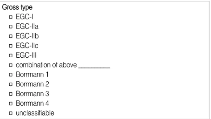
해설: 조기위암은 일본의 분류법을 따르며, 진행위암은
Bor-rmann의 분류법을 따른다.
Unclassifiable은 일본의 분류법에 따르면 Borrmann 5형에 해
당한다.
해설: WHO의 분류법(2000)을 따른다.
선암종에서 두 가지 이상의 분화도가 섞여 나올 때는 간질을
제외한 암세포의 면적이 가장 많은 유형으로 분류한다.
원주세포로 구성된 뚜렷한 선구조는 well differentiated로 분류
하고, 입방형 세포로 구성된 작은 선구조는 moderately
differ-entiated로 분류한다(Fig. 2).
분화가 나쁜 관상 선암종에서 내강을 형성하는 선구조는
mod-erately differentiated로, 내강을 형성하지 않을 때는 poorly
dif-ferentiated로 분류한다.
선구조나 편평상피 분화가 없는 경우에는 undifferentiated
carcinoma로 분류한다.
Others에는 carcinoid, adenocarcinoid, parietal cell
carcino-ma, choriocarcinocarcino-ma, endodermal sinus tumor, embryonal
car-cinoma, Paneth cell rich-adenocarcinoma 등이 포함된다.
Malignant lymphoma, stromal tumor 등은 포함하지 않는다.
해설: Intestinal type은 주로 장 상피를 닮은 선구조로 구성
된 종양이다.
Diffuse type은 종양세포가 작고 둥글며, 내강을 형성하는 선
구조가 거의 없는 종양이다.
Mixed type은 intestinal type과 diffuse type이 각각 50%씩
보이는 경우다.
Indeterminate type은 분화가 나빠서 어느 쪽으로도 분류하기
곤란한 경우다.
Adenocarcinoma (signet-ring cell carcinoma 포함)만 분류
한다.
Gross type □ EGC-I □ EGC-IIa □ EGC-IIb □ EGC-IIc □ EGC-III □ combination of above __________ □ Borrmann 1 □ Borrmann 2 □ Borrmann 3 □ Borrmann 4 □ unclassifiable Histologic type □ papillary adenocarcinoma□ tubular adenocarcinoma, well differentiated □ tubular adenocarcinoma, moderately differentiated □ tubular adenocarcinoma, poorly differentiated □ mucinous adenocarcinoma
□ signet-ring cell carcinoma □ adenosquamous carcinoma □ squamous cell carcinoma □ small cell carcinoma □ hepatoid adenocarcinoma □ undifferentiated carcinoma □ other ______________________
Fig. 1.Schematic view of tumor location. Stomach is longitudinally divided into three equal parts, and cross-sectionally divided into four parts. 식도 식도 * * * 소만 위 전벽 후벽 대만 상부 중부 하부 십이지장
Histologic type by Lauren classification
□ intestinal □ diffuse □ mixed □ indeterminate
해설: 종양의 크기는 가장 긴 축의 길이와 이에 수직인 길이
의 곱으로 표시하며, 종양의 깊이는 가장 두꺼운 곳에서 현미경
으로 측정한다.
해설: 일본의 분류에서는 carcinoma in situ (CIS)를 인정하
지 않는다.
궤양에 의해 근육층이 소실된 부위에 종양이 있으면 subserosa
침범으로 판정한다.
근육층을 직접 침범하지 않았더라도 점막하층-근육층의 경계
에 해당하는 가상의 선 아래로 침윤하면 근육층 침범으로 판정
한다(Fig. 3).
Omentum 내의 침윤은 pT2b로 분류한다.
암세포가 mesothelial cell과 붙어 있거나, 복강으로 노출되어
있는 경우는 pT3로 분류한다(Fig. 4).
암세포가 pancreas capsule을 관통하면 pT4로 분류한다.
Duodenum, esophagus는 adjacent structure가 아니다.
Adjacent structure 침범 시에는 침범 장기를 기록한다.
해설: EMR검체는 핀을 박아 고정판에 고정한 후, 폭 0.2 cm
의 연속평행절편을 만든다. Submucosal invasion이 있는 경우에
는 점막하층 내에 침윤된 깊이를 소수점 두 자리까지 적는다.
[EMR only]
□ carcinoma in situ (pTis)
□ invades lamina propria of mucosa (pT1a) □ invades muscularis mucosa (pT1a) □ invades submucosa (pT1b)
depth of submucosal invasion: _____ cm
Tumor size ___×___×___ cm
Depth of invasion
□ carcinoma in situ (pTis)
□ invades lamina propria of mucosa (pT1a) □ invades muscularis mucosa (pT1a) □ invades up to 1/3 of submucosa (sm1) (pT1b) □ invades up to 2/3 of submucosa (sm2) (pT1b) □ invades greater than 2/3 of submucosa (sm3) (pT1b) □ invades proper muscle (pT2a)
□ invades subserosa (pT2b) □ penetrates serosa (pT3)
□ directly invades adjacent structure (pT4)
Fig. 3.Involvement of proper muscle layer. The cancer cells do not invade smooth muscle, but invlove the imaginary line between submucosa and proper muscle layer. (Inlet) High magnification of tumor cells.
Fig. 4.When the tumor cells (arrow) touched the mesothelial cells (left) or exposed to the serosal surface (right), the tumor is classi-fied as T3.
Fig. 2.The histologic features of (A) well differentiated tubular, (B) moderately differentiated tubular, (C) poorly differentiated tubular adenocarcinoma, and (D) signet-ring cell carcinoma.
A
C
B
D
Fig. 5.The tumor cells present in subserosal layer as an endolym-phatic emboli. In this case, depth of invasion is as follow: invades into proper muscle layer (invades subserosa by lymphatic emboli).
해설: Lymphatic or vascular space 내에 국한된 암세포는 침
윤 깊이에 해당하지 않으며, 별도로 괄호 안에 기록한다(Fig. 5).
(이 부분은 소위원회에서 가장 논란이 된 부분 중 하나다. UICC
에서는 liver, kidney 이외의 모든 종양에 이러한 원칙을 적용하
는 반면, 일본의 규약집에서는 lymphatic or venous invasion도
침윤 깊이에 포함시키고 있다. 우리는 UICC의 기준을 적용하기
로 하였으나, 추후 이러한 증례를 추적한 결과에 따라 재분류해
야 할 것이라고 생각한다.)
해설: 치료 후의 침윤 깊이는 ypT로 기록한다.
(치료 전의 침윤 깊이를 알 수 있다면 기록하면 되겠지만, 치료
전의 상태를 정확히 평가할 수 없기 때문에 치료 후 침윤 깊이를
표시한다. 이 경우에 치료하지 않은 증례와 구별하기 위해서 별
도의 표시를 한다.)
해설: 재발 암의 침윤 깊이는 rpT로 기록한다.
해설: Involved by carcinoma인 경우에는 safety margin을
0 cm으로 적는다.
해설: 방향이 표시되어 있지 않은 경우에는 가장 가까운
lat-eral margin과 deep margin만 기록한다.
절제연이 involved by adenoma인 경우에는 free from
car-cinoma로 표시하고, pre-existing adenoma 항목에서 절제연 침
범을 기록한다.
pN0: no metastasis
pN1: metastasis in 1-6 LN
pN2: metastasis in 7-15 LN
pN3: metastasis in more than 15 LN
해설: Isolated tumor cells는 전이 숫자에 포함하지 않는다.
(전이된 종양의 크기가 0.2 mm 이하면 isolated tumor cells로,
0.2-2.0 mm면 micrometastasis로 정의하고 있다. 그러나 크기가
0.2 mm 이하더라도 헤마톡실린에오신 염색으로 발견되는 림프절
전이의 대부분은 malignant activity (gland formation, stromal
reaction, proliferation)가 동반되어 있으므로 micrometastasis로
분류하는 것이 옳다. 면역염색으로 발견되는 isolated tumor cells
의 대부분은 헤마톡실린에오신 염색에서는 암세포인지 아닌지 전
혀 알 수 없다.)
해설: 별도로 표시되어 접수된 림프절의 결과는 별도로 기록
한다.
해설: 작은 vessel 침범은 lymphatic invasion으로, 근육층이
있는 큰 vessel 침범은 venous invasion으로 간주한다.
(헤마톡실린에오신 염색으로 lymphatic vessel과 blood vessel을
구별할 수 없는 경우가 흔하다. 그러므로 크기 및 근육층 유무를
기준으로 감별한다.)
해설: Intraneural invasion은 별도로 구별하지 않고
perineu-ral invasion에 함께 기록한다.
해설: Carcinoma in adenoma에 해당하는 경우에만 기록하며,
암종과 분리된 선종은 separate lesion에 기록한다.
위선종은 WHO의 종양 분류에 따르면 intraepithelial
neopla-sia-adenoma다. 일본에서는 융기형, 평탄형, 함몰형을 모두 선종
이라 한다. 서양에서는 경계가 분명한 융기형을 선종이라 하고 나
머지 모두를 epithelial dysplasia라고 한다. Histology는 tubular,
tubulovillous, villous로 나눈다. Grade는 low grade dysplasia와
high grade dysplasia로 나누며, 핵의 길이가 세포 길이의 1/2 이
하인 경우 low grade dysplasia로 분류하고, 인접한 세 개 이상의
선구조가 high grade dysplasia (핵의 길이가 세포 길이의 1/2
이상)의 소견을 보일 경우 high grade dysplasia로 분류한다.
예) Pre-existing adenoma: Tubular adenoma with high grade
dysplasia (3.4×2.5×0.4 cm, involvement of proximal
resec-tion margin)
Regional lymph node metastasis
□ no metastasis in all ____ regional lymph nodes □ metastasis to ___ out of ___ regional lymph nodes
pN___
[Lymph node groups]
(예) LN#4,3/5; LN#5,0/4; …………
[recurrent cancer]
(예) invades subserosa, recurred cancer (rpT2b)
Resection margin
□ involved by carcinoma □ free from carcinoma
safety margin, proximal ____ cm
distal _____ cm Perineural invasion
□ not identified □ present Lymphatic invasion □ not identified □ present Venous invasion □ not identified □ present [EMR only] □ involved by carcinoma □ free from carcinoma
safety margin, proximal ____ cm distal _____ cm anterior ____ cm posterior ____ cm
deep ____ cm (sm invasion only)
Pre-existing adenoma (describe when present)
histology, grade __________________
size and involvement of resection margin _________
[lymphatic or vascular invasion]
(예) invades proper muscle (pT2a), (invades subserosa by lymphatic invasion)
[after treatment]
해설: 병변이 있을 때는 기록하고, 없을 때는 기록하지 않는다.
해설: 병변이 있을 때는 기록하고, 없을 때는 기록하지 않는다.
절제 위의 선택 기재사항
해설: 선택 기재사항은 병원의 특성에 따라 필요한 항목을 선
택하여 사용할 수 있다.
해설: Adjuvant chemotherapy 또는 radiation therapy에 의
한 치료효과를 조직학적으로 판정하는 데 사용하며, 아직 국제적
으로 공인된 기준이 없으므로 일본의 규약집을 따르기로 한다.
(Viable cells include cells having eosinophilic cytoplasm with
vacuolation and swollen nuclei.)
Grade는 다음과 같이 분류한다.
grade 0: no effect
grade 1: viable cells account for 1/3 or more
grade 1a: viable cells 2/3 or more
grade 1b: viable cells 1/3-2/3
grade 2: viable cells account for less than 1/3
grade 3: no viable cells evident
해설: Gastric type 은 muc5AC (gastric foveolar)와 muc6
(pyloric gland), 그리고 intestinal type은 muc2 (goblet cell)와
CD10 (brush border)의 염색 결과에 따라 분류한다
Gastric type과 intestinal type은 한 가지만 10% 이상이면
해당형, 두 가지가 10% 이상이면 mixed, 두 가지가 10% 미만
이면 unclassified로 판정한다.
해설: 0.2 mm 이하의 전이에서 malignant activity가 없을
때 (no gland formation, no stromal reaction, no proliferation,
usually in venous or in lymphatic) isolated tumor cell로 판
정하고, 전이 숫자에 포함하지 않지만, 별도로 기록한다.
0.2-2 mm 크기거나 그보다 작더라도 malignant activity가
있으면 micrometastasis로 판정하고, 전이 숫자에 포함하며, 별
도로 기록한다.
해설: Type I은 식도암으로 분류한다.
Chronic gastritis Graded variables H. pylori densitypolymorphonuclear neutrophilic activity chronic inflammation glandular atrophy intestinal metaplasia Non-graded variables erosion lymphoid follicle foveolar hyperplasia pseudopyloric metaplasia pancreatic metaplasia endocrine cell hyperplasia
Associated findings
Ulceration Perforation
Mesenteric seeding (M1) Metastasis in other sites (M1)
specify _________________________________________ Separate lesions Peptic ulcer Adenoma GIST Polyp specify _________________________________________ Lymphatic invasion □ mural or □ extramural □ intratumoral or □ peritumoral □ minimal or □ moderate or □ marked
Venous invasion
□ mural or □ extramural □ intratumoral or □ peritumoral □ minimal or □ moderate or □ marked
Perineural invasion
□ perineural or □ intraneural or □ both □ mural or □ extramural
□ intratumoral or □ peritumoral □ minimal or □ moderate or □ marked
Tumor border by Ming’s classification
□ expanding □ infiltrative
Stromal reaction
□ absent □ desmoplasia
□ lymphocytes (□ absent, □ mild, □ moderate, □ severe) □ eosinophils □ neutrophils □ granulomatous Mucin phenotype □ gastric type □ intestinal type □ mixed □ unclassified Therapeutic efficacy grade __________
Isolated tumor cells and micrometastasis
(예) Regional lymph node metastasis:
Metastasis to 2 out of 20 lymph nodes, including 1 micrometastasis (pN1)
Isolated tumor cells in 5 lymph nodes (예) Regional lymph node metastasis:
No metastasis in all (20) lymph nodes Isolated tumor cells in 2 lymph nodes (pN0[i+])
Subclassification of GE junction cancer
□ type I (adenocarcinoma of distal esophagus) :
epicenter of the tumor is between 1 cm and 5 cm above the GE junction
□ type II (true cardia cancer of the stomach)
epicenter of the tumor is located within 1 cm oral and 2 cm aboral of the GE junction
□ type III (subcardia cancer of the stomach)
epicenter of the tumor is located between 2 cm and 5 cm aboral of the GE junction
해설: 분류는 Sydney classification에 준한다.
Graded variables는 negative, mild, moderate, marked로 나
눈다.
Non-graded variables는 관찰되는 경우에만 기록한다.
내시경 생검의 표준 기재사항
위암 판독용 양식
위암 병리보고서 작성 예
참고문헌
1. Ministry of Health and Welfare, Republic of Korea. Korea Central Cancer Registry. 2002 Annual Report of the Korea Central Cancer
Location
□ cardia □ fundus □ body □ antrum/ □ lesser curvature □ greater curvature □ anterior wall □ posterior wall
Specimen type
□ endoscopic biopsy
Histologic type
□ papillary adenocarcinoma
□ tubular adenocarcinoma, well differentiated □ tubular adenocarcinoma, moderately differentiated □ tubular adenocarcinoma, poorly differentiated □ mucinous adenocarcinoma
□ signet-ring cell carcinoma □ adenosquamous carcinoma □ squamous cell carcinoma □ small cell carcinoma □ hepatoid adenocarcinoma □ undifferentiated carcinoma □ other______________________
Stomach, (subtotal, total, proximal) gastrectomy, (endoscopic mucosal
resection):
. (Multiple, Early, Advanced) gastric carcinoma
1. Location : [ ] esophagus, [ ] upper third, [ ] middle third, [ ] lower third, [ ] duodenum
Center at (cardia, fundus, body, antrum)
(lesser curvature, greater curvature, anterior wall, posterior wall, circle) 2. Gross type : EGC type (I, IIa, IIb, IIc, III, _____________),
Borrmann type (1, 2, 3, 4, unclassifiable) 3. Histologic type : papillary adenocarcinoma,
tubular adenocarcinoma, (well, moderately, poorly) differentiated, mucinous adenocarcinoma, signet-ring cell carcinoma, small cell carcinoma, undifferentiated carcinoma, other ______________________
4. Histologic type by Lauren : (intestinal, diffuse, mixed, indeterminate) 5. Size : ____×____×____ cm
6. Depth of invasion : carcinoma in situ (pTis),
invades mucosa (lamina propria, muscularis mucosa) (pT1a), invades submucosa (sm1, sm2, sm3, sm) (pT1b),
[depth of sm invasion : _____ cm]
invades proper muscle (pT2a), invades subserosa (pT2b), penetrates serosa (pT3), directly invades adjacent structure (pT4) 7. Resection margin: (involved by carcinoma, free from carcinoma)
safety margin: distal ___ cm, proximal ___ cm
[anterior ___ cm, posterior ___ cm, deep ___ cm (sm only)] 8. Lymph node metastasis : no metastasis in ___ regional lymph nodes (pN0)
metastasis to __ out of __ regional lymph nodes (pN_)
(lesser curvature / , greater curvature / , LN / , ) 9. Lymphatic invasion : (not identified, present)
10. Venous invasion : (not identified, present) 11. Perineural invasion : (not identified, present)
12. Pre-existing adenoma : histology, grade, size, resection margin _________________________
13. Associated findings : (ulceration, perforation, mesenteric seeding, metas-tasis)
14. Separate lesions : (peptic ulcer, adenoma, GIST, polyp, etc.) (이탤릭체는EMR검체에서만 사용하는 항목임.)
Stomach, subtotal gastrectomy:
. Advanced gastric carcinoma
1. Location : lower third and middle third, Center at antrum and lesser curvature
2. Gross type : Borrmann type 2
3. Histologic type : tubular adenocarcinoma, moderately differentiated 4. Histologic type by Lauren : intestinal
5. Size : 5.2×3.5×1.5 cm
6. Depth of invasion : invades proper muscle (pT2a) 7. Resection margin : free from carcinoma
safety margins: distal 1.5 cm, proximal 6.6 cm
8. Lymph node metastasis : no metastasis in 39 regional lymph nodes (pN0) (lesser curvature 0/10, greater curvature 0/10, LN3 0/2, LN4 0/14,
‘‘LN’’ 0/3)
9. Lymphatic invasion : present 10. Venous invasion : not identified 11. Perineural invasion : not identified
12. Pre-existing adenoma : tubular adenoma with high grade dysplasia (2.5×1.5×0.1 cm, resection margin : free from adenoma)
Stomach, endoscopic mucosal resection:
. Early gastric carcinoma
1. Location : antrum, lesser curvature 2. Gross type : EGC type IIb+IIc
3. Histologic type : tubular adenocarcinoma, moderately differentiated 4. Histologic type by Lauren : intestinal
5. Size : 2.2×1.5×0.15 cm
6. Depth of invasion : invades submucosa (depth of sm invasion : 0.03 cm) (pT1b)
7. Resection margin : free from carcinoma
safety margin: distal 1.0 cm, proximal 0.6 cm, anterior 0.4 cm, posterior 0.7 cm, deep 0.05 cm
8. Lymphatic invasion : present 9. Venous invasion : not identified 10. Perineural invasion : not identified
Stomach, antrum, lesser curvature, endoscopic biopsy:
Registry. 2003.
2. Connolly JL, Fletcher CD. What is needed to satisfy the American College of Surgeons Commission on Cancer (COC) requirements for the pathologic reporting of cancer specimens? Hum Pathol 2003; 34: 111.
3. Kempson RL. The time is now. Checklists for surgical pathology reports. Arch Pathol Lab Med 1992; 116: 1107-8.
4. Campton CC. College of American Pathologists. Reporting on cancer specimen: Case summaries and background documentation. North-field, IL, USA: 2003. (www.cap.org)
5. Association of Directors of Anatomic and Surgical Pathology.
Rec-ommendations for the reporting of major tumor types. Gastric car-cinoma. Ver 1-1, 2003. (www.panix.com/~adasp)
6. The Korean gastric cancer association. Korean classification of gas-tric cancer. 1st ed. Seoul: Medlang, 2002.
7. Japanese Research Society for Gastric Cancer. Japanese Classificaiton of Gastric Carcinoma. 2nd ed. Tokyo: Kanehara Co, 2001. 8. Hamilton SR, Aaltonen LA. WHO Classification of Tumours.
Pathol-ogy and Genetics. Tumours of the Digestive System. Lyon: IARC-Press, 2000 .
9. Frita A, Percy C, Jack A, et al. WHO International Classification of Diseases for Oncology (ICD-O). 3rd ed. Genova: 2000.