2001;1:103-120.
Mol Cancer Ther
Wenhu Duan, Anupama Rangan, Hariprasad Vankayalapati, et al.
1
Biological Effects
Updated version
http://mct.aacrjournals.org/content/1/2/103
Access the most recent version of this article at:
Cited Articles
http://mct.aacrjournals.org/content/1/2/103.full.html#ref-list-1
This article cites by 62 articles, 18 of which you can access for free at:
Citing articles
http://mct.aacrjournals.org/content/1/2/103.full.html#related-urls
This article has been cited by 11 HighWire-hosted articles. Access the articles at:
E-mail alerts
Sign up to receive free email-alerts
related to this article or journal.
Subscriptions
Reprints and
.
pubs@aacr.org
Department at
To order reprints of this article or to subscribe to the journal, contact the AACR Publications
Permissions
.
permissions@aacr.org
Department at
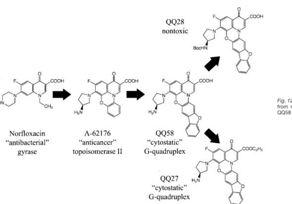
Design and Synthesis of Fluoroquinophenoxazines That
Interact with Human Telomeric G-Quadruplexes and
Their Biological Effects
1
Wenhu Duan,2,3
Anupama Rangan,2
Hariprasad Vankayalapati,2,3
Mu-Yong Kim,3
Qingping Zeng,4
Daekyu Sun, Haiyong Han, Oleg Yu. Fedoroff,5
David Nishioka, Sun Young Rha,6
Elzbieta Izbicka,
Daniel D. Von Hoff, and Laurence H. Hurley3,7,8
College of Pharmacy, The University of Texas, Austin, Texas 78712 [W. D., A. R., M-Y. K., Q. Z., O. Y. F., L. H. H.]; Institute for Drug Development, San Antonio, Texas 78245 [D. S., S. Y. R., E. I., D. D. V. H.]; Department of Biology, Georgetown University, Washington, DC 20057 [D. N.]; and Arizona Cancer Center, Tucson, Arizona 85724 [H. H., D. D. V. H., L. H. H.]
Abstract
In this study we have identified a new structural motif for a ligand with G-quadruplex interaction that results in biological effects associated with G-quadruplex-interactive compounds. Fluoroquinolones have been reported to possess weak telomerase inhibitory activity in addition to their better known bacterial gyrase poisoning. Starting with a
fluoroquinobenzoxazine, which has modest potency in a human topoisomerase II assay, we have designed a more potent inhibitor of telomerase that has lost its topoisomerase II poisoning activity. This fluoroquinophenoxazine (FQP) interacts with G-quadruplex structures to inhibit the progression of Taq polymerase in a G-quadruplex polymerase stop assay. In addition, we demonstrate by1H NMR
studies that this compound interacts with telomeric G-quadruplex structures by external stacking to the G-tetrad with both the unimolecular fold-over and the parallel G-quadruplex structures. A photocleavage assay confirms the FQP interaction site, which is located off
center of the external tetrad but within the loop region. Molecular modeling using simulated annealing was performed on the FQP-parallel G-quadruplex complex to determine the optimum FQP orientation and key molecular interactions with the telomeric G-quadruplex structure. On the basis of the results of these studies, two additional FQP analogues were synthesized, which were designed to test the importance of these key interactions. These analogues were evaluated in the Taq polymerase stop assay for G-quadruplex interaction. The data from this study and the biological evaluation of these three FQPs, using cytotoxicity and a sea urchin embryo system, were in accord with the predicted more potent telomeric G-quadruplex interactions of the initial lead compound and one of the analogues. On the basis of these structural and biological studies, the design of more potent and selective telomeric G-quadruplex-interactive compounds can be envisaged.
Introduction
Fluoroquinolones are best known therapeutically as bacterial gyrase inhibitors (1, 2) but more recently have also been demonstrated to be active against eukaryotic topoisomerase II, either as catalytic inhibitors or topoisomerase II poisons (3), or as telomerase inhibitors (4). One such group of com-pounds, the quinobenzoxazines, typified by A-62176 (Fig. 1B), has served as a starting point for the design of more potent topoisomerase II inhibitors using a structure-based approach (5). The extended aromatic conjugation system of A-62176 suggested to us that quinobenzoxazines may also intercalate with the more expansive system of G-quadruplex DNA and thereby be telomerase inhibitors in parallel with other G-quadruplex-interactive compounds, such as the anthraqui-nones (6 –10), cationic porphyrins (11–13),9 piperazines (14),
ethidium compounds, (15), and a pentacyclicacridinium com-pound (16). After we started this project, an independent report that bacterial quinolones may be weak telomerase inhibitors (4) also supported the idea that a ring-extended fluoroquinolone would be a more potent telomerase inhibitor.
G-quadruplexes (Fig. 1A) were first identified in 1962 (17); however, their possible biological significance was only sug-gested starting in the late 1980s (18 –23), and they were first proposed to be potential targets for drug design in 1989 (24). The existence of chaperone proteins that facilitate their for-mation (25–27), G-quadruplex binding proteins that
recog-Received 8/17/01; revised 9/21/01; accepted 10/2/01.
The costs of publication of this article were defrayed in part by the payment of page charges. This article must therefore be hereby marked
advertisement in accordance with 18 U.S.C. Section 1734 solely to
indi-cate this fact.
1Supported by NIH Grants CA88310 and CA67760. 2These authors contributed equally to this paper.
3Present address: College of Pharmacy, The University of Arizona,
Tuc-son, AZ 85721.
4Present address: Monsanto Co., St. Louis, MO 63167.
5Present address: Department of Biomolecular Sciences, University of
Manchester Institute of Science and Technology, Manchester M60 1QD, England.
6Present address: Cancer Metastasis Research Center, Yonsei University
College of Medicine, Seoul, Korea.
7Present address: College of Pharmacy and Department of Chemistry,
The University of Arizona, Tucson, AZ 85721 and Arizona Cancer Center, 1515 North Campbell Avenue, Tucson, AZ 85724.
8To whom requests for reprints should be addressed, at Arizona Cancer
Center, 1515 N. Campbell Ave., Tucson, AZ 85724. Phone: 520-626-5622; Fax: 520-626-5623; E-mail: hurley@pharmacy.arizona.edu.
9D-F. Shi, R. T. Wheelhouse, D. Sun, and L. H. Hurley.
Quadruplex-interactive agents as telomerase inhibitors: synthesis of porphyrins and structure-activity relationship for the inhibition of telomerase. J. Med. Chem., in press, 2001.
nize these structures (25, 28 –31), and G-quadruplex-selec-tive helicases (32–34) that unwind these structures make their existence in vivo in human cells a real possibility. Re-cently their in vivo existence in ciliates has been supported by antibody studies (35).
G-quadruplex-interactive compounds have aroused more interest recently because it has been demonstrated that these compounds not only inhibit telomerase in cell-free (6, 9) and in in vitro systems (36) but also cause telomere shortening and cell crisis in cancer cells Fig. 1. A, G-tetrad and
G-quadru-plexes. Panel a, four guanine residues forming a planar structure G-tetrad through Hoogsteen hydrogen bond-ing. Panel b, a parallel G-quadruplex model. Panel c, an intermolecular anti-parallel G-quadruplex model. Panel d, an intramolecular basket G-quadru-plex model. Each parallelogram in
panels b– d represents a G-tetrad. B, structures of the topoisomerase
II-interactive compound (A-62176) and G-quadruplex-interactive com-pounds QQ58, QQ27, QQ28, and TMPyP4.
(14).10A number of reviews on G-quadruplex and their targeting
by small molecules have appeared recently (37– 45).
In this contribution we demonstrate that QQ58 (Fig. 1B), a FQP11that lacks bacterial gyrase or topoisomerase II
poi-soning activity, is able to bind to G-quadruplex structures and, by doing so, inhibit telomerase. Structural insight into the interactions of this compound with both intermolecular and intramolecular G-quadruplex structures has been obtained by one-dimensional1H NMR molecular modeling and a
photo-cleavage assay. We also demonstrate that this compound induces chromosomal effects and slowing of the division of sea urchin embryos typical of G-quadruplex-interactive compounds. On the basis of molecular modeling studies, two additional analogues of this compound were designed and synthesized (QQ27 and QQ28; Fig. 1B), and their interactions with G-quadruplex structures and biological effects were ex-amined. The maintenance or loss of biological potency paral-leled their interactions with the G-quadruplex structure, and this was predicted on the basis of molecular modeling.
Materials and Methods
Synthesis of Extended Fluoroquinolones (QQ58, QQ27, and QQ28 in Figure 2)
General. All of the melting points were recorded on a
Tho-mas-Hoover capillary melting point apparatus and are un-corrected.1H and13C data were obtained on a Varian Unity
300 MHz NMR spectrometer. The chemical shifts are relative to the trace proton, carbon, or fluorine signals of the deuter-ated solvent. Coupling constants, J, are reported in Hz and refer to apparent peak multiplicity rather than coupling con-stants. Mass spectroscopic experiments were performed by the Mass Spectroscopy Center at The University of Texas, Austin, TX. Elemental analysis of C, H, and N was done by Desert Analytics, Tucson, AZ. Flash column chromatography was performed on silica gel 60, 230 – 400 mesh, purchased from Spectrum. All of the starting materials were obtained from commercial sources unless otherwise specified.
Ethyl 3-(2-hydroxydibenzofuran-3-amino)-2-[(2,3,4,5,-tetrafluorophenyl)carbonyl] Prop-2-enoate (2). A solution
of ethyl 2,3,4,5-tetrafluorobenzoylacetate (1.1 g; 4.18 mmol) in triethyl orthoformate (1 ml; 6.06 mmol) and acetic anhy-dride (1.8 ml; 19 mmol) was heated and stirred at 130°C for 4 h. During the process, the formed ethyl acetate was re-moved. The mixture was then distilled under vacuum to yield an orange oil, which was dissolved in 30 ml of dichlorometh-ane. 3-Amino-2-hydroxydibenzofuran hydrobromide (4.07 g; 14.6 mmol), premixed with two equivalents of pyridine, was added to the dichloromethane solution, and the mixture was stirred overnight at room temperature. After the solvent was removed under reduced pressure, the remaining residue was absorbed on silica gel and subjected to flash column
chro-matography (ethyl acetate:hexane⫽ 2:1), yielding a bright yellow powder (3.81 g; 83%).1H NMR (CDCl
3)␦ (mixture of
isomers; multiplet, m; singlet, s) 1.17–1.26 (m, 3H), 2.95 (s, br, 1H), 4.10 – 4.25 (m, 2H), 7.05–7.20 (m, 1H), 7.33 (m, 2H), 7.43 (m, 1H), 7.52 (m, 3H), 7.85 (m, 1H), 8.69 (s, 0.7H), 8.74 (s, 0.3H); 19F NMR ␦ ⫺156.2 (m, 1F), ⫺154.5 (m, 0.75F),
⫺135.4 (m, 0.25F), ⫺140.3 (m, 0.75F), ⫺139.4 (m, 1F), 139.3 (m, 0.25F); and MS (CI): m/z 474 (M⫹ H).
Ethyl 1,2-difluoro-4-oxo-4H-pyrido[3,2,1-k,l ]-(3-amino- 2-hydroxydibenzofuranyl)[1,2-g]phenoxazine-5-carboxy-late (3). Ethyl
3-(2-hydroxydibenzofuran-3-amino)-2-[(2,3,4,5,-tetrafluorophenyl)carbonyl] prop-2-enoate (2.32 g; 4.9 mmol) and NaH (60% in mineral oil; 0.45 g; 11.3 mmol) were mixed with freshly distilled tetrahydrofuran and stirred at⫺78°C for 15 min. The mixture was allowed to warm to room temperature gradually and then heated at 65°C for 30 min. The excess NaH was quenched by addition of 30 ml of methanol. The solution was evaporated to dryness, and the product was purified through flash column chromatography, yielding a yellow pow-der (1.84 g; 89%): mp ⬎229°C (decomposed); 1H NMR ␦ (CDCl3) 9.01 (s, 1H), 7.90 (d, 1H, J⫽ 7.8 Hz), 7.80 (dd, 1H, J ⫽ 7.8, 2.1 Hz), 7.39 (t, 1H, J⫽ 6.9 Hz), 4.48 (q, 2H, J ⫽ 7.2 Hz), 1.49 (t, 3H, J⫽ 7.2 Hz);19F NMR⫺133.6 (dd, 1F, J ⫽ 23, 10 Hz), ⫺1561.5 (dd, 1F, J ⫽ 21, 9 Hz); and MS (CI): m/z 434 (M ⫹ H). Ethyl (R)-1-{3-[(tert-butoxy)carbonylamino]pyrrolidin-1-yl}-2-fluoro-4-oxo-4H-pyrido[3,2,1-k,l ]-(3-amino-2- hydroxydibenzofuranyl)[1,2-g]phenoxazine-5-carboxy-late (4). Ethyl 1,2-difluoro-4-oxo-4H-pyrido[3,2,1-k,l
](3- amino-2-hydroxydibenzofuranyl)[1,2-g]phenoxazine-5-carboxylate (0.232 g; 0.54 mmol) and (R)-3-(tert-butoxycarbonylamino)pyrrolidine (0.302 g; 1.62 mmol) were dissolved in 30 ml of pyridine, and the residue was purified using flash column chromatography (dichloro-methane:ethylacetate⫽ 2:3), yielding a yellow powder (0.27 g, 84%): mp:⬎261°C (decomposed).1H NMR␦ 8.90 (s, 1H), 7.90 (d, 1H, J⫽ 7.5 Hz), 7.64 (m, 3H), 7.57 (s, 1H), 7.51 (t, 1H, J ⫽ 6.9 Hz), 7.39 (t, 1H, J⫽ 6.9 Hz), 5.35 (s, br, 1H), 4.44 (q, 2H, J ⫽ 7.2 Hz), 3.70 (m, 1H), 3.61 (m, 1H), 3.40 (m, 2H), 2.29 (m, 1H), 2.00 (m, 1H), 1.49 (s, 9H), 1.46 (t, 3H, J⫽ 7.2 Hz);19F⫺119.8 (d, 1F, J⫽ 13 Hz); and MS (CI): m/z 600 (M⫹1). (R)-1-(3-aminopyrrolidin-1-yl)-2-difluoro-4-oxo-4H- pyrido[3,2,1-k,l]-(3-amino-2-hydroxydibenzofuranyl)[1,2-g]phenoxazine-5-carboxylic Acid HCl Salt (5; QQ58).
Ethyl (R)-1-{3-[(tert-butoxy)carbonylamino]pyrrolidin-1-yl}-2- fluoro-4-oxo-4H-pyrido[3,2,1-k,l]-(3-amino-2-hydroxydiben-zofuranyl)[1,2-g]phenoxazine-5-carboxylate (0.228 g; 0.526 mmol) was mixed with 10 ml of ethanol and 3 ml 1NKOH. The mixture was refluxed for 30 min. HCl (9 ml of 2N) and 6 ml of ethanol were then added to the mixture, and it was refluxed for 4 h. After the reaction mixture was cooled down slowly, a yellow powder was obtained by filtration, washing with water and ethanol, and drying (250 mg, 95%): mp: ⬎250°C (decomposed); 1H NMR (DMSO-d 6) 9.24 (s, 1H), 8.49 (m, 1H), 8.12 (s, 1H), 8.05 (d, 1H, J⫽ 7.5 Hz), 7.69 (d, 1H, J⫽ 8.4 Hz), 7.54 (t, 1H, J ⫽ 7.2 Hz), 7.47 (t, 1H, J ⫽ 13.5 Hz), 7.40 (t, 1H, J⫽ 7.5 Hz), 3.80–4.05 (m, 5H), 2.31 (m, 1H), 2.08 (m, 1H);19F NMR⫺119.1 (d, 1F, J ⫽ 15 Hz); and MS (CI): m/z 472 (M⫹ H). Anal. (C26H18N3FO5.HCl.7/4H2O) C, H, N (Cal. C 62.09 H3.96 N8.35; Found C62.21 H3.75 N 8.13).
10M. A. Shammas, R. B. Batchu, J. Y. Wang, L. H. Hurley, R. J. S. Reis,
and N. Munshi. Telomerase inhibition and cell growth arrest following porphyrin treatment of multiple myeloma cells, submitted for publication.
11The abbreviations used are: FQP, fluoroquinophenoxazine; NMR,
nu-clear magnetic resonance; CI, confidence interval; SA, simulated anneal-ing; PIPER, N,N ⬘-bis[2-(-1-piperidino)ethyl]-3,4,9,10-perylenetetracar-boxylic diimide; PMC, primary mesenchyme cell; VP, vegetal plate.
Ethyl (R)-1-(3-aminopyrrolidin-1-yl)-2-fluoro-4-oxo-4H- pyrido[3,2,1-k,l]-(3-amino-2-hydroxydibenzofuranyl)[1,2-g]phenoxazine-5-carboxylate (6; QQ28). To a mixture of ethyl (R)-1-{3-[(tert-butoxy)carbonylamino]pyrrolidin-1-yl}-2- fluoro-4-oxo-4H-pyrido[3,2,1-k,l]-(3-amino-2-hydroxydiben-zofuranyl)[1,2-g]phenoxazine-5-carboxylate (50 mg; 0.008 mmol) and methylene chloride (2 ml) was added trifluoroace-tic acid (0.5 ml) at room temperature. After addition, the reaction mixture was stirred at room temperature for 2 h. Solvent was removed to give a residue and to this residue was added methanol (5 ml) and triethylamine (0.5 ml), and the yellow solid was collected to give a yellow powder (30 mg; 72%): mp: ⬎210°C (decomposed); 1H NMR (DMSO-d 6) ␦ 8.79 (s, 1H), 8.05 (s, 1H), 8.03 (s,1H), 7.81 (s, 1H), 7.61 (d, 1H, J⫽ 8.0 Hz), 7.47 (t, 1H, J ⫽ 8.0 Hz), 7.36 (t, 1H, J ⫽ 8.0 Hz), 7.21 (d, 1H, J⫽ 8.0 Hz), 4.23 (q, 2H, J ⫽ 6.8 Hz), 3.78 (m, 2H), 3.67 (m,1H), 3.54 (m, 2H), 2.00 (m, 1H), 1.68 (m, 1H), 1.32 (t, 3H, J⫽ 6.8 Hz); MS (FAB): 500.6 (M ⫹ H); HRMS (FAB) calc. for C28H23FN3O5 500.1622, found 500.1623; Anal.
(C28H22FN3O5.H2O) C, H, N (Cal. C64.98 H4.67 N8.12: Found C65.10 H4.81 N7.90). (R)-1-{3-[(tert-butoxy)carbonylamino]pyrrolidin-1-yl}- 2-fluoro-4-oxo-4H-pyrido[3,2,1-k,l]-(3-amino-2-hydroxy-dibenzofuranyl)[1,2-g]phenoxazine-5-carboxylic acid (7; QQ27). Ethyl (R)-1-{3-[(tert-butoxy)carbonylamino]pyrroli- din-1-yl}-2-fluoro-4-oxo-4H-pyrido[3,2,1-k,l]-(3-amino-2-hydroxydibenzofuranyl)[1,2-g]phenoxazine-5-carboxylate (100 mg; 0.17 mmol) was dissolved in methanol (6 ml), a solution of NaOH (37 mg; 0.67 mmol) in water (3 ml) was added, and the reaction mixture was stirred at room temper-ature for 2 h. Methanol was evaporated off, clear solution was neutralized with 10% HCl to pH⫽ 2.0, and precipitate was collected and dried with a vacuum pump to give a yellow powder (91 mg, 95%): MP: 235–237°C;1H NMR (DMSO-d 6) ␦ 9.15 (s, 1H), 8.40 (s,1H), 8.04 (d, 1H, J ⫽ 7.5 Hz), 7.94 (s, 1H), 7.66 (d, 1H, J⫽ 8.2 Hz), 7.51 (t, 1H, J ⫽ 7.5 Hz), 7.39 (m, 2H), 4.12 (m, 1H), 3.95 (m, 1H), 3.89 (m, 1H), 3.77 (m, 1H), 3.56 (m, 1H), 2.11 (m,1H), 1.89 (m, 1H),1.41 (m,1H); MS (FAB): 572.6 (M ⫹ H); HRMS (FAB) (M ⫹ H) calc. for C31H27FN3O7 572.1838, found 572.3338; Anal.
(C31H26FN3O7.7/4H2O) C, H, N (Cal. C61.73 H4.63 N7.13; Found C61.97 H4.50 N6.96).
Drug and Oligonucleotide Preparation. Drug solutions
were prepared as 5 mMstock solutions in DMSO and stored at ⫺20°C. These stock solutions were diluted to working concentrations in distilled water immediately before use. Oligonucleotides were synthesized on a PerSeptive Biosys-tems Expedite 8909 automatic DNA synthesizer. The sam-ples for NMR experiments were purified by reverse phase high-performance liquid chromatography on a C18 column (Dynamax-300A) and dialyzed extensively against 10 mMKCl or 20 mMNaCl solutions followed by deionized water. Solid supports and phosphoramadites were purchased from Glen Research and PerSeptive Biosystems. Oligonucleotides for polymerase extension assay and photocleavage reactions were purified by denaturing PAGE, diluted to required con-centrations, and dispensed into small aliquots.
Decatenation Assay. Kinetoplast DNA (0.25g) was
in-cubated with various concentrations of QQ58 or A-62176 in
10l of reaction buffer [50 mMTris-HCl (pH 8.0), 120 mMKCl, 10 mMMgCl2, 0.5 mMATP, and 0.5 mMDTT) for 10 min. Two
units of human topoisomerase II (TopoGEN Inc.) were added to the mixture, which was then incubated at 37°C for 30 min. The reaction was terminated with 0.1 volume of stop buffer (5% sarkosyl, 0.025% bromophenol blue, and 50% glycerol). The decatenation products were analyzed on 1% agarose gels run with 0.5g/ml ethidium bromide.
Preparation and End-labeling of Oligonucleotides for Topoisomerase II Cleavage Reaction. DNA oligomers 5⬘-d
[CGATGGGGAAGATCGGGCTCGTATACATTGATACGGGG-CTCATGAGCGCTTGTTTCGGCG]-3⬘ (A1) and 5⬘-d[CGCC-GAAACAAGCGCTCATGAGCCCCGTATCAATGTATACGAG CCCGATCTTCCCCATCG]-3⬘ (A2; Ref. 46) were synthesized as above. The 5⬘ end-labeled single-strand oligonucleotide was obtained by incubating A1 with T4 polynucleotide kinase and [␥-32P]ATP at 37°C for 1 h. Labeled DNA was purified
with a Bio-Spin 6 chromatography column (Bio-Rad) after inactivating T4 polynucleotide kinase by heating for 8 min at 70°C. The labeled strand was then annealed with the com-plementary strand (A2) and purified on an 8% native poly-acrylamide gel.
Topoisomerase II Cleavage Reaction. The labeled
dou-ble-stranded DNA was incubated with 20 units of human topoisomerase II in 20l of reaction buffer [30 mMTris-HCl (pH 7.6), 3 mMATP, 15 mM-mercaptoethanol, 8 mMMgCl2, and 60 mM NaCl] at 30°C for 10 min in the presence of various concentrations of QQ58 or A-62176. Reactions were terminated by adding SDS to 1% of the final concentration, and topoisomerase II was removed by proteinase K digestion (100g/ml) at 42°C for 1 h followed by phenol-chloroform extraction and ethanol precipitation. Samples were loaded onto a 12% denaturing sequencing gel. The dried gels were exposed on a phosphor screen. Imaging and quantification were performed using a PhosphorImager (Storm 820) and ImageQuant 5.1 software from Molecular Dynamics.
Telomerase Assay. Telomerase assays were run as
described previously (47) using a primer extension assay in which a 5⬘-biotinylated primer consisting of three telo-meric repeats was used, and the incorporation of32P-labeled
GTP was measured. In brief, reaction mixtures (20 l) contained 4 l of cell extracts (60 g total cell protein), 50 mMTris-OAc (pH 8.5), 50 mMK-OAc, 1 mMMgCl2, 5 mM
BME, 1 mMspermidine, 1M5⬘-biotinylated telomere primer (TTAGGG)3, 1.2M[␣-32P]-dGTP (800 Ci/mmol), 1 mMdATP, and 1 mMdTTP, and were incubated at 37°C for 30 min. Reactions were terminated by adding 20l of Streptavidin-Dynabeads, which bind selectively to the desired targets (5⬘-biotinylated primer), forming a magnetic bead-target complex. This complex was separated from the suspension using a magnet (Dynal MPC) and washed several times with washing buffer (2 M NaCl) to eliminate [␣-32P]-dGTP
back-ground. Telomerase reaction products were separated from the magnetic beads by protein denaturation with 5.0 M guanidine-HCl at 90°C for 30 min. After recovery of these products, analysis was performed by 8% denaturing PAGE. Telomerase activities were quantified by densitometric analyses of an autoradiogram using ImageQuant software.
All of the reactions were carried out in amber Eppendorf tubes under subdued lighting to avoid photocleavage.
Polymerase Stop Assay. The DNA primer 5
⬘-d[TAATAC-GACTCACTATAG]-3⬘ and template DNA
5⬘-d[TCCAAC-TATGTATAC(TTGGGG)4
TTAGCGGCACGCAATTGCTATA-GTGAGTCGTATTA]-3⬘ were synthesized and purified as before (48). About 200 ng of the primer DNA was 5 ⬘-end-labeled with32P using T4 polynucleotide kinase and
subse-quently purified by denaturing PAGE. The assay was carried out as described previously (48). Briefly, 24 nMof labeled primer and 12 nMof template DNA were annealed in a 1⫻ reaction buffer [10 mMTris-HCl (pH 8.0) and 5 mMKCl] by heating to 95°C and slowly cooled to room temperature. MgCl2, dNTP, and Taq DNA polymerase
(Boeringher-Manheim) were added, and the mixture was incubated at 55°C for 20 min. The polymerase extension was stopped by adding 2⫻ stop buffer (10 mMEDTA, 10 mMNaOH, 0.1% xylene cyanol, and 0.1% bromphenol blue) and loaded onto a 12% sequencing gel. After electrophoresis, the gels were dried and visualized on a PhosophorImager and quantitated using ImageQuant software.
Photo-mediated Strand Cleavage Reaction. The
se-quence of the single-stranded DNA (G-quadruplex) used was 5⬘-d[CATGGTGGTTT(GGGTTA)4CCAC]-3⬘. About 400 ng of
G-quadruplex DNA was 5⬘-end-labeled with32P using T4
polynucleotide kinase (New England Biolabs) and subse-quently purified by denaturing PAGE. This 5⬘-labeled G-quadruplex DNA was stored in Tris-EDTA buffer [10 mM Tris-HCl, 1 mMEDTA (pH 7.5)] at a concentration of 5 ng/l and⬃3000 cpm/l. For each photocleavage reaction, 30 l of G-quadruplex DNA was mixed with 30l of a 200 mMKCl solution, placed in a water bath at 95°C for 4 min, and slowly cooled to room temperature. A control set was run in distilled water instead of KCl solution. QQ58 (6l) at varied concen-trations was added to each sample and transferred to a 24-well Titretek microtiter plate (ICN). This plate was placed on top of a Pyrex glass shield and irradiated for 2 h with an 85-W xenon lamp placed under the Pyrex glass. (Pyrex glass was used to filter the UV light under 300 nm, thereby elimi-nating DNA damage caused directly by UV irradiation.) Dur-ing the irradiation, the Titretek plate was rotated three times to eliminate light heterogeneity. Reactions were terminated by the addition of 10 g of calf thymus DNA, followed by phenol-chloroform extraction and ethanol precipitation. The resulting samples were subjected to treatment with 0.1 M piperidine. The samples were then loaded onto a 16% se-quencing gel and electrophoresed. The dried gels were ex-posed to a phosphor screen. Imaging and quantification were performed using a PhosophorImager and ImageQuant software.
NMR Spectroscopy. NMR experiments were performed
on a Varian UNITY plus 500 MHz spectrometer. All of the titration experiments were carried out at 30°C in a 90% H2O/10% D2O solution containing 150 mM KCl, 25 mM
KH2PO4, and 1 mMEDTA (pH 7.0). A standard 1–1 echo pulse
sequence with a maximum excitation centered at 12 ppm was used for water suppression. Thirty-two scans were ac-quired for each spectrum with a relaxation delay of 2 s. Two-dimensional Nuclear Overhauser Effect Spectroscopy
spectra of exchangeable protons were collected at 30°C in time proportional phase incrementation mode with a mixing time of 200 ms using 2048 and 1024 complex points in t2and
t1experiments, respectively. NMR data were processed and analyzed using the FELIX program (Molecular Simulations Inc.). Nearly all of the nonexchangeable DNA protons of the 5⬘-d[A(G3T2A)3G3]-3⬘ and 5⬘-d[TAG3T2A]4-3⬘ in a free form or
in a complex with ligand, except for several H5⬘/H5⬙ pairs, were assigned as described (49).
Model Building and Molecular Dynamics Simulations.
The initial coordinates used in the model building process were those published for the NMR-based model of the 1:1 PIPER:5⬘-d[TAG3T2A]4-3⬘ complex (49) and the antiparallel human telomeric sequence d[AG3(T2AG3)3] (50). The
neces-sary replacements and the addition of hydrogens were car-ried out using INSIGHT II (Ref. 51; Molecular Simulations Inc.), and position was refined by energy minimization (1000 cycles of steepest descent and 2⫻ 1000 cycles of conjugate gradient) while constraining the positions of heavy atoms. Finally, the entire structure was subjected to conjugate gra-dient minimization until convergence was reached, at which time constraints were gradually removed. Molecular dynam-ics simulations (100 ps) at 300 K and mechandynam-ics (2⫻ 1000 cycles of conjugate gradient minimization) were performed using DISCOVER with the consistent force field. This struc-ture subsequently served as the starting strucstruc-ture for addi-tional energy refinement, docking, molecular dynamics, and complex formation.
To explore the molecular interactions of QQ58 and its analogues, QQ27 and QQ28, with the G-quadruplex struc-tures, molecular models were built and energy minimized using a protocol similar to that described for G-quadruplex structures. The obtained low energy structures were manu-ally docked into the intercalation site, and energies were computed using the AFFINITY (52) program. To clarify the orientation of the ligands in the intercalation site, the elec-trostatic potentials at the van der Waals surface of the G-quadruplex were determined by using solvent surface cal-culations. The SA docking with 100 fs/stage duration (50 SA stages) was then performed to find the most favorable ori-entation. Thus, orientation with low intermolecular potential energy was obtained while moving ligand and ligand-binding G-tetrad residues. The resulting ligand-G-quadruplex com-plex trajectories were energy minimized using 1000 cycles of conjugate gradient minimizer, and the interaction energies were computed.
Cytotoxicity Assay. Human breast carcinoma
MDA-MB-231 and prostate carcinoma DU-145 cell lines were pur-chased from American Tissue Culture Collection (Rockville, MD) and cultured according to the supplier’s instructions. Exponentially growing cells (1–2 ⫻ 103 cells) in 0.1 ml of
medium were seeded on day 0 in a 96-well microtiter plate. On day 1, 0.1-ml aliquots of medium containing graded concentrations of the investigational compound were added to the cell plates. At days 7–10, the cell cultures were incu-bated with 50l of 3-(4,5-dimethylthiazol-2-yl)-2,5-diphenyl-tetrazolium bromide (1 mg/ml in Dulbecco’s PBS) for 4 h at 37°C. The resulting formazan precipitate was solubilized with 200l of 0.04MHCl in isopropyl alcohol (53). For
determi-nation of the IC50values, the absorbance readings at 570 nm were fitted to the four-parameter logistic equation.
Sea Urchin Embryo Culture. Lytechinus pictus sea
ur-chins (Marinus Inc., Long Beach, CA) were maintained at 15°C in refrigerated aquaria containing Instant Ocean artifi-cial sea water. Spawning, fertilization, drug treatment, and embryo processing were done as described (54). Briefly, 10 min after insemination, the fertilized eggs were allowed to settle, and the supernatant was aspirated and replaced with fresh artificial sea water. The embryos were cultured at 18°C. Stock solutions (10 mM) of the tested agents were prepared in DMSO. Twenty min after fertilization, the agents were added to 1% embryo suspensions to a final concentration of 1M. Equivalent amounts of DMSO were added to control egg suspensions. At measured times after insemination (10 and 24 h), the embryos were pelleted by centrifugation. Video images of the embryos were captured with a Zeiss standard research microscope interfaced with a Javelyn video camera and a Panasonic time-lapse video recorder. The nuclei were stained by the Feulgen reaction, and the chromatin was visualized and photographed with an Olym-pus BH2 photomicroscope equipped with fluorescence op-tics, as described elsewhere (55).
Results
Chemistry. The synthetic procedure (Fig. 2) was based on a
published method for the synthesis of the quinobenzoxazines, with some modification (56). The synthesis was started with ethyl 2,3,4,5-tetrafluorobenzoylacetate (Fig. 2, 1), which was prepared according to published procedure (5). Treatment of 2,3,4,5-tetrafluorobenzoylacetate with triethyl orthoformate in acetic anhydride followed by 2-hydroxy-3-amino-dibenzofuran hydrobromide generated the enamino keto acid intermediate (Fig. 2, 2). This intermediate was treated with sodium hydride at ⫺78°C and then heated to reflux to complete the double annulation (Fig. 2, 3). Regioselective nucleophilic substitution of fluoride with 3-(R)-(tert-butoxycarbonylamino)pyrrolidine was carried out to give the key intermediate (Fig. 2, 4). Final target molecules 5, 6, and 7 (Fig. 2) were prepared by hydrolysis and deprotection of 4, hydrolysis of 4, and deprotection of 4, respectively, in overall yields of 59, 59, and 44.7%, respectively.
Comparison of the Effects of A-62176 and QQ58 on Topoisomerase II and Telomerase Activity
On the basis of our previous insights into structural re-quirements (extended planarity, cationic interactions) for ligand binding with G-quadruplex structures, the fluoro-Fig. 2. Condition: (a) (i) Ac2O, CH(OC2H5)3, (ii)
3-amino-4-hydroxydibenzofuran hydrobromide; (b) THF, NaH; (c) pyridine, (R) t-Boc-3-aminopyrroli-dine; (d) OH⫺/H⫹; (e) OH⫺; (f) CF3COOH.
quinolones seemed to be good candidates for structural modification. Our objective was to convert a topoisomer-ase II poison (A-62176) to a G-quadruplex-interactive compound. As this work progressed, a paper was pub-lished (4) that demonstrated that quinolones were telom-erase inhibitors, suggesting to us that quinolones might interact with G-quadruplex structures. Our first objective was to compare the topoisomerase II inhibitory activities and G-quadruplex interactions of A-62176 and QQ58 to determine whether it would be possible to separate these activities.
Whereas A-62176 Is Both a Topoisomerase II Poison and a Catalytic Inhibitor, QQ58 Is Only a Catalytic Inhib-itor. Experiments were designed to compare the effects of
A-62176 and QQ58 on the activity of human topoisomerase II. The ability to cleave and religate double-stranded DNA is central to the physiological functions of topoisomerase II. Kinetoplast DNA is a massive network consisting of thou-sands of interlocked, closed circular DNA molecules called minicircles. Topoisomerase II introduces a double-strand break in DNA to release one minicircle from the network, which is called a decatenation reaction (57, 58). The in vitro effects of A-62176 and QQ58 were determined by using a decatenation assay. As shown in Fig. 3, A and B, QQ58 had about a 5-fold lower decatenation activity than A-62176. To additionally characterize the properties of A-62176 and
QQ58, the contrasting effect on the DNA cleavage reaction of human topoisomerase II of QQ58 was determined. As shown in Fig. 3, C and D, the intensity of the topoisomerase II-mediated cleavage at site A decreased as the concentration of QQ58 was increased, whereas the DNA cleavage was initially enhanced at low concentrations of A-62176 and then decreased at high concentrations. These results show that the two fluoroquinolones act differently on human topoi-somerase II: QQ58 acts as a catalytic inhibitor that inhibits the activity of topoisomerase II without trapping the cleaved complex, and A-62176 is a topoisomerase II poison that interferes with the breakage-rejoining reactions of topoi-somerase II by trapping the enzyme covalent reaction inter-mediate, known as the cleaved complex.
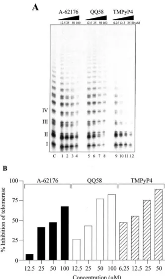
QQ58 Is a More Potent Inhibitor of Telomerase than A-62176, but Is Less Potent than the Cationic Porphyrin TMPyP4. The telomerase inhibitory activity of QQ58 was
compared with A-62176 and the cationic porphyrin TMPyP4 (Fig. 1B), a known inhibitor of telomerase because of its ability to sequester the single-stranded DNA primer required for the reverse transcriptase activity of telomerase into a G-quadruplex structure (Ref. 6, 59; Fig. 4A). The IC50values for the inhibition of telomerase assay in the direct assay for A-62176, QQ58, and TMPyP4 were 58, 28, and 8 M, re-spectively. These were determined from the histogram plots shown in Fig. 4B. It is important to note that the telomerase Fig. 3. A, inhibition of decatenation of human topoisomerase II by QQ58 and A-62176. Catenated KDNA was treated with 2 units of human topoisomerase
II in the presence of QQ58 or A-62176 followed by SDS/proteinase K and then analyzed on agarose gel. Lane 1 contains catenated KDNA only. Lane 2 contains KDNA and topoisomerase II without compounds. Lanes 3–7 contain 2, 5, 10, 20, and 50Mof QQ58, respectively. Lanes 8 –12 contain 0.5, 1, 2, 5, and 10Mof A-62176, respectively. B, intensity of decatenated KDNA was quantitated from the gel using ImageQuant software. C, autoradiogram of a 12% denaturing polyacrylamide gel showing the topoisomerase II-mediated cleavage pattern in the presence of QQ58 or A-62176. The cleavage reaction was done in a reaction buffer as described in “Materials and Methods.” Lane 3 contains DNA only. Lane 4 contains DNA and topoisomerase II without QQ58 or A-62176. Lanes 5–10 and 11–16 contain 0.2, 0.4, 1, 2, 5, and 10MQQ58 and A-62176, respectively. D, strand breakage products indicated by the arrow in C were quantitated using a PhosphorImager and ImageQuant software. The intensity of the DNA cleavage produced by QQ58 or A-62176 was determined from the volume of the bands normalized by the total radioactivity in each lane.
inhibitory potencies are⬃10-fold less than in the more fa-miliar PCR-based telomeric repeat amplification protocol as-say. Thus, by extension of the quinobenzoxazine fused ring system of A-62176 (Fig. 1B) to the quinophenoxazine ring system in QQ58 (Fig. 1B), QQ58 shows an⬃2-fold increase in potency in inhibition of telomerase over A-62176, but this is still less than TMPyP4.
Physical and Biochemical Characterization of the Interaction of QQ58 with the Intramolecular Antiparallel and Intermolecular Parallel G-Quadruplex Structures
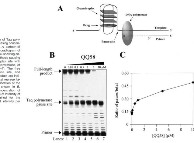
QQ58 Stabilizes an Intramolecular G-Quadruplex Suf-ficiently to Cause Polymerase Pausing. The polymerase
stop assay (48) can be used to evaluate the interaction of ligands with the intramolecular G-quadruplex structures. The principle of this assay is shown in the cartoon in Fig. 5A. This assay was originally developed to study the effect of
G-quadruplex secondary structures on polymerase processiv-ity (60). It has also been adopted to aid in the identification of the compounds that bind to and stabilize G-quadruplex structures (48). QQ58 causes a sequence-specific inhibition of Taq polymerase at the G-quadruplex site on the template DNA (Fig. 5B). As expected, there is polymerase pausing as the enzyme extends primer along the template, even in the absence of QQ58 in lane 1 of Fig. 5B (attributable to the presence of potassium in the reaction buffer that facilitates G-quadruplex formation). However, as the concentration of QQ58 is increased, there is enhanced pausing at the G-quadruplex site on the template DNA (Fig. 5B). The ratio of the intensity of the bands at the G-quadruplex pause site to the total intensity per lane increases by⬃3-fold as the con-centration of QQ58 increases from 0 to 10M(Fig. 5C). This indicates that QQ58 interacts with the G-quadruplex suf-ficiently to stabilize the G-quadruplex structure and inhibit the polymerase stop assay. In this same assay, A-62176 had little effect on the pausing of Taq polymerase at the G-quadruplex structure site.12
1H NMR Studies Demonstrate That QQ58 Forms a
Sta-ble Complex with a Parallel-stranded G-Quadruplex in Which the Drug Stacks at the GT Step. We chose the
d[TAG3T2A]4intermolecular G-quadruplex (“b” in Fig. 1A) for
these studies on the basis of its relevance to the human telomeric TTAGGG repeat and our previous success in de-termining the solution NMR structure of its complex with another telomerase inhibitor (49). The one-dimensional 1H
NMR spectra recorded during the titration of d[TAG3T2A]4
with QQ58 are shown in Fig. 6A. The1H NMR signals of the
Hoogsteen-bound guanine imino protons (10 –12 ppm range) gradually disappeared during the titration, and a new set of reciprocal upfield-shifted broadened imino proton reso-nances appeared, as shown at the QQ58:DNA ratio of 1:1 (Fig. 6A). The more pronounced upfield shift of the G5 imino proton relative to G4 and G3 infers that the site of QQ58 binding is between G5 and T6. Separate proton resonances for the free DNA and the drug:G-quadruplex DNA complex point to the slow exchange rate on the NMR time scale between the free and bound DNA. The spectra are very comparable with the spectra that were obtained for the ti-tration of PIPER with the same oligonucleotide (49). These NMR results indicate a strong and specific binding of QQ58 with parallel G-quadruplex DNA. Although attempts to obtain a two-dimensional NOESY spectra were made, the quality of the spectra impaired spectral assignment. The drug proton resonances were difficult to assign despite the complete assignment of the protons of the G-quadruplex in both the unbound and drug-bound states. Nevertheless, the identical changes in the DNA spectra on titration with either QQ58 or PIPER argue for a common geometry of the drug-G-quad-ruplex complexes. Because the structure of the PIPER-G-quadruplex complex has already been determined (49), it was used as a reference point for additional molecular mod-eling studies. A proposed model of the QQ58-intermolecular
12A. Rangan, unpublished observations.
Fig. 4. Inhibition of human telomerase by A-62176, QQ58, and TMPyP4.
A, autoradiogram showing telomerase inhibition with increasing
concen-trations of A-62176, QQ58, and TMPyP4. Lanes 1– 4, 5– 8, and 9 –12 contain 12.5, 25, 50, and 100 Mof A-62176, QQ58, and TMPyP4, respectively. B, graphical representation of data from A.
four-stranded G-quadruplex complex was prepared.13The
drug molecule binds to the G-quadruplex at the GT step by stacking of the aromatic moiety to the surface of the 3 ⬘-terminal G-tetrad. The binding site and QQ58 molecule ori-entation in regard to DNA bases are identical to those of the PIPER-G-quadruplex complex. This explains the similarity in the pattern of the chemical shift changes.
QQ58 Binds to the Intramolecular G-Quadruplex Struc-ture by Stacking between the Diagonal Loop and Exter-nal G-Tetrad. The intramolecular basket conformation of a
monomeric G-quadruplex DNA (“d” in Fig. 1A) is likely to be involved in the interaction with proteins and ligands in vivo (38). To examine the interaction of QQ58 with the intramolecular G-quadruplex structure,1H NMR studies were carried out, with
the established basket G-quadruplex consisting of the 22-mer human telomeric sequence d[AG3(T2AG3)3] (50). The
one-di-mensional NMR titration of 22-mer DNA with QQ58 is shown in Fig. 6B. The spectra of the imino protons in the region of 10 –12 ppm demonstrate the shift of only one particular Hoogsteen bped guanine, namely, the G22 residue that is a part of the G-tetrad adjacent to the diagonal loop. This suggests that the interaction of QQ58 with intramolecular G-quadruplex is in the proximity of this G-tetrad. The complexity of the one-dimen-sional spectra in the imino proton region, arising because of either strand aggregation or dynamics within the NMR time scale used, made the interpretation of the two-dimensional
NOESY spectra rather complex. Thus, to additionally charac-terize the intramolecular QQ58-G-quadruplex complex, photo-cleavage studies were performed.
Photo-mediated DNA Cleavage Reactions Confirm the Proximity of QQ58 to the External G-Tetrad. To
addition-ally characterize the binding mode of QQ58 to the intramo-lecular G-quadruplex structures, a photo-mediated cleavage reaction of the QQ58-G-quadruplex complex was carried out. A DNA sequence that forms an intramolecular G-quad-ruplex under conditions standardized previously was used (12). The photo-reactive property of the FQPs leads to strand cleavage in the QQ58-G-quadruplex complex in the pres-ence of light of a specific wavelength. This in turn gives important information about the drug localization in the com-plex. The photocleavage pattern produced by QQ58 on the intramolecular G-quadruplex is shown in Fig. 7A. With in-creased concentrations of the drug, there is enhanced cleav-age at G1, G6, and G12, which correspond to specific gua-nines involved in the G-quadruplex. These guagua-nines, G1, G6, and G12, correspond to the residues that form the lowermost G-tetrad (Fig. 7B), with G12 and G6 being the most reactive residues. This photocleavage data confirms that the drug is bound external to this G-tetrad. On the basis of the NMR studies and the photocleavage experiments, a cartoon (Fig. 7B) is proposed for the binding of QQ58 with the se-quence that forms an intramolecular G-quadruplex structure (Ref. 12; Fig. 7B). G12 in this structure corresponds to G22 in the previous two-dimensional1H NMR study, providing
con-13O. Fedoroff, unpublished observations.
Fig. 5. Inhibition of Taq poly-merase with increasing concen-trations of QQ58. A, cartoon of the assay. B, autoradiogram of the sequencing gel showing en-hanced DNA synthesis pausing at the G-quadruplex site with increasing concentrations of QQ58 (Lanes 1–7). The free primer, the pause site, and the full-length product are indi-cated. C, graphical representa-tion of the quantificarepresenta-tion of the sequencing gel shown in B, showing the concentration of QQ58 to the ratio of intensity of the bands obtained for the pausing site/total intensity per lane.
firmation of the location of QQ58 in the intramolecular fold-over G-quadruplex structure.
Molecular Modeling Predicts That QQ58 Binds to the Human Intramolecular Telomeric Repeat d[AG3(T2AG3)3].
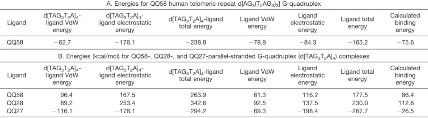
Docking simulations and analysis revealed that QQ58 binds to the human intramolecular telomeric repeat d[AG3(T2AG3)3] G-quadruplex with a binding energy of⫺75.6 kcal/mol (Table 1A). Although this binding energy is lower than that of QQ58 with the parallel-stranded G-tetraplex (see later), the favor-able interactions in the T2A loop suggest that it is a human telomeric G-quadruplex-interactive agent. The aminopyrroli-dine-NH2is oriented to interact with G10 within the proposed
intercalation site. Furthermore, the central ring nitrogen is positioned into the guanine carbonyl channel, and the car-boxylic acid functional group extends into the minor groove. The extended phenoxazine ring exhibits stacking interac-tions with the G14 and G10 bases. This orientation was confirmed as the most stable docking model by binding energy calculations.
Comparison of the Biochemical and Biological Effects of QQ58 and Its Amino (QQ27) and Carboxyl-modified (QQ28) Analogues
On the basis of NMR and photocleavage data, initial molecular modeling results suggested a complex in which QQ58 stacks to the external tetrad in both the intramolecular antiparallel and parallel G-quadruplex structures, as illustrated for the former in Fig. 7B. Molecular modeling was carried out on the human telomeric antiparallel (Fig. 8A) and parallel-stranded (Fig. 8B) G-quadruplex structures with QQ58 to gain additional insight into these structures, and on the basis of the results from the parallel-stranded G-quadruplex, two synthetically accessible molecules (QQ27 and QQ28 in Fig. 1B) were designed and synthesized, which were predicted to effect specific H-bonding interactions between the FQP and the G-quadruplex structures, resulting in much less favorable interactions of QQ28 with the intermolecular parallel-stranded G-quadruplex structure than either QQ27 or QQ58. A parallel series of biochemical and biological experiments were carried out to test these ideas.
Molecular Modeling Predicts That QQ58 Has a Higher Binding Interaction with the G-Quadruplex Structure.
Molecular modeling and SA docking experiments revealed that QQ58 exhibited the highest binding interactions with the four-stranded parallel G-quadruplex (d[TAG3T2A]4; Table 1B) and is intercalated at the G5-T6 nucleotide step approximately be-tween two pairs of guanines and thymines, with the positively charged side chain located in the groove (Fig. 8B). A strong intermolecular hydrogen bond interaction of the N7 atom of G5 with the carboxylic proton of QQ58 (N7 to COOH is 2.38 Å) was observed (Fig. 8C). The QQ58 aminopyrrolidine amino group takes part in a hydrogen bonding interaction with the 5⬘ phos-phate group of G5 (Fig. 8C). QQ58 also shows favorable stack-ing interactions between the G5 guanine tetrad and two of the 6-thymine residues. These electrostatic (⫺167.5 Kcal/mol) and van der Waals (⫺96.4 Kcal/mol) interactions give rise to a cal-culated binding energy of ⫺86.4 Kcal/mol (Table 1B). In the case of compound QQ28, such interactions were not observed, presumably because of the strong steric clash of the tert-butoxy carbonyl group of the aminopyrrolidine with G5 at the intercalation site. This in turn leads to the shift of the carboxylic acid functionality from its H-bonding site at G5. Analysis of all of the dynamic trajectories reveals that the tert-butoxy carbonyl group is rigidly positioned and oriented. This results in dramatic loss of both electrostatic and van der Waals energies relative to QQ58. Similarly, the presence of an ethyl ester group in the case of QQ27 restricts the electrostatic and hydrogen bonding interactions with the G5 nucleotide region of the intercalation site but to a much lesser extent than in QQ58. The calculated binding energies for QQ27 and QQ28 with the parallel-stranded complexes are⫺26.5 and 112.6 Kcal/mol, respectively. Fig. 6. Effect of QQ58 on the1
H NMR spectra of G-quadruplex struc-tures. A, one-dimensional1
H NMR titration of d[TAG3T2A]4with QQ58 to
obtain a 1:1 QQ58:DNA ratio. The imino proton and aromatic proton regions of the 500 MHz NMR spectra are shown with increasing amounts of ligand added at 30°C. G3, G4, and G5 represent the imino proton resonances of the free DNA, whereas G3*, G4*, and G5* represent the imino proton resonances of the ligand-bound DNA. B, one-dimensional1
H NMR titration of d[AG3(T2AG3)3] with QQ58 to obtain a 1:1 QQ58:DNA
ratio. The imino proton regions of the 500 MHz NMR spectra are shown with increasing amounts of ligand added at 30°C. The arrow points to the imino proton resonance of G22, which shifts on addition of QQ58.
The Polymerase Stop Assay Demonstrates That QQ28 Has Only a Minimal Stabilizing Effect on G-Quadruplex Structure, Whereas QQ27 Has an Intermediate Effect.
On the basis of molecular modeling results, we predicted that the steric bulk of the N-Boc group on the aminopyrrolidine of QQ28 should interfere with intercalation into the G-quadruplex structure and prevent any ionic interaction between the posi-tively charged amino group of QQ28 and the anionic backbone of DNA. However, the ethyl ester of QQ58 should not dramat-ically affect the binding of QQ27. A polymerase stop assay was used to evaluate the comparative abilities of QQ58, QQ27, and QQ28 to stabilize the intramolecular fold-over G-quadruplex structure. The results shown in Fig. 9, A and B, demonstrate that, as predicted, QQ58 and QQ27 have approximately the same potency in inhibition of the Taq polymerase, whereas QQ28 was much less potent.
Comparative Cytotoxicity Measurements of QQ58, QQ27, and QQ28 Demonstrate That QQ58 and QQ27 Are More Potent Than QQ28. The IC50values of QQ58, QQ27, and QQ28 were determined against human breast and pros-tate carcinoma tumor cell lines. As shown in Table 2, the
cytotoxicity in both cell lines followed the same pattern, with the IC50 potency values increasing in the order QQ28 ⬎
QQ27 ⬎ QQ58. For QQ28 and QQ58, the cytotoxicity in DU-145 was higher than in MDA-MB-231. We have observed a similar high sensitivity to other quinolones in DU-145 cells in comparison with MDA-MB-231.14
The Antiproliferative and Chromosomal Effects of QQ58, QQ27, and QQ28 Show That QQ58 and QQ27 Are More Potent Than QQ28. Nonmammalian organisms are
attractive supplements to conventional in vivo models, be-cause the key cellular pathways are conserved throughout the animal kingdom. We have examined early stage sea urchin embryos as a novel in vivo model that allows rapid assessment of drug effects over many cell cycles in much less time than in other animal models or in cultured human tumor cells. To date we have evaluated the in vivo effects of anticancer agents from diverse groups, such as alkylators, DNA-interactive natural products, and telomere- and
telom-14E. Izbicka, manuscript in preparation.
Table 1 Computed interaction energies (kcal/mol)
A. Energies for QQ58 human telomeric repeat d[AG3(T2AG3)3] G-quadruplex
Ligand d[TAG3T2A]4 -ligand VdW energy d[TAG3T2A]4 -ligand electrostatic energy d[TAG3T2A]4-ligand total energy Ligand VdW energy Ligand electrostatic energy Ligand total energy Calculated binding energy QQ58 ⫺62.7 ⫺176.1 ⫺238.8 ⫺78.9 ⫺84.3 ⫺163.2 ⫺75.6
B. Energies (kcal/mol) for QQ58-, QQ28-, and QQ27-parallel-stranded G-quadruplex (d[TAG3T2A]4) complexes
Ligand d[TAG3T2A]4 -ligand VdW energy d[TAG3T2A]4 -ligand electrostatic energy d[TAG3T2A]4-ligand total energy Ligand VdW energy Ligand electrostatic energy Ligand total energy Calculated binding energy QQ58 ⫺96.4 ⫺167.5 ⫺263.9 ⫺61.3 ⫺116.2 ⫺177.5 ⫺86.4 QQ28 89.2 253.4 342.6 92.5 137.5 230.0 112.6 QQ27 ⫺116.1 ⫺178.1 ⫺294.2 ⫺69.3 ⫺198.4 ⫺267.7 ⫺26.5
Fig. 7. QQ58 photo-mediated strand cleavage reaction of intramolecular G-quadruplex. A, auto-radiogram of a photocleavage sequencing gel with increasing concentrations of QQ58. Lanes 1 and 2 are the AG/TC Maxam-Gilbert sequencing reactions, and Lanes 4 – 8 are increasing concen-trations of QQ58 (1–25M). The enhanced cleav-age sites are indicated by G1, G6, and G12. G7 is in the same tetrad but is not cleaved excessively.
B, diagrammatic representation of the proposed
external binding of QQ58 to the G-quadruplex below the lower-most G-tetrad based on the pho-tocleavage results with the ligand.
erase-interactive agents, including a telomere mimic. We determined the effects of these agents on cellular prolifera-tion using a new quantitative assay and on chromosome morphology. The specific and reproducible effects of many of these drugs correlated with the detection of similar effects in tumor cells (54, 55, 61– 63).
In this study, we evaluated the in vivo effects of 1Mof QQ28, QQ27, and QQ58 in developing sea urchin embryos. Preliminary dose-dependence tests have shown that these concentrations are subtoxic and compatible with the assays. Antiproliferative effects of the agents were assessed by ex-amining the morphology of the embryos cultured for 10 and 24 h in the presence of the three quinolones. Photomicro-graphs (Fig. 10A) show that at 10 h all of the embryos are still within fertilization coats. The control group and QQ28-treated embryos appear normal with well-defined blasto-coels. Cleavage is delayed in QQ27- and QQ58-treated
em-bryos, so there are fewer cells, and a blastocoel has not yet formed. At 24 h (Fig. 10B) all of the embryos have hatched and are swimming, which suggests lack of major toxicity of the examined compounds. The control group and QQ28-treated embryos appear normal with well-defined blasto-coels into which PMCs have ingressed at the vegetal pole (seen as the thicker layer of cells in the bottom of the em-bryos). Invagination of the vegetal pole has begun in the control group and the QQ28-treated embryos. Because of the cleavage delay in the QQ27- and QQ58-treated embryos, the embryos are smaller and there are fewer cells. Addition-ally, the blastocoels appear to be abnormally filled with cells, and there does not appear to be a concentration of PMCs at a well-defined vegetal pole. The antiproliferative effect of QQ58 is apparently greater than that of QQ27, and this difference is more noticeable at 10 h than at the later time point at 24 h.
Fig. 8. Molecular modeling of the d[AG3(T2AG3)3]-QQ58 (A) and d[TAG3T2A]4-QQ58 (B and C) complexes. A, structure of the d[AG3(T2AG3)3]-QQ58
complex in the final model after energy minimization. The G-quadruplex structure is shown as a stick model with compound QQ58 in color by atom type Corey-Pauling Koltun model. The proposed intercalation sites (yellow) are labeled. B, structure of the d[TAG3T2A]4-QQ58 complex in the final model after
energy minimization. Coloring is as in A. C, stereo representation of the proposed binding mode of QQ58 in complex with parallel-stranded G-quadruplex shown in color by atom type. The white dotted lines show the hydrogen bonding distances in Å. The G-quadruplex complex is clipped to afford a better view of the intercalation site.
Chromosome-specific effects of the agents were deter-mined using high-power fluorescence microscopy. Al-though a single focal plane is exceeded at magnifications greater than⫻400, resolution of mitotic chromosomes in all phases of mitosis is possible. Both the nuclei and chromosomes may be visualized in bright red over a dark background with fluorescence optics (green excitation), with very little photobleaching. We have used this method to assess chromosome-specific effects of QQ28, QQ27, and QQ58 in 10-h embryos cultured in the continuous presence of the compounds. The 10-h embryos have been chosen as more representative than the 24-h ones be-cause at 10 h the embryos are actively dividing and there is the most chromosomal activity. Fig. 11 (top left panel)
shows representative mitotic chromosomes in a control (nontreated) embryo at 10 h after fertilization. The con-densed mitotic chromosomes stain more deeply than the less condensed interphase nuclei in the background. Many metaphase chromosomes are observed in typical linear arrangements on metaphase plates. During an-aphase the chromosomes separate and move to opposite poles cleanly with very little “tailing” of chromosome arms on the mitotic spindle. Telophase chromosomes are ob-served as two small, dense concentrations of mitotic chro-mosomes at opposite poles, where they will decondense into interphase nuclei. As additionally shown in Fig. 11, not only the control but also QQ28-treated embryos show the normal “tight” mitotic chromosomes among a larger num-ber of nuclei. On the other hand, the QQ27- and QQ58-treated embryos show a reduced number of nuclei, con-sistent with a growth delay. The nuclei are larger and more diffuse. The mitotic chromosomes are more diffuse and segregate abnormally, often to multiple poles, and end-to-end fusions are often observed. These results are con-sistent with the cleavage delay, and fewer cells are seen in the live embryos.
Discussion
In this study we have identified a new structural motif for a ligand with G-quadruplex interaction that results in bio-logical effects associated with G-quadruplex-interactive compounds. The initial lead compound was a quinoben-zoxazine, which derived its structural origin from the well-known fluoroquinolone antibiotics (see Fig. 12), typified by norfloxacin. Researchers at Abbott Laboratories, Abbott Park, IL demonstrated that this quinobenzoxazine com-pound could be converted into a mammalian cytotoxic agent by expansion of the dicyclic quinolone to the tetra-cyclic quinobenzoxazine (A-62176), which was shown to be a topoisomerase II poison (64). The same structural elements (planarity, amphoteric nature) can then be used as a basis to design an even more expanded ring system (quinophenoxazine) that is now a G-quadruplex-interac-tive drug (QQ58) that causes biological effects associated with G-quadruplex-interactive compounds.
Comparison of Complexes of A-62176 and QQ58 with the Topoisomerase II-DNA Complex and the G-Quadru-plex Structure. Whereas both norfloxacin and A-62176
tar-get gyrase and topoisomerase II-DNA complexes to produce poisoned complexes in which these drugs stabilize the co-valently linked protein-DNA complexes, QQ58 has lost this property (see Fig. 3) but is instead able to recognize and bind quite specifically to G-quadruplex structures. The quinoben-Table 2 Cytotoxicity of novel quinolones against human tumor cell lines Compound IC50(M) MDA-MB-231 DU-145 QQ58 0.15 0.07 QQ27 1.25 1.86 QQ28 ⬎20 5.77
Fig. 9. Differential effects of QQ27, QQ28, and QQ58 on DNA synthesis arrest by G-quadruplex structure. A, autoradiogram of the sequencing gel showing Taq polymerase primer extension arrest at the G-quadruplex site in the presence of QQ58, QQ27, and QQ28. Lanes G, C, A, and T are DNA sequencing lanes. B, quantitation of the sequencing gel shown in A. The drug concentrations are plotted against the normalized ratios of the band intensity of the G-quadruplex pause site and the band intensity of the full-length product.
zoxazine molecule A-62176 requires divalent cations such as Mg2⫹ to form a stable complex with DNA, and we have proposed a 2:2 drug:Mg2⫹model for the complex with both duplex DNA and the topoisomerase II-DNA complex (65). In contrast to this requirement for divalent cation, the FQP does not require metals for its interaction with either the intra- or intermolecular G-quadruplex structure, and we have more direct and precise structural information on the structure of the complex in which only one molecule of the FQP binds to a single G-quadruplex molecule, in contrast to the two in the model of the A-62176-DNA–topoisomerase II complex (65). It seems likely that QQ58, like A-62176, will bind nonspe-cifically by an intercalative mechanism to duplex DNA, i.e., similar to that in which divalent cations are not involved. An important objective is to design G-quadruplex-interactive compounds belonging to the FQP group that clearly differ-entiate between duplex and G-quadruplex DNA.
Molecular Recognition of the G-Quadruplex Structure by QQ58. Despite the contrasts in the molecular targets
(duplex topoisomerase II versus G-quadruplex structures) of A-62176 and QQ58, there are common molecular features of both molecules that appear critical for recognition of unique features of the two different receptors. These are the am-photeric nature of both molecules, the intramolecular sepa-ration distance of the positive and negative charges, and the planar nature of both the quinobenzoxazine and FQP mole-cules. For the FQP molecule QQ58, the carboxylic acid and amino group of the pyrrolidine are both involved in donor-acceptor interactions with N7 of guanine in the external tetrad and the anionic backbone of the G-quadruplex struc-ture, respectively. The charge separation of the amphoteric groups on QQ58 is precisely that required for in-register interaction with the complementary groups on the G-quad-ruplex structure. This observation provides the insight into why even norfloxacin shows some modest telomerase inhi-bition. However, because norfloxacin and A-62176 have minimal stacking interactions within the G-tetrad-thymine step of the G-quadruplex structures, there is presumably insufficient- interaction to adequately stabilize the com-plex. The relative importance of the charge-charge interac-tions and the stacking is illustrated by the comparative mod-eling and biological studies of QQ58, QQ27, and QQ28. Whereas steric disruption of the two in-register donor-acceptor pairs in the QQ28-G-quadruplex interactions al-most completely eliminates G-quadruplex binding and bio-logical activity, the sterically tolerated ethylester of QQ27 still permits one of two H-bonds and may partially make up for the post-electrostatic interactions by increasing the van der Waals interactions, thus permitting residual G-quadruplex binding and biological activity that is almost as good as QQ58. Most important, our QQ58-G-quadruplex model, which is based on1H NMR and photocleavage data, along
with molecular modeling, accounts fully for the modulation of Fig. 10. Effects of FQPs on proliferation dynamics of early sea urchin
embryos. The embryos were cultured in the presence of FQPs or DMSO (control) for 10 h (A) or 24 h (B), pelleted by centrifugation, and video micrographed as described in “Materials and Methods.” A, 10 h: the thin surrounding coat elevated away from the surface of the embryo is the fertilization coat (FC), which is present in all embryos. The thick layer below the FC is the cellular layer of the early blastula embryo. At 10 h, there are 400 –500 cells in it. These layers are fully formed in control and QQ28-treated embryos. There are fewer and larger cells in the QQ27- and QQ58-treated embryos, so the layer is thicker and not fully formed. The cellular layer surrounds the blastocoel, the fluid-filled cavity of the early embryo into which cells of the cellular layer will move during the formation of the larval gut. B, 24 h: no FCs are present in any of the embryos. This demonstrates that toxicity is not found at 1Mbecause to hatch out of the FCs the embryos must synthesize a specific protease, hatching enzyme that degrades the FCs. Hatching occurs at⬃15 h in these embryos. In addition, all embryos produce cilia and begin to swim at the normal time, shortly before hatching, thus demonstrating their viability, although there are fewer cells in the QQ27- and QQ28-treated cells. In control and QQ28-treated embryos, development is normal. There is a thin cellular layer composed of⬃1000 cells that surrounds the blastocoel. Cells from the cellular layer have moved into the blastocoels. They are the PMCs. At 7:00 in the control embryo and at 9:00 in the QQ28-treated embryo the cellular layer has begun to invaginate to form the larval gut. At this stage it can be labeled the VP. In the QQ27- and QQ58-treated embryos, cell
division has proceeded since 10 h, and there are obviously more cells. However, the embryos lack definitive blastocoels because cells seem to abnormally fill the blastocoels. Well-organized PMCs and obvious VPs are not present, although the thickened, flattened cellular layer in the QQ58-treated embryo at 3:00 could be a VP.
activity of QQ27 and QQ28 and also provides an important starting point for the design of more potent compounds.
Biological Effects of the FQPs. The three quinolones
QQ28, QQ27, and QQ58 examined in this study exhibit dif-ferent modes of interaction with G-quadruplexes and effects on topoisomerase II activity. Interestingly, QQ28, which nei-ther interacts with G-quadruplex nor poisons topoisomerase II, showed the lowest cytotoxicity in two model human tumor cell lines in vitro and had no major antiproliferative effect on developing sea urchin embryos in vivo. The G-quadruplex-interactive compounds QQ58 and QQ27, which do not poi-son topoisomerase II, showed increased cytotoxicity in hu-man tumor cells and sea urchin embryos. In both systems, the antiproliferative effects of QQ58 were more potent than those of QQ27.
Although QQ27 and QQ58 significantly delayed growth of the embryos at the early stages, the development of the
embryos was not largely impaired, which is consistent with a lack of major cytotoxic effects. Under subtoxic conditions, we were able to observe chromosome end fusions induced only by these compounds. The chromosomal abnormality exhibited specifically by embryos treated with QQ27 and QQ58 is an apparent end-to-end adherence of anaphase and telophase chromosomes. The end-to-end adhesions are manifested in the form of anaphase chromosomes that often extend from pole to pole of the mitotic spindle. These effects are very similar to the chromosomal fusions that result from a mutation in the telomerase template in Tetrahymena (66) and a dominant negative mutation in the telomere binding protein TRF2 in HTC75 cells (67).
The end-to-end adherence of chromosomes is observed in embryos treated with structurally dissimilar telomere- and telomerase-interactive agents designed to target very differ-ent sites in the telomere-telomerase complex. We have Fig. 11. Chromosomal effects
of FQPs in early sea urchin em-bryos. The embryos were cul-tured in the presence of FQPs or DMSO (control) for 10 h and pel-leted by centrifugation. The nu-clei were stained by Feulgen re-action and the chromatin was visualized as described under “Materials and Methods.” Chro-mosomal bridges are marked with arrows.
found that the G-quadruplex-interactive porphyrin TMPyP4 showed the most potent chromosome destabilizing proper-ties with no significant inhibition of DNA synthesis (54). Be-cause the isomeric porphyrin TMPyP2, which does not show stacking interactions with G-quadruplex, has not demon-strated similar effects in sea urchins and other systems ex-amined to date, the telomere effects of TMPyP4 might be related to stabilization and/or de novo formation of G-quad-ruplex in cells. This specific chromosomal effect has also been induced by an oligonucleotide telomere mimic but not the scrambled sequence (54), and by other agents, of which the mechanisms of action might involve regulation of te-lomere maintenance (62, 63).
By analogy, the chromosome destabilizing activity of QQ27 and QQ58 suggests that these compounds might also affect telomere integrity. The results suggest that the G-quadruplex interactions are linked with the ability of FQP to induce chromosome destabilization. Poisoning of topoi-somerase II does not apparently play a role in the process, because QQ58 and QQ27, which do not poison topoisomer-ase II, stimulated end-to-end adherence of chromosomes. We have demonstrated15that the quinolones can stabilize,
but do not stimulate, de novo formation of G-quadruplex. Thus, the present results imply that G-quadruplex-like struc-tures may well exist in vivo. Additional investigation of this issue is warranted.
Acknowledgments
We thank the other members of the National Cooperative Drug Design Group team in Austin and San Antonio for valuable discussions. We also
thank Dr. David Bishop, College of Pharmacy, The University of Arizona, Tucson, AZ for preparing, proofreading, and editing the final version of the manuscript, figures, schemes, and tables.
References
1. Wang, J. C. DNA topoisomerases. Annu. Rev. Biochem., 65: 635– 692, 1996.
2. Reece, R. J., and Maxwell, A. DNA gyrase: structure and function. CRC Crit. Rev. Biochem. Mol. Biol., 26: 335–375, 1991.
3. Nitiss, J. L. Investigating the biological functions of DNA topoisomer-ases in eukaryotic cells. Biochim. Biophys. Acta, 1400: 63– 81, 1998. 4. Yamakuchi, M., Nakata, M., Kawahara, K., Kitajima, I., and Maruyama, I. New quinolones, ofloxacin and levofloxacin, inhibit telomerase activity in transitional cell carcinoma cell lines. Cancer Lett., 119: 213–219, 1997. 5. Zeng, Q., Kwok, Y., Kerwin, S. M., Mangold, G., and Hurley, L. H. Design of new topoisomerase II inhibitors based upon a quinobenzox-azine self-assembly model. J. Med. Chem., 41: 4273– 4278, 1998. 6. Sun, D., Thompson, B., Cathers, B. E., Salazar, M., Kerwin, S. M., Trent, J. O., Jenkins, T. C., Neidle, S., and Hurley, L. H. Inhibition of human telomerase by a G-quadruplex-interactive compound. J. Med. Chem., 40: 2113–2116, 1997.
7. Perry, P. J., Read, M. A., Davies, R. T., Gowan, S. M., Reszka, A. P., Wood, A. A., Kelland, L. R., and Neidle, S. 2,7-Disubstituted amidoflu-orenone derivatives as inhibitors of human telomerase. J. Med. Chem., 42: 2679 –2684, 1999.
8. Harrison, R. J., Gowan, S. M., Kelland, L. R., and Neidle, S. Human telomerase inhibition by substituted acridine derivatives. Bioorg. Med. Chem. Lett., 9: 2463–2468, 1999.
9. Read, M. A., Wood, A. A., Harrison, R. J., Gowan, S. M., Kelland, L. R., Dosanjh, H. S., and Neidle, S. Molecular modeling studies on G-quadru-plex comG-quadru-plexes of telomerase inhibitors: structure-activity relationships. J. Med. Chem., 42: 4538 – 4546, 1999.
10. Read, M., Harrison, R. J., Romagnoli, B., Tanious, F. A., Gowan, S. H., Reszka, A. P., Wilson, W. D., Kelland, L. R., and Neidle, S. Structure-based design of selective and potent G-quadruplex-mediated telomerase inhibitors. Proc. Natl. Acad. Sci. USA, 98: 4844 – 4849, 2001.
15A. Rangan, unpublished observations.
Fig. 12. Evolution of QQ58 from norfloxacin and effects of QQ58 structural modification.
![Fig. 8. Molecular modeling of the d[AG 3 (T 2 AG 3 ) 3 ]-QQ58 (A) and d[TAG 3 T 2 A] 4 -QQ58 (B and C) complexes](https://thumb-ap.123doks.com/thumbv2/123dokinfo/5072529.71952/13.907.100.815.132.700/fig-molecular-modeling-ag-ag-qq-tag-complexes.webp)