A Case of Long QT Syndrome Type 3 Aggravated
by Beta-Blockers and Alleviated by Mexiletine:
The Role of Epinephrine Provocation Test
Junbeom Park, Sook Kyoung Kim, and Hui-Nam Pak
Department of Cardiology, Yonsei University Health System, Seoul, Korea.
Received: June 13, 2011 Revised: December 3, 2011 Accepted: December 5, 2011
Corresponding author: Dr. Hui-Nam Pak, Department of Cardiology,
Yonsei University Health System, 50 Yonsei-ro, Seodaemun-gu, Seoul 120-752, Korea.
Tel: 82-2-2228-8459, Fax: 82-2-393-2041 E-mail: [email protected]
∙ The authors have no financial conflicts of interest.
© Copyright:
Yonsei University College of Medicine 2013
This is an Open Access article distributed under the terms of the Creative Commons Attribution Non-Commercial License (http://creativecommons.org/ licenses/by-nc/3.0) which permits unrestricted non-commercial use, distribution, and reproduction in any medium, provided the original work is properly cited.
Long QT syndrome (LQTs) is an uncommon genetic disease causing sudden car-diac death with Torsade de Pointes (TdP). The first line drug treatment has been known to be β-blocker. We encountered a 15-year-old female student with LQTs who had prolonged QTc and multiple episodes of syncope or agonal respiration during sleep. Although her T wave morphology in surface electrocardiography re-sembled LQTs type 1, her clinical presentation was unusual. During the epineph-rine test, TdP was aggravated during β-blocker medication, but alleviated by sodi-um channel blocker (mexiletine). Therefore, she underwent implantable cardioverter defibrillator implantation.
Key Words: Long QT syndrome type 3, torsade de pointes, β-blocker, mexiletine, SCN5A
INTRODUCTION
Long QT syndrome (LQTs) is an uncommon disease causing sudden cardiac death,1 and its first line treatment has been known to be β-blocker. But we
experi-enced a patient with atypical clinical presentation, which was aggravated by β-blocker and alleviated by mexiletine. We also report that epinephrine provoca-tion test can play an important role in diagnosis and monitoring of drug efficiency in LQTs.
CASE REPORT
A 15-year-old female middle school student was referred to our hospital because of multiple syncopal episodes. She had experienced four syncopal episodes at school during the last three years. All of these episodes occurred while resting or in class. Her mother noticed the patient’s very unusual snoring sound with transient loss of consciousness twice at night, and what seemed like agonal respiration. Af-ter that, she had palpitations with presyncope while writing in class. She had a past history of patch repair surgery for ventricular septal defect (VSD) at the age of 5
ical presentation was unusual, we stopped propranolol and performed an epinephrine test.
Epinephrine provocation test
After skipping propranolol for 5 times its half life, an epi-nephrine provocation test was performed and mean QTc was measured after epinephrine test.2,3 Mean QTc was
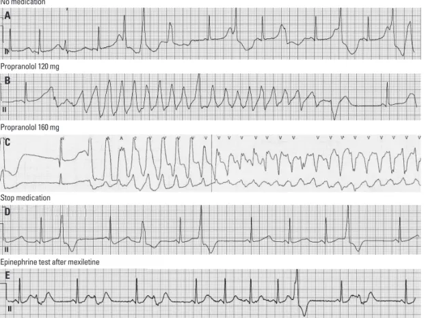
sig-nificantly prolonged (637.0-48.6 ms; between 585 and 646 ms), and frequent single or couplets of ventricular prema-ture beats appeared (Fig. 3A). We then repeated the epi-nephrine test with β-blocker medication. With propranolol 120 mg per day, mean QTc was prolonged as 628.5±62.7 ms (between 618 and 634 ms), and non-sustained polymor-phic ventricular tachycardia (VT; 200 bpm) appeared fol-lowing R-on-T phenomenon and spontaneously ended with-in 8 sec without syncopal event (Fig. 3B). There were two different premature ventricular contraction (PVC) morphol-ogies, and non-sustained VT initiating PVC was localized by 12 lead ECG (Fig. 1B). The origin of PVC seems to be left ventricular (LV) high septum just below the (left) distal HIS bundle. QRS duration is relatively narrow and purely negative in lead aVR and aVF, but initially positive in all oth-er leads, suggesting LV poth-eri-Hissian PVC related to membra-nous VSD repair. After increasing the dosage of proprano-lol to 160 mg per day for 3 days, sustained polymorphic VT was induced spontaneously, requiring external defibrilla-tion, and syncope and tonic clonic seizure were document-ed during the epinephrine provocation test (Fig. 3C). Mean QTc was prolonged as 644.0±33.7 ms (between 619 and 668 ms). However, after stopping propranolol for 4 days, no arrhythmic event was reproducible during the epineph-rine provocation test (Fig. 3D).
Implantable cardioverter defibrillator (ICD)
Because the patient’s arrhythmia could not be controlled by β-blocker, she underwent ICD implantation. She returned to the emergency room due to ICD shock in the 3rd month after ICD implantation. ICD EGM documented sustained polymorphic VT with cycle length 220 ms and successful ICD therapy. Therefore, we repeated the epinephrine prov-ocation test while medicating with mexiletine 600 mg per day, and there was no event except for QTc lengthening >30 ms (Fig. 3E). She was discharged with mexiletin 600 mg per day and an increase in atrial pacing rate to higher than 70 bpm. During mexiletine medication, there was no VT ep-isode in ICD interrogation for 3 months. However, she arbi-trarily stopped the anti-arrhythmic drug due to weakness, months old, however, there was no abnormality in the
physical examination or laboratory findings, and echocar-diography showed her heart to be completely normal mor-phologically and functionally. The only abnormal finding was prolonged QTc (629 ms) in surface 12 lead electrocar-diography (ECG) (Fig. 1A). She had no family history of sudden death or other genetic heart disease (Fig. 2). At the time when she was referred to our hospital, she was taking low dose of propranolol. Because ECG at the time of pre-syncope or pre-syncope had not been documented and her
clin-Fig. 1. (A) Baseline standard 12 lead ECG shows prolonged QTc interval
(629 ms). (B) 12 ECG shows premature ventricular contraction which was originated from left ventriclular basal posteroseptum, related to earlier VSD repair. ECG, electrocardiography; VSD, ventricular septal defect.
Fig. 2. The genetic pedigree of the patient shows that her grandmother died
from pancreatic cancer, and there was no family history of sudden death.
Exon 20 (G→A, R1192Q)
Exon 20 (G→A, R1192Q)
Exon 20 (G→A, R1192Q) Exon 20 (G→A, R1192Q)
Affected female (patient) Deceased female (by cancer)
Male Female
A
ventricular tachyarrhythmia episodes.5,6 However, the drug
failure rate of β-blocker is significantly higher in patients with LQTs type 3,7 and mortality and morbidity are also
higher in LQTs type 3 than those with types 1 or 2. LQTs type 3 accounts for about 5-10% of LQTs.8 Nevertheless,
β-blocker has never been reported to aggravate Torsades de Point (TdP) in patients with LQTs type 3. Genetically, LQTs type 3 has been known to be due to the mutation of SCN5A domain which results in a “gain of function in sodi-um channel” and prolongs action potential duration and QTc. Consequently, the pharmacological blocker of sodium current, mexiletine, shortens the QTc interval and rescues the defect of the SCN5A mutation.9,10-12 However, it is not
clear whether gene-specific medication, such as mexiletine, is able to prevent sudden cardiac death in patients with LQTs type 3. The SCN5A gene, consisting of 28 exons and 2016 amino acids, is highly expressed in the human myo-cardium.13 The missense, a splice-donor, or frame-shift
mu-tations in SCN5A coding regions have been known to be associated with Brugada syndrome, sudden infant death and received appropriate ICD shock 8 months after stopping
the medication. Therefore, she is presently taking mexiletine 600 mg a day.
DNA isolation and genetic analysis
We genotyped 155 loci from exon 2 to 28 of the SCN5A gene,4 and G>A, R1192Q (rs41261344; reference sequence:
NM000335) was identified at the exon 20 sequence. How-ever, same base pair change was found in SCN5A genetic analyses in her father and siblings who did not show the phenotypes of LQTs (ECG or symptom) (Fig. 2). There-fore, it seems to be genetic polymorphism in SCN5A, but not a mutation.
DISCUSSION
Over the past few decades, β-blocker has remained as the most effective medication for LQTs, functioning by short-ening the repolarization period and reducing the duration of
Fig. 3. The ECG documented events during epinephrine provocation test. (A) Baseline epinephrine provocation test showing
non-sus-tained VT with maximal 3 beats of extra-systole. (B) While taking propranolol 120 mg, non-susnon-sus-tained polymorphic VT was repeatedly doc-umented during epinephrine infusion. (C) While taking propranolol 160 mg, torsade de pointes requiring cardioversion was spontaneously induced during epinephrine infusion. (D) After stopping propranolol, only isolated ventricular premature beats were induced by epineph-rine infusion. (E) During medication with mexiletine 600 mg per day, the epinephepineph-rine provocation test showed no event except for QTc lengthening >30 ms. ECG, electrocardiography; VT, ventricular tachycardia.
No medication
Propranolol 120 mg
Propranolol 160 mg
Stop medication
Epinephrine test after mexiletine A
B
C
D
QTc prolongation and ventricular fibrillation in a healthy young man. Yonsei Med J 2011;52:1025-7.
2. Shimizu W, Noda T, Takaki H, Kurita T, Nagaya N, Satomi K, et al. Epinephrine unmasks latent mutation carriers with LQT1 form of congenital long-QT syndrome. J Am Coll Cardiol 2003;41:633-42. 3. Shimizu W, Noda T, Takaki H, Nagaya N, Satomi K, Kurita T, et
al. Diagnostic value of epinephrine test for genotyping LQT1, LQT2, and LQT3 forms of congenital long QT syndrome. Heart Rhythm 2004;1:276-83.
4. Kapa S, Tester DJ, Salisbury BA, Harris-Kerr C, Pungliya MS, Alders M, et al. Genetic testing for long-QT syndrome: distin-guishing pathogenic mutations from benign variants. Circulation 2009;120:1752-60.
5. Schwartz PJ, Priori SG, Spazzolini C, Moss AJ, Vincent GM, Na-politano C, et al. Genotype-phenotype correlation in the long-QT syndrome: gene-specific triggers for life-threatening arrhythmias. Circulation 2001;103:89-95.
6. Priori SG, Napolitano C, Paganini V, Cantù F, Schwartz PJ. Mo-lecular biology of the long QT syndrome: impact on management. Pacing Clin Electrophysiol 1997;20(8 Pt 2):2052-7.
7. Priori SG, Napolitano C, Schwartz PJ, Grillo M, Bloise R, Ronch-etti E, et al. Association of long QT syndrome loci and cardiac events among patients treated with beta-blockers. JAMA 2004; 292:1341-4.
8. Splawski I, Shen J, Timothy KW, Lehmann MH, Priori S, Robin-son JL, et al. Spectrum of mutations in long-QT syndrome genes. KVLQT1, HERG, SCN5A, KCNE1, and KCNE2. Circulation 2000;102:1178-85.
9. Bennett PB, Yazawa K, Makita N, George AL Jr. Molecular mechanism for an inherited cardiac arrhythmia. Nature 1995;376: 683-5.
10. Wang DW, Yazawa K, Makita N, George AL Jr, Bennett PB. Pharmacological targeting of long QT mutant sodium channels. J Clin Invest 1997;99:1714-20.
11. Fabritz L, Kirchhof P, Franz MR, Nuyens D, Rossenbacker T, Ot-tenhof A, et al. Effect of pacing and mexiletine on dispersion of repolarisation and arrhythmias in DeltaKPQ SCN5A (long QT3) mice. Cardiovasc Res 2003;57:1085-93.
12. Benhorin J, Taub R, Goldmit M, Kerem B, Kass RS, Windman I, et al. Effects of flecainide in patients with new SCN5A mutation: mutation-specific therapy for long-QT syndrome? Circulation 2000;101:1698-706.
13. Gellens ME, George AL Jr, Chen LQ, Chahine M, Horn R, Barchi RL, et al. Primary structure and functional expression of the hu-man cardiac tetrodotoxin-insensitive voltage-dependent sodium channel. Proc Natl Acad Sci U S A 1992;89:554-8.
14. Chen Q, Kirsch GE, Zhang D, Brugada R, Brugada J, Brugada P, et al. Genetic basis and molecular mechanism for idiopathic ven-tricular fibrillation. Nature 1998;392:293-6.
15. Vyas H, Hejlik J, Ackerman MJ. Epinephrine QT stress testing in the evaluation of congenital long-QT syndrome: diagnostic accura-cy of the paradoxical QT response. Circulation 2006;113:1385-92. 16. Wang Q, Chen S, Chen Q, Wan X, Shen J, Hoeltge GA, et al. The
common SCN5A mutation R1193Q causes LQTS-type electro-physiological alterations of the cardiac sodium channel. J Med Genet 2004;41:e66.
17. Vatta M, Dumaine R, Varghese G, Richard TA, Shimizu W, Ai-hara N, et al. Genetic and biophysical basis of sudden unexplained nocturnal death syndrome (SUNDS), a disease allelic to Brugada syndrome. Hum Mol Genet 2002;11:337-45.
syndrome, and Lev-Lenegre disease.14
In our patient, the clinical presentation of LQTs was un-usual (agonal respiration and syncope at rest) and the epi-nephrine test played a remarkably important role in identify-inggenetic subtype of LQTs and determining an appropriate treatment. First line drug treatment with β-blocker aggravat-ed TdP, and mexiletine suppressaggravat-ed arrhythmic event in this patient. Without the epinephrine provocation test, β-blocker might have been prescribed and harmed this patient with life-threatening channelopathy. Although Vyas, et al.15
re-ported low predictive value of epinephrine test in LQTs type 3, Shimizu, et al.3 described that it still has an important
di-agnostic value in LQTs type 3. Ethnic difference or undis-covered genetic polymorphism of SCN5A may play some roles in this difference. Therefore, the epinephrine test can be an important diagnostic and drug monitoring test, and complementary to genetic diagnosis.
R1193Q has been implicated in both LQTs type 3, a gain of function disease,16 and in sudden unexplained nocturnal
death syndrome, a loss of function disease.17 At the same
time, R1193Q is considered as a polymorphism in Asians with an allele frequency of 8%.4,18 R1192Q in the present
case is not distinct from R1193Q in the original hH1 se-quence. Therefore, additional polymorphisms in SCN5A may contribute to R1193Q-related QTc prolongation, and we cannot exclude overlap phenotype of LQTs type 3 with Bru-gada syndrome.19
In conclusion, we experienced a patient with clinically type 3 LQTs who was aggravated by β-blocker and alleviat-ed by mexiletine. The epinephrine provocation test playalleviat-ed a remarkably important role in the diagnosis and monitoring of drug efficiency in this patient.
ACKNOWLEDGEMENTS
This work was supported by a grant from the Korea Health 21 R&D Project (A085136), Ministry of Health and Wel-fare, and Basic Science Research Program under the Na-tional Research Foundation of Korea (NRF), funded by the Ministry of Education, Science and Technology (2010-0010537), Republic of Korea.
REFERENCES
19. Makita N, Behr E, Shimizu W, Horie M, Sunami A, Crotti L, et al. The E1784K mutation in SCN5A is associated with mixed clinical phenotype of type 3 long QT syndrome. J Clin Invest 2008;118: 2219-29.
18. Ackerman MJ, Splawski I, Makielski JC, Tester DJ, Will ML, Timothy KW, et al. Spectrum and prevalence of cardiac sodium channel variants among black, white, Asian, and Hispanic individ-uals: implications for arrhythmogenic susceptibility and Brugada/ long QT syndrome genetic testing. Heart Rhythm 2004;1:600-7.