Recurrence of Adenoid Cystic Carcinoma
in the Breast After Lumpectomy and
Adjuvant Therapy
Ji Hyun Youk, MD, Min Jung Kim, MD, Eun-Kyung Kim, MD, Ji Young Lee, MD, Ki Keun Oh, MD, Byeong Woo Park, MD
denoid cystic carcinoma (ACC) of the breast is very rare, accounting for only 0.1% of all breast carcinomas.1–4This unusual neoplasm of the breast has certain histopathologic and biological characteristics that distinguish it from more common histologic types. For example, the prognosis of ACC appears to be very favorable. The incidence of axillary lymph node involvement is lower, and distant metastases are uncommon, although they can occur without pre-vious lymph node involvement. Clinical experience has shown that this entity has a low propensity for local recurrence after excision.4,5We present a case of local recur-rence of ACC in the breast detected by ultrasonography after successful treatment by lumpectomy followed by adjuvant chemotherapy and radiotherapy.
Received January 5, 2006, from the Department of Diagnostic Radiology (J.H.Y., M.J.K., E.-K.K., J.Y.L., K.K.O.), Research Institute of Radiological Science, and Department of Surgery (B.W.P.), Yonsei University College of Medicine, Seoul, Korea. Revision requested February 7, 2006. Revised manuscript accepted for publication February 22, 2006.
Address correspondence to Eun-Kyung Kim, MD, Department of Diagnostic Radiology, Yonsei University College of Medicine, Seodaemun-ku, Shinchon-dong 134, Seoul 120-752, Korea.
E-mail: [email protected] Abbreviations
ACC, adenoid cystic carcinoma
Case Report
A 48-year-old woman came to our hospital because her screening mammography showed dense mammary tis-sue with a poorly defined mass in the upper center of the right breast. Adjunctive breast ultrasonography per-formed at our hospital showed a mass indicative of malignancy in the same location (Figure 1). An ultra-sonographically guided 14-gauge core needle biopsy was taken, and pathologic diagnosis confirmed ACC. The patient underwent lumpectomy of the right breast and dissection of right axillary lymph nodes. The speci-men of breast measured 2.7 × 5 × 3.4 cm and contained a poorly defined, pinkish tan, round mass measuring 1.3 × 1.2 cm. The surgical margins were free of tumor, and all 14 dissected lymph nodes were negative for tumor. The final pathologic report was ACC, and the estrogen recep-tor, progesterone receprecep-tor, and c-erb-B2 status of the tumor were all negative. The patient received adjuvant chemotherapy with a 6-cycle regimen of cyclophos-phamide-methotrexate-5-fluorouracil and radiotherapy to the right breast in a daily fraction of 1.8 Gy for a total of
© 2006 by the American Institute of Ultrasound in Medicine • J Ultrasound Med 2006; 25:921–924 • 0278-4297/06/$3.50
A
Case Report
59.4 Gy (50.4 Gy to the whole breast and then a 9-Gy boost to the tumor bed area). The patient tol-erated the treatment well and had no evidence of recurrence on the follow-up mammography or ultrasonography until 6 months after the adju-vant therapies.
The patient had localized tenderness in the periareolar aspect of the right breast 1 year after lumpectomy of the ipsilateral breast. Physical examination found only localized tenderness without any evidence of a focal mass at the ten-der site. Follow-up mammography and targeted
ultrasonography of the tender area showed no abnormalities (Figure 2). After 6 months, there was aggravated tenderness at the lumpectomy site, and ultrasonographic examination showed a 6-mm, round, microlobulated, hypoechoic nodule at the lumpectomy site. The tenderness was aggravated as the probe compressed the nod-ule (Figure 3). The patient underwent an ultra-sonographically guided 14-gauge core needle biopsy, and ACC was diagnosed again. A simple mastectomy was performed, and the recurrence of ACC was confirmed pathologically.
922 J Ultrasound Med 2006; 25:921–924
Recurrence of Adenoid Cystic Carcinoma
Figure 1. Mammograms and sonogram obtained 18 months before recurrence. A, Mediolateral oblique (left) and craniocaudal (right) mammograms
show a poorly defined, round, hyperdense mass in the upper center of the right breast (arrows). No abnormalities are seen in the left breast.
B, Sonogram reveals a 1.3-cm, round, microlobulated, hypoechoic mass with posterior acoustic enhancement in the 12-o’clock location.
A B
Figure 2. Follow-up mammograms and sonogram obtained 1 year after lumpectomy and adjuvant therapy. A, Right mediolateral oblique (left) and
craniocaudal (right) mammograms show postoperative changes of the breast deformity, distortion, and increased densities due to fibrotic scar forma-tion. Therefore, it is difficult to differentiate them from any possible recurring lesion. B, Sonogram of the area of localized tenderness. The upper medi-al portion of the right breast depicts no abnormmedi-ality.
A B
Discussion
As with all types of breast cancer, treatment options have evolved for patients with ACC of the breast and depend on the circumstances of a given patient. Wide excision alone could be cura-tive in most cases because of the favorable bio-logical behavior of this disease.6 The case presented here is notable as a local recurrence of ACC in the breast even after successful treatment with lumpectomy, adjuvant chemotherapy, and radiotherapy. Few articles have described adju-vant radiotherapy after initial local excision, and, to our knowledge, there has been only 1 case of local recurrence of ACC in the breast after lumpectomy followed by adjuvant radiotherapy, suggesting that radiation after lumpectomy may play a complementary role for local control.4,5,7 Little mention is made in the literature of adju-vant systemic therapy for this diagnosis, and clear indications for the role of adjuvant systemic ther-apy remain to be defined.
Because of the rarity of this entity, there is little information about the characteristic features of ACC on mammography or ultrasonography. Santamaria et al8reported that on mammogra-phy this tumor often appears as moderately cir-cumscribed, lobulated nodules that are similar to other types of benign and malignant tumors. In 2 cases of ACC described by Sheen-Chen et al,9 ultrasonography showed nodules with well-defined margins and heterogeneous echogenici-ty without the characteristic changes of an infiltrating carcinoma. Therefore, a benign-look-ing breast lesion in radiologic examinations can-not completely exclude the existence of this disease9; some tumors cannot be identified on mammography because of increased density of the breast.8,9 Ultrasonography can identify and help characterize a lesion in the breast with a predominance of dense fibroglandular tissue that could be missed on mammography.10
In this case, the first targeted ultrasonography for localized tenderness failed to depict a recur-ring lesion. According to Leung et al,11most of the ultrasonographic examinations performed to evaluate focal breast pain yielded negative findings, and no cancer was identified. They con-cluded that the inappropriate use of targeted ultrasonography for focal breast pain should be a reason for concern in the absence of evidence-based data that would justify its use. In a study by McClenathan and de la Roza,12however, pain was
a prominent symptom of patients with ACC, sev-eral of whom were monitored for localized pain in the breast for more than 1 year before a mass or radiographic abnormality was detected. Therefore, if localized tenderness or pain occurs with or with-out a mass, ACC of the breast should be included in a differential diagnosis. In our case, localized pain was the first sign of recurrence of ACC with-out previous symptoms of localized pain or ten-derness related to the primary lesion. Despite little information about the recurrence of ACC, local-ized pain could be a new symptom, regardless of previous symptoms. If there is a painful sensation as the probe compresses the lesion on follow-up ultrasonography after treatment of ACC, this unusual sign could be indicative of its recurrence, whether there was localized pain related to prima-ry ACC. Suppose additional magnetic resonance imaging was performed when localized pain pre-sented but targeted ultrasonography did not show a recurring lesion; in such a case, it might be help-ful for detection of a recurring lesion. Until now, to our knowledge, the findings of ACC in the breast on magnetic resonance imaging have not been reported, and performance of magnetic resonance imaging has been uncertain for the detection of recurrence of ACC. Further study regarding this is needed. Local recurrence, although rare and generally occurring long after the initial event, should be considered in a patient with a history of ACC in the breast, and appropriate follow-up and clinical correlations could be helpful for early detection of a possible recurrence.
J Ultrasound Med 2006; 25:921–924 923 Youk et al
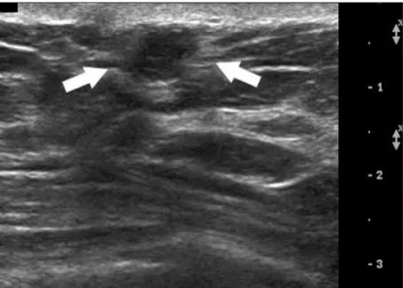
Figure 3. Sonogram obtained 6 months after image in Figure 2 shows a new
pal-pable nodule just at the tender site of the right breast (arrows).
References
1. Anthony PP, James PD. Adenoid cystic carcinoma of the breast: prevalence, diagnostic criteria, and histogenesis. J Clin Pathol 1975; 28:647–655.
2. Lamovec J, Us-Krasovec M, Zidar A, Kljun A. Adenoid cys-tic carcinoma of the breast: a histologic, cytologic, and immunohistochemical study. Semin Diagn Pathol 1989; 6:153–164.
3. Miliauskas JR, Leong AS. Adenoid cystic carcinoma in a juvenile male breast. Pathology 1991; 23:298–301. 4. Arpino G, Clark GM, Mohsin S, Bardou VJ, Elledge RM.
Adenoid cystic carcinoma of the breast: molecular markers, treatment, and clinical outcome. Cancer 2002; 94:2119– 2127.
5. Leeming R, Jenkins M, Mendelsohn G. Adenoid cystic car-cinoma of the breast. Arch Surg 1992; 127:233–235. 6. Sumpio BE, Jennings TA, Sullivan PD, Merino MJ. Adenoid
cystic carcinoma of the breast: data from the Connecticut tumor registry and a review of the literature. Ann Surg 1987; 205:295–301.
7. Millar BA, Kerba M, Youngson B, Lockwood GA, Liu FF. The potential role of breast conservation surgery and adjuvant breast radiation for adenoid cystic carcinoma of the breast. Breast Cancer Res Treat 2004; 87:225–232.
8. Santamaria G, Velasco M, Zanon G, et al. Adenoid cystic carcinoma of the breast: mammographic appearance and pathologic correlation. AJR Am J Roentgenol 1998; 171: 1679–1683.
9. Sheen-Chen SM, Eng HL, Chen WJ, Cheng YF, Ko SF. Adenoid cystic carcinoma of the breast: truly uncommon or easily overlooked? Anticancer Res 2005; 25:455–458. 10. Dinkel HP, Clees M, Gassel AM. Role of sonography in
detecting recurrence in adenoid cystic carcinoma of the breast. AJR Am J Roentgenol 1999; 173;497–498. 11. Leung JW, Kornguth PJ, Gotway MB. Utility of targeted
sonography in the evaluation of focal breast pain. J Ultrasound Med 2002; 21:521–526.
12. McClenathan JH, de la Roza G. Adenoid cystic breast can-cer. Am J Surg 2002; 183:646–649.
924 J Ultrasound Med 2006; 25:921–924
Recurrence of Adenoid Cystic Carcinoma