INTRODUCTION
Most patients with neurofibromatosis type II (NF-II) will ev-entually encounter bilateral deafness, creating an obstacle to social life. In patients with NF-II, sound amplification with he-aring aids does not provide an effective solution, as speech dis-crimination scores (SDSs) usually remain low in the presence of retrocochlear lesions. Certain NF-II patients with mild to moderate hearing loss might benefit slightly from the use of hearing aids; however, in most cases, their hearing loss is pro-gressive and often inevitably exceeds a level that can be reha-bilitated by hearing aids.
In the past, no effective methods for rehabilitating patients with severe to profound hearing loss existed; consequently, lip-reading and sign language have been the only means of communication. In 1979, House and Hitselberger successfully
performed a single-channel auditory brainstem implant (ABI) for the hearing rehabilitation of a patient with NF-II. Subse-quently, the principles and concepts of treatment for patients with severe to profound hearing loss have changed.
Advancements in electronic and medical devices have pro-vided satisfactory treatment results in patients with severe to profound hearing loss. Otologists should be fully aware of the diverse indications and potential benefits of every possible hearing rehabilitation method in order to provide active sup-port for patients with NF-II.
MECHANISMS OF HEARING LOSS
IN NEUROFIBROMATOSIS TYPE II
NF-II is an autosomal dominant neoplastic syndrome. It has an incidence of 1 in 25000 people and a penetrance of nearly 100% at 60 years of age.1 Clinical manifestations include
cen-tral and peripheral nervous system tumors, and the hallmark of NF-II is the development of bilateral vestibular schwanno-mas (VSs), which present in 90% to 95% of NF-II patients.2
Though VSs are benign tumors, they cause significant hearing loss, with binaural hearing loss occurring in nearly all NF-II patients. However, the mechanisms of hearing loss in NF-II are not truly understood.
The most frequent hypothesis regarding the source of hear-ing loss in NF-II is that it is caused by direct compression of the cochlear nerve. Previous studies have reported that the pres-ence of hearing loss is correlated with larger tumor volumes.3
Hearing Restoration in Neurofibromatosis
Type II Patients
Jeon Mi Lee
1, Jin Woo Chang
2, Jae Young Choi
1, Won Seok Chang
2, and In Seok Moon
11Department of Otorhinolaryngology, Yonsei University College of Medicine, Seoul;
2Department of Neurosurgery, Brain Research Institute, Yonsei University College of Medicine, Seoul, Korea.
Patients with neurofibromatosis type II will eventually succumb to bilateral deafness. For patients with hearing loss, modern medical science technology can provide efficient hearing restoration through a number of various methods. In this article, several hearing restoration methods for patients with neurofibromatosis type II are introduced.
Key Words: Neurofibromatosis type II, auditory brain stem implantation, cochlear implantation, hearing aids, vestibular schwan-noma
pISSN: 0513-5796 · eISSN: 1976-2437
Received: February 29, 2016 Revised: March 30, 2016 Accepted: April 1, 2016
Co-corresponding authors: Dr. Won Seok Chang, Department of Neurosurgery,
Brain Research Institute, Yonsei University College of Medicine, 50-1 Yonsei-ro, Seodaemun-gu, Seoul 03722, Korea.
Tel: 82-2-2228-2176, Fax: 82-2-393-9979, E-mail: [email protected] and Dr. In Seok Moon, Department of Otorhinolaryngology, Yonsei University College of Medicine, 50-1 Yonsei-ro, Seodaemun-gu, Seoul 03722, Korea.
Tel: 82-2-2228-3626, Fax: 82-2-393-0580, E-mail: [email protected] •The authors have no financial conflicts of interest.
© Copyright: Yonsei University College of Medicine 2016
This is an Open Access article distributed under the terms of the Creative Com-mons Attribution Non-Commercial License (http://creativecomCom-mons.org/licenses/ by-nc/3.0) which permits unrestricted non-commercial use, distribution, and repro-duction in any medium, provided the original work is properly cited.
Yonsei Med J 2016 Jul;57(4):817-823 http://dx.doi.org/10.3349/ymj.2016.57.4.817
However, this hypothesis cannot explain hearing loss when tumors are small, progressive hearing loss in the presence of non-growing tumors, or unpredictable onset of hearing loss.4,5
In 2012, Roosli, et al.6 reviewed the histopathology of
cochle-ae in patients with VSs and observed loss of inner and outer hair cells, atrophy of the stria vascularis, loss of cochlear neu-rons, and the presence of endolymphatic hydrops. The non-tumorous cochleae did not present with such changes. These structural changes were not correlated with tumor size or dis-tance from the cochlea.6 These findings indicate that hearing
loss in VSs is more likely due to end organ failure than proxi-mal cochlear nerve functions. It was recently reported that el-evated levels of intralabyrinthine protein as observed via fluid attenuated inversion recovery magnetic resonance imaging (FLAIR MRI) is closely associated with hearing loss in NF-II.3
Intralabyrinthine protein elevation is caused by cochlear ap-erture obstruction or destruction by VSs and explains the end organ failure in NF-II. It was also demonstrated that increases in intralabyrinthine protein obtained from perilymph aspira-tion could accurately identify the presence of VSs.7
HEARING AIDS
The pattern of hearing loss is unpredictable in NF-II. Sudden, relapsing, or progressive hearing loss may occur regardless of tumor size or time from initial diagnosis. However, the natural history of hearing loss in NF-II has previously been studied.4
In this study, 108 ears were examined; the pure-tone average (PTA) at baseline was 22.2±21.8 dB with a mean SDS of 93.9± 13.1%. Without treatment, the follow-up PTA at 2 years de-creased to 37.0±31.4 dB, and SDS also dropped to 83.9±28.4%. NF-II is likely to be diagnosed before any loss of hearing, indi-cating that in most cases, the baseline hearing is normal. This decrease is faster and more severe than that of general age-re-lated hearing loss,8 and of note, a decrease in SDS severely
re-duces the effectiveness of hearing aids. While hearing aids can be helpful for any patient with hearing loss, those with mo-derate levels of loss receive the most potential benefit. Severe or profound loss can limit the usefulness of even the most pow-erful hearing aids. In many cases of profound hearing loss, a hearing aid can only provide environmental awareness or a de-gree of rudimentary perception of speech.9
Contralateral routing of signals (CROS) hearing aids have been introduced to provide audiologic benefits by routing the sound from the hearing-impaired side to the intact ear. Use of CROS hearing aids is the easiest method for rehabilitation in patients with asymmetric hearing loss without surgery; how-ever, there are several limitations, including poor cosmetics due to the use of two hearing aids with a connection wire, dis-comfort from the occlusion of the better ear, and most of all, degradation of speech intelligibility in certain demanding sit-uations10 and significant deficit in noisy environments.11
De-spite its convenience and simplicity, cosmetic inferiority and discomfort from the occlusion effect in the normal ear has pre-cluded the widespread use of CROS as a conventional type of hearing aid.
BONE-CONDUCTION HEARING IMPLANT
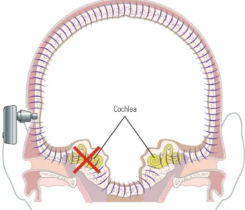
For patients with single-sided deafness, occurring either sp-ontaneously or after tumor removal, a bone-conduction hear-ing implant (BCHI) can be a good hearhear-ing rehabilitation op-tion. In this option, the speech processor receives the auditory signal and transforms it to a vibration, after which the BCHI transmits the vibration to both the ipsilateral and contralateral cochleae as it passes through the skin and skull. Conductive hearing loss, mixed hearing loss, and single-sided deafness are all good candidates for BCHI. In cases of patients with sin-gle-sided deafness, the vibration generated from the deaf side is transferred to the contralateral normal cochlea, where it can be recognized. However, BCHI is not as applicable to binaural hearing, as it does not offer many of the advantages of binaural hearing: summation effects, binaural squelch, and sound lo-calization cannot be attained (Fig. 1). Despite these limitations, patients’ satisfaction with BCHI is usually quite high.12,13From its first introduction in 1976,14 BCHIs have developed
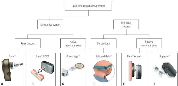
at a remarkable pace, and various products have been com-mercialized or are in clinical trials. BCHIs can be classified ac-cording to transmit system: one is a direct-drive system that transmits the vibration through the skull, and the other is a skin-drive system that transmits the vibration through the skin (Fig. 2).
Examples of direct-drive systems include Ponto® (Oticon
Me-dical, Smørum, Denmark), Baha® BP100 (Cochlear Bone
An-Fig. 1. Mechanisms of a bone-conduction hearing implant. Vibrations gen-erated from the device are transferred to the contralateral cochlea and recognized on the contralateral side.
chored Solutions AB, Molnlycke, Sweden), and BonebridgeTM
(MED-EL, Innsbruck, Austria). The Ponto® and Baha® BP100
systems use screws to attach the speech processor to the skull, so that the vibrations generated from the device are directly transmitted to the skull. They are known to achieve a sufficient hearing gain, as the vibration directly transmits via the screw, and a degree of binaural squelch effect has also been report-ed.15 Despite the above-mentioned benefits, recipients often
suffer from various skin problems and loosening of the screws due to their fixation methods.16 To overcome these limitations,
active transcutaneous devices were introduced. In the case of the BonebridgeTM system, active BCHIs are implanted into the
temporal bone and vibrate within the temporal bone. Speech processors are attached to the scalp through magnets. This promotes sufficient hearing gain and is not associated with any skin problems. However, larger internal devices are re-quired, which has made it difficult to perform such implanta-tions in children or in patients who have undergone previous mastoidectomy. Various methods are followed to overcome these limitations, such as the use of a lift system or the retrosi-gmoid approach (RSA).17
A skin-drive system generates the vibration outside of the skull. The vibration must pass through the skin to reach the skull. Softband Baha® (Cochlear Bone Anchored Solutions AB,
Molnlycke, Sweden), Sophono® (Medtronic, Louisville, CO,
USA), and Baha® Attract (Cochlear Bone Anchored Solutions
AB, Molnlycke, Sweden) use this system. Softband Baha® uses
elastic headbands around the head, instead of screws, to fix the device to the skull. It can be used with children who are too young to undergo implant surgery.18 In addition, it is quite effective at
simulating and predicting the outcomes of BCHIs and can thus
be used with patients who are planning to undergo BCHI sur-geries.19 However, for secure fixation, the pressure that is
requ-ired may cause deformations of the skin and subcutaneous tissues, and tension headaches. This problem can be solved by using magnets, which is how the speech processors of Sopho-no® and Baha® Attract are attached to the scalp. However, thick
skin could weaken the magnetic forces and reduce the fixation; thus, the thickness of skin should be less than 5 mm.20 A
skin-drive system tends to have less hearing gain than a direct-skin-drive system due to the attenuation of the mechanical energy as it pass-es through skin;21 however, theoretically it can be overcome by
fixing the speech processor in the proper location with ade-quate pressure.22
As all BCHIs contain permanent magnets, MRI compatibil-ity is a challenging concern. Computed tomography (CT) sh-ould be used as a first choice for the safety of both patients and devices; however, certain clinical conditions require an MRI scan in order to obtain proper image information. In cases of NF-II, regular MR imaging is required. BonebridgeTM and
So-phono® are certified to be conditionally safe up to 1.5 and 3
Tesla,23 respectively, although there will be an image artifact in
the region near the implant. Manufacturers have specified the size of the corresponding MRI artifact, specifically, a sphere of 15 cm in diameter for BonebridgeTM, a distance of 5–10 cm
from the Sophono® implant, and a distance of 11.5 cm from the
center of the Baha® Attract implant.24,25 Steinmetz, et al.26 also
reported a 29-year-old patient with VS who was scanned with a BonebridgeTM implant on the contralateral side; due to the
image artifact, the tumor was concealed. If an MRI absolutely needs to be performed with a focus on structures in the skull near the implant, the implant must be explanted to eliminate
ar-Fig. 2. Categorization of bone-conduction hearing implants. (A) Ponto®. (B) Baha® BP100. (C) BonebridgeTM. (D) Softband Baha®. (E) Baha® Attract. (F)
So-phono®. Photos provided courtesy of Oticon Medical (A), Cochlear Bone Anchored Solutions AB (B, D, and E), MED-EL (C), and Medtronic (F).
Ponto® Baha® BP100 BonebridgeTM Softband Baha® Baha® Attract Sophono®
Percutaneous transcutaneousActive Conventional
Direct-drive system Skin-drive
system Bone-conduction hearing implant
Passive transcutaneous
tifacts. Therefore, in patients with NF-II, it is important to select the proper implant for such an event.
COCHLEAR IMPLANT
In the early days of their use, patients with ABIs were only able to recognize environmental sounds or to detect sounds with a further reliance on lip-reading, and they were not capable of discriminating speech sounds.27 For better hearing
rehabilita-tion, a cochlear implant (CI) is applied to patients with NF-II. CIs offer many advantages over ABIs, and the most important benefit is that CIs can provide better speech understanding. As intracochlear electrode placement permits reliable tonotop-ic stimulation, better auditory performance is generally ex-pected.28 A CI can be attempted either with the tumor
remain-ing in place or after the tumor has been removed.
Two decades ago, the first CI was performed simultaneous-ly with the removal of a tumor in a patient with NF-II.29 CI was
also attempted on patients who were non-surgically treated, such as those who underwent stereotactic radiotherapy [or a gamma knife surgery (GS)] or refused to undergo surgery. GS is an alternative to surgery for tumor control and has been pro-posed to allow hearing preservation, though it is not without risks. Prasad, et al.30 reported the outcomes of 200 cases of VSs
that were treated with GS, and 25% demonstrated either an increase or no changes of volume, while hearing deterioration was found in 60% of the patients over a 6-year period. Out-comes for CI after GS for VS are varied in that some were able to achieve good post-implantation speech perception, while others were only capable of detecting environmental sounds.31
However, regardless of the variable outcomes, the problem is that tumors can grow at any time without complete tumor re-moval, and larger tumors are correlated with hearing loss.3
Additionally, the function of CI after GS for VS is likely to de-crease over time, and malignant transformation is also a con-cern. There have been reported cases of malignant transfor-mation of tumors in NF-II following GS, and reports suggest that up to 50% of all malignant transformations occur in NF-II patients.32 It is important to remember that regular follow-up
via MRI is critical when patients are non-surgically treated. If such imaging is obtained with the magnetic device implanted, many adverse effects could occur. MRI performed on patients with an implanted magnetic device could potentially result in a migration of the device. Demagnetization and malfunction of the devices could also occur, and the heating of such internal devices could damage the surrounding tissues. These effects can be avoided by removing the magnets before performing MRI scans or by using low-Tesla (T) MRI. Current US Food and Drug Administration (FDA) guidelines have approved the use of 0.2 to 1.5 T with the magnet in place.33 Generally, MRI
in patients with a magnetic device is reported to be safe with elastic head bands placed around their heads.34 Furthermore,
heat from the CI during 1.5-T MRI is reported to be lower than 0.1°C;34 hence, usage of 1.5-T MRI with the magnetic device is
relatively safe. However, image quality obtained from the MRI scan is another issue. Ipsilateral soft tissues within 7 to 8 cm from the magnet are poorly visualized, while the contralateral side image has no distortion.35 Several otologists recommend
simultaneous high-resolution computed tomography (HRCT) imaging and a comparison of the images for better expression.
If CI is to be performed simultaneously with tumor removal, it is necessary to preserve the cochlear nerve while removing the tumor. VSs mostly originate from vestibular nerves; there-fore, preserving the cochlear nerve while removing the tumor is possible in certain cases.36 Sporadic VSs tend to grow and
only compress the nearby cochlear nerve. However, VSs in pa-tients with NF-II directly invade into the cochlear nerve37 and
are quite adherent to the cochlear nerve, making safe dissec-tion of the cochlear nerve from the tumor difficult.29 Although
anatomically well-separated, histological injuries such as mi-nor bleeding or an axonal injury might decline the function of the cochlear nerve.38 However, though an injured cochlear
nerve cannot function with the auditory signal, the nerve can function with a direct electrical signal under the conditions of the CI. Response to an electrical signal can be evaluated by stimulating the cochlear promontory with an electrode during the operation.
Various approach techniques can be applied; however, a translabyrinthine approach (TLA) is known to be the best ap-proach for accomplishing both complete tumor removal and successful CI. TLA, which provides the otologist with a famil-iar surgical view, offers early identification of the cochlear nerve in the auditory canal during the surgery and eliminates any need for cerebellar retraction.39 Cole, et al.40 compared the
RSA with TLA for VS resection and observed that TLA was as-sociated with a lower risk of cochlear nerve injury, which is essential for performing CI, dysphagia, and dysrhythmia.
There are many reports of performing CIs after tumor remov-al in either sporadic or NF-II-associated VSs.31,41-44 Generally,
outcomes for sound field and speech perception in post-lingual CI patients and in patients with tumors have been similar, and half of the patients with tumors were able to communicate on telephones.45 It is not certain what the appropriate time
inter-val is between implantation and tumor remointer-val. Theoretically, simultaneous implantation would reduce the possibility of failure, as the chance of fibrosis or ossification of the cochlea could be avoided. Furthermore, distortion of the anatomy due to tumor removal can be minimized if the implantation is per-formed simultaneously. The time required for fibrosis or ossi-fication of the cochlea to occur is unknown; however, there have been reports of CI failure occurring due to cochlear ossi-fication when performed 1 year after removing a tumor via TLA.43 On the other hand, there is also a report of a successful
implantation being performed 3 months after the tumor re-moval, via the same approach.42
AUDITORY BRAINSTEM IMPLANT
After the first successful implantation in 1979, ABIs were origi-nally the only way to restore hearing in patients with NF-II. Ini-tially, ABIs were developed for patients who could not benefit from a CI, i.e., patients with non-functioning cochlear nerves. Patients with NF-II are typical candidates; however, the indi-cations of ABI are becoming wider, such that patients with a co-chlear anomaly or a coco-chlear ossification following meningitis are now also good candidates.46
The operation principle of an ABI is similar to that of a CI. In cases of CIs, the external auditory processor receives the audi-tory signal and transforms it into an electrical signal. This sig-nal is then transmitted to the electrode that was inserted into the cochlea, and the stimulation is delivered to the auditory nerve. Finally, the auditory signal is recognized in the brain.
In cases of ABIs, the electrode is inserted in the cochlear nu-cleus, which is located at the brainstem, proximal to the audi-tory nerve. The transformed signal is then transmitted to the electrode, and the brain recognizes the signal (Fig. 3).
The cochlear nucleus is located on the dorsal lateral side of the lateral recess of brainstem and can be accessed via the Fo-ramen of Luschka. For successful implantation, inserting the electrode into the exact location of the cochlear nucleus is key to the procedure. However, detecting the exact location is dif-ficult, as these structures are covered with flocculus and are not easily observed in a natural state due to the occurrence of anatomical distortions from either the tumor itself or the op-eration. TLA provides the otologist with a familiar and direct surgical view, providing a solution to this anatomical prob-lem. Furthermore, TLA is free from cerebellar retraction and is advantageous when performing ABI. A total of 90% to 95%
Fig. 3. Concurrent tumor removal and auditory brainstem implant via the translabyrinthine approach. (A) Implantable internal device. (B) Diagram of auditory brainstem implant via translabyrinthine approach. The tumor, which originated from the cochleovestibular nerve, was resected with the nerve. A flat elec-trode was inserted, which stimulated the dorsal cochlear nucleus. Photos provided courtesy of Cochlear Headquarters (A). The figure was created by Dong-Su Jang, medical illustrator (B).
A B
Fig. 4. Comparison of the retrosigmoid approach and the translabyrinthine approach for tumor removal. (A) Severe cerebellar retraction is needed to ex-pose the tumor; however, tumors in the internal auditory canal are not readily removable via the retrosigmoid approach. (B) In the translabyrinthine ap-proach, tumors in the internal auditory canal are well exposed without cerebellar retraction. Arrows indicate the direction of visual field.
of cases of NF-II have bilateral VSs.2 If a contralateral VS is too
large, cerebellar retraction is hardly feasible; thus, RSA would not be able to provide the proper surgical views. Moreover, VSs in NF-II are likely to grow into the internal auditory canal,47
preventing complete tumor resection via RSA (Fig. 4). For these reasons, TLA is recommended by experienced surgeons.48
In-tra-operative monitoring is also strongly recommended for bet-ter detection of the exact implant location.49
ABIs provide effective hearing rehabilitation for patients who cannot benefit from CI. Since the first ABI in 1979, House Ear Institute (Los Angeles, CA, USA) surgeons have performed more than 230 ABIs. In their patient series, 85% were able to detect the auditory signal, and 93% showed great improvement in understanding sentences with the assistance of lip-reading.27
In 2002, two implantees were reported to have the ability to communicate on the phone.50 Vincenti, et al.43 compared the
outcomes of hearing rehabilitation in patients with NF-II who underwent tumor removal and received either a CI or ABI at a single institute. In closed-set conditions, patients with a CI showed outstanding results compared to patients with an ABI. However, in open-set conditions, the two groups did not show significant differences. CIs in cases of NF-II need to preserve the cochlear nerve while completely removing the tumor, which is a difficult task. Remaining tumor tissue lowers the CI func-tion and makes it difficult to follow-up via MRI. With ABIs, on the other hand, it is possible to perform the surgery without preserving the cochlear nerve, making it easier to completely remove the tumor and follow-up via MRI.
CONCLUSION
VSs, especially in cases of NF-II, were once considered to be a life-threatening disease. However, with early diagnosis, treat-ment with a multidisciplinary approach, and advancetreat-ments in electronic and medical devices, not only have survival rates improved but the quality of life has also substantially been en-hanced with options for successful hearing rehabilitation.
ACKNOWLEDGEMENTS
The authors would like to thank Dong-Su Jang, MFA (Medical Illustrator, Medical Research Support Section, Yonsei Univer-sity College of Medicine, Seoul, Korea), for his help with the il-lustrations.
This research was supported by the Basic Science Research Program through the National Research Foundation of Korea (NRF), funded by the Korean government (2014R1A1A2058141).
REFERENCES
1. Evans DG, Moran A, King A, Saeed S, Gurusinghe N, Ramsden R. Incidence of vestibular schwannoma and neurofibromatosis 2 in
the North West of England over a 10-year period: higher incidence than previously thought. Otol Neurotol 2005;26:93-7.
2. Evans DG, Huson SM, Donnai D, Neary W, Blair V, Newton V, et al. A clinical study of type 2 neurofibromatosis. Q J Med 1992;84:603-18.
3. Asthagiri AR, Vasquez RA, Butman JA, Wu T, Morgan K, Brewer CC, et al. Mechanisms of hearing loss in neurofibromatosis type 2. PLoS One 2012;7:e46132.
4. Masuda A, Fisher LM, Oppenheimer ML, Iqbal Z, Slattery WH; Natural History Consortium. Hearing changes after diagnosis in neurofibromatosis type 2. Otol Neurotol 2004;25:150-4.
5. Graamans K, Van Dijk JE, Janssen LW. Hearing deterioration in pa-tients with a non-growing vestibular schwannoma. Acta Otolar-yngol 2003;123:51-4.
6. Roosli C, Linthicum FH Jr, Cureoglu S, Merchant SN. Dysfunction of the cochlea contributing to hearing loss in acoustic neuromas: an underappreciated entity. Otol Neurotol 2012;33:473-80. 7. Silverstein H. Labyrinthine tap as a diagnostic test for acoustic
neu-rinoma. Otolaryngol Clin North Am 1973;6:229-44.
8. Gates GA, Cooper JC Jr, Kannel WB, Miller NJ. Hearing in the elder-ly: the Framingham cohort, 1983-1985. Part I. Basic audiometric test results. Ear Hear 1990;11:247-56.
9. Flint PW, Haughey BH, Lund V, Niparko JK, Robbins KT, Thomas JR, et al. Cummings otolaryngology. Philadelphia, PA: Elsevier; 2015. 10. Lotterman SH, Kasten RN. Examination of the CROS type hearing
aid. J Speech Hear Res 1971;14:416-20.
11. Lin LM, Bowditch S, Anderson MJ, May B, Cox KM, Niparko JK. Amplification in the rehabilitation of unilateral deafness: speech in noise and directional hearing effects with bone-anchored hearing and contralateral routing of signal amplification. Otol Neurotol 2006;27:172-82.
12. Laske RD, Röösli C, Pfiffner F, Veraguth D, Huber AM. Functional results and subjective benefit of a transcutaneous bone conduc-tion device in patients with single-sided deafness. Otol Neurotol 2015;36:1151-6.
13. Snapp H, Angeli S, Telischi FF, Fabry D. Postoperative validation of bone-anchored implants in the single-sided deafness population. Otol Neurotol 2012;33:291-6.
14. Berger KW. Early bone conduction hearing aid devices. Arch Oto-laryngol 1976;102:315-8.
15. Kompis M, Kurz A, Pfiffner F, Senn P, Arnold A, Caversaccio M. Is complex signal processing for bone conduction hearing aids use-ful? Cochlear Implants Int 2014;15 Suppl 1:S47-50.
16. Dun CA, Faber HT, de Wolf MJ, Mylanus EA, Cremers CW, Hol MK. Assessment of more than 1,000 implanted percutaneous bone conduction devices: skin reactions and implant survival. Otol Neu-rotol 2012;33:192-8.
17. Lassaletta L, Sanchez-Cuadrado I, Muñoz E, Gavilan J. Retrosig-moid implantation of an active bone conduction stimulator in a pa-tient with chronic otitis media. Auris Nasus Larynx 2014;41:84-7. 18. Hol MK, Cremers CW, Coppens-Schellekens W, Snik AF. The BAHA
Softband. A new treatment for young children with bilateral con-genital aural atresia. Int J Pediatr Otorhinolaryngol 2005;69:973-80. 19. Doshi J, McDermott AL. Bone anchored hearing aids in children.
Expert Rev Med Devices 2015;12:73-82.
20. Siegert R. Partially implantable bone conduction hearing aids with-out a percutaneous abutment (Otomag): technique and prelimi-nary clinical results. Adv Otorhinolaryngol 2011;71:41-6.
21. Heywood RL, Patel PM, Jonathan DA. Comparison of hearing thresholds obtained with Baha preoperative assessment tools and those obtained with the osseointegrated implant. Ear Nose Throat J 2011;90:E21-7.
understand-ing with a new implant technology: a comparative study with a new nonskin penetrating Baha system. Biomed Res Int 2014;2014: 416205.
23. Reinfeldt S, Håkansson B, Taghavi H, Eeg-Olofsson M. New devel-opments in bone-conduction hearing implants: a review. Med De-vices (Auckl) 2015;8:79-93.
24. Jansson KJ, Håkansson B, Reinfeldt S, Rigato C, Eeg-Olofsson M. Magnetic resonance imaging investigation of the bone conduction implant - a pilot study at 1.5 Tesla. Med Devices (Auckl) 2015;8:413-23.
25. Rha MS, Jeong SW, Seo YW, Moon IS. Hearing rehabilitation with Sophono® in patients with unilateral hearing loss after
meningio-ma removal. Korean J Otorhinolaryngol-Head Neck Surg 2015;58: 514-9.
26. Steinmetz C, Mader I, Arndt S, Aschendorff A, Laszig R, Hassep-ass F. MRI artefacts after Bonebridge implantation. Eur Arch Oto-rhinolaryngol 2014;271:2079-82.
27. Toh EH, Luxford WM. Cochlear and brainstem implantation. 2002. Neurosurg Clin N Am 2008;19:317-29, vii.
28. Carlson ML, Breen JT, Driscoll CL, Link MJ, Neff BA, Gifford RH, et al. Cochlear implantation in patients with neurofibromatosis type 2: variables affecting auditory performance. Otol Neurotol 2012;33:853-62.
29. Hoffman RA, Kohan D, Cohen NL. Cochlear implants in the man-agement of bilateral acoustic neuromas. Am J Otol 1992;13:525-8. 30. Prasad D, Steiner M, Steiner L. Gamma surgery for vestibular
schwannoma. J Neurosurg 2013;119 Suppl:745-59.
31. Lustig LR, Yeagle J, Driscoll CL, Blevins N, Francis H, Niparko JK. Cochlear implantation in patients with neurofibromatosis type 2 and bilateral vestibular schwannoma. Otol Neurotol 2006;27:512-8. 32. Evans DG, Birch JM, Ramsden RT, Sharif S, Baser ME. Malignant
transformation and new primary tumours after therapeutic radia-tion for benign disease: substantial risks in certain tumour prone syndromes. J Med Genet 2006;43:289-94.
33. Majdani O, Leinung M, Rau T, Akbarian A, Zimmerling M, Lenarz M, et al. Demagnetization of cochlear implants and temperature changes in 3.0T MRI environment. Otolaryngol Head Neck Surg 2008;139:833-9.
34. Crane BT, Gottschalk B, Kraut M, Aygun N, Niparko JK. Magnetic resonance imaging at 1.5 T after cochlear implantation. Otol Neu-rotol 2010;31:1215-20.
35. Kim BG, Kim JW, Park JJ, Kim SH, Kim HN, Choi JY. Adverse events and discomfort during magnetic resonance imaging in cochlear implant recipients. JAMA Otolaryngol Head Neck Surg 2015;141: 45-52.
36. Kim JW, Moon IS. Simultaneous translabyrinthine tumor removal and cochlear implantation in vestibular schwannoma patients. Yonsei Med J 2016; In press.
37. Linthicum FH Jr, Brackmann DE. Bilateral acoustic tumors. A diag-nostic and surgical challenge. Arch Otolaryngol 1980;106:729-33. 38. Lambert PR, Ruth RA, Thomas JF. Promontory electrical
stimula-tion in postoperative acoustic tumor patients. Laryngoscope 1992; 102:814-9.
39. Chamoun R, MacDonald J, Shelton C, Couldwell WT. Surgical ap-proaches for resection of vestibular schwannomas: translabyrin-thine, retrosigmoid, and middle fossa approaches. Neurosurg Fo-cus 2012;33:E9.
40. Cole T, Veeravagu A, Zhang M, Azad T, Swinney C, Li GH, et al. Ret-rosigmoid versus translabyrinthine approach for acoustic neuroma resection: an assessment of complications and payments in a lon-gitudinal administrative database. Cureus 2015;7:e369.
41. Neff BA, Wiet RM, Lasak JM, Cohen NL, Pillsbury HC, Ramsden RT, et al. Cochlear implantation in the neurofibromatosis type 2 patient: long-term follow-up. Laryngoscope 2007;117:1069-72. 42. Arístegui M, Denia A. Simultaneous cochlear implantation and
translabyrinthine removal of vestibular schwannoma in an only hearing ear: report of two cases (neurofibromatosis type 2 and uni-lateral vestibular schwannoma). Otol Neurotol 2005;26:205-10. 43. Vincenti V, Pasanisi E, Guida M, Di Trapani G, Sanna M. Hearing
rehabilitation in neurofibromatosis type 2 patients: cochlear ver-sus auditory brainstem implantation. Audiol Neurootol 2008;13: 273-80.
44. Tran Ba Huy P, Kania R, Frachet B, Poncet C, Legac MS. Auditory rehabilitation with cochlear implantation in patients with neuro-fibromatosis type 2. Acta Otolaryngol 2009;129:971-5.
45. Celis-Aguilar E, Lassaletta L, Gavilán J. Cochlear implantation in patients with neurofibromatosis type 2 and patients with vestibular schwannoma in the only hearing ear. Int J Otolaryngol 2012;2012: 157497.
46. Choi JY, Song MH, Jeon JH, Lee WS, Chang JW. Early surgical re-sults of auditory brainstem implantation in nontumor patients. La-ryngoscope 2011;121:2610-8.
47. Bernardeschi D, Peyre M, Collin M, Smail M, Sterkers O, Kalama-rides M. Internal auditory canal decompression for hearing main-tenance in neurofibromatosis type 2 patients. Neurosurgery 2015 Nov 16 [Epub]. http://dx.doi.org/10.1227/neu.0000000000001125. 48. Monsell EM, McElveen JT Jr, Hitselberger WE, House WF. Surgical
approaches to the human cochlear nuclear complex. Am J Otol 1987;8:450-5.
49. Waring MD. Intraoperative electrophysiologic monitoring to assist placement of auditory brain stem implant. Ann Otol Rhinol Laryn-gol Suppl 1995;166:33-6.
50. Lenarz M, Matthies C, Lesinski-Schiedat A, Frohne C, Rost U, Illg A, et al. Auditory brainstem implant part II: subjective assessment of functional outcome. Otol Neurotol 2002;23:694-7.