Image Processing Algorithms for Non-destructive Testing
1)SangBock Lee*
Abstract
In this study, an image processing algorithm was developed to increase readability of the images of specific parts of a KTX train acquired by using a mobile digital radiographic testing device in a situation where a running train is stopped. The image processing algorithm was realized by using a Visual C++ development tool.
The algorithm developed in this study allows to select an interested region in the acquired images when the interested region is suspected to cause a problem, and applies a thinning process based Sobel operators to the selected region. The experimental results show that the readability of defect parts that are not visible to naked eyes was increased through edge detector. Application of the algorithm developed in this study may help to accurately read non-destructive inspection images.
▸Keyword : Non-destructive test, Image processing, Algorithms, Sobel operator
I. Introduction
Due to the economic development and enhanced industrialization, Korea’s high precision industry is rapidly growing. A representative example of Korea’s high precision industry is the Gyeongbu KTX railroad which has been operated for about 10 years as well as the Honam KTX railroad which has started to be operated from 2015, both of them are considered as a collection of high precision instruments. Therefore, safe operation of high precision instruments is necessary, and the demand for non-destructive inspection will continuously increase as continued inspection is required to seek safety.
Conventional methods of non-destructive inspection include magnetic particle testing, ultrasonic testing, radiographic testing, liquid penetrant testing, leak test, and so on. Radiographic testing, which is a method of detecting defect regions by applying penetrating radiation on recording media such as film, has excellent detection
power with respect to spherical combination, dissimilar metal, and discontinuous regions in parallel with irradiation direction. Ultrasonic testing, which is a method of detecting discontinuous regions on the basis of acoustic changes caused by irradiating ultrasonic wave to combined part regions, has excellent detection power with respect to planar defect such as cracks, lack of penetration, and incomplete fusion. Magnetic particle testing, which is a method of using magnetic materials (magnetic particles) and leaked magnetic field to detect defects on or under a welding joint surface of a steel material, is employed to detect defects such as cracks.
Liquid penetrant testing, which is a method of penetrating a penetrant including a fluorescent or a dye and developing the penetrant to visualize opening defects on a welding joint surface, has excellent detection power with respect to open defects on a surface. Leak testing is a method of testing whether to leak gas or liquid from water tight or oil tight tank, or pipe.
In particular, radiographic testing, ultrasonic testing, magnetic particle testing, and leak testing should be
∙First Author: SangBock Lee, Corresponding Author: SangBock Lee
*SangBock Lee([email protected]), Dept. of Radiology, Nambua University
∙Received: 2016. 07. 11, Revised: 2016. 08. 02, Accepted: 2016. 08. 29.
∙This research was carried out J. W. Yoon with me and with the support of the National Research Foundation of Korea.
carried out according to the classification society’s requirements provided in documented testing plans and procedures, and testers who implement these tests should have knowledge and experience in relation to each of the tests and their qualification should be certified. Even when a qualified tester carries out testing and reading, issue of precision instrument readability may be raised.
In this study, an image processing algorithm was developed to increase readability of the images of specific parts of a KTX train acquired by using a mobile digital radiographic testing device in a situation where a running train is stopped. Application of the algorithm developed in this study may help to accurately read non-destructive inspection images.
II. Image Processing Algorithm
1. Non-destructive Testing
Radiography, which is mostly applied in medical fields, is actively applied in industrial areas in recent times[1-4].
The radiation used for radiography is usually X-ray. In industrial areas, high voltage X-ray radiography is usually used since it allows to effectively inspect the internal structure of thick or high-density specimen[5]. An X-ray image is formed as the information about X-ray photons attenuated while penetrating a specimen is turned into shadow. If all the X-ray photons penetrate the specimen, the image will be turned out black. Whereas, if all the X-ray photons are attenuated, no image will be found.
Therefore, the attenuation difference depending on materials composing the specimen lead to the contrast of an image thereby producing the image[6].
The X-ray used in industrial areas is convenient to inspect a relatively large part composed of high-density materials such as a metal. Recently, testing of semiconductor parts is performed by using a digital radiography instrument which has higher resolution and the testing includes a method of inspecting micro-sized defects of a printed circuit board (PCB)[7]. In this case, relatively small parts on a PCB should be precisely inspected, sampling inspection is often used in quality control rather than total inspection. However, in most electronic parts whose inner side consists of metals, while the outer side consists of synthetic resins
protecting the inner side, low voltage X-ray radiography may show the outer synthetic regions, but not the inner metal regions.
Therefore, high voltage X-ray radiography is performed to inspect the inner side, while low voltage X-ray radiography is performed to inspect the outer side.
Hence, for an effective inspection, dual energy level radiography may be employed for imaging the inner side and the outer side at the same time, and a subtraction method may be used to find out the difference between the two sides.
In this study, we developed an image processing algorithm for digital radiographic test images of defects of electronic parts of KTX train.
2. Image Processing Algorithm
Since the electronic parts consist of PCB substrate, Visual C++ development tool was used in order to realize the Sobel operator algorithm which detects the perpendicular and parallel edges on the substrate. The arithmetic operation is performed by multiplying the pixels in an input image with the pixels on the corresponding positions of a mask, and then summing up all the pixels and allocating the value on the central pixel.
The project performed by using the Visual C++
development tools included following steps:
(1) A resource editor was used to realize the program.
A pop-up menu "EdgeDetect" was created and then a sub-menu “Sobel" was created by using the resource editor.
(2) [ClassWizard] was selected by right-clicking the created “Sobel" sub-menu.
(3) [Add Function] was pressed to add the OnSobel() function to the CTestView class.
(4) After adding the function, the [Edit Code] button was pressed to encode the OnSobel() function of the CtestView class, as shown in Fig. 1.
Fig. 1. The Coding to Call The Sobel Function
(5) In the OnSobel() function of the CtestView class, the Sobel() function of the called CTestDoc class was created. The Add Member Function was created by right-clicking the CTestDoc class.
(6) After moving to the CTestDoc class, the Sobel() function was encoded as shown in Fig. 2.
Fig. 2. The Sobel Function Coding
(7) The algorithm was built and run[8].
(a) Original Image (b) Interested Region on Image
(c) Image After Processing
Fig. 3. Step-by-Step Experimental Image After Building
III. Experimental Methods
The images of the electronic parts were acquired by using DGSF-80 Generator. The specimen was an instrument including a failed PCB substrate. The specimen was put on the IP, and the tube and the table were located at the height of 100 cm. The radiation field size was adequately adjusted. The imaging conditions were 100 kVp, 50 mA, and 0.1 sec.
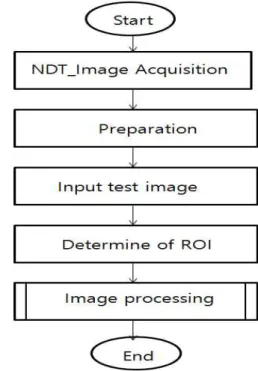
To process the obtained images, the images were pre-processed to raw file formant and then input to the program realized by using the Visual C++ language. The imaging process is shown in Fig. 4.
Fig. 4. Experimental Procedure
To obtain the images, the specimen of which inner board was damaged was located on the image plate as shown in Fig. 5. The distance between the X-ray tube and the specimen was 1 m.
Fig. 5. Experimental sample
The specimen was located on the image plate to prepare imaging as shown in Fig. 6.
Fig. 6. Prepare to Shoot
Fig. 7. Obtained Image
The obtained image shown in Fig. 7 was input to the realized program as shown in Fig. 8.
Fig. 8. Image Input
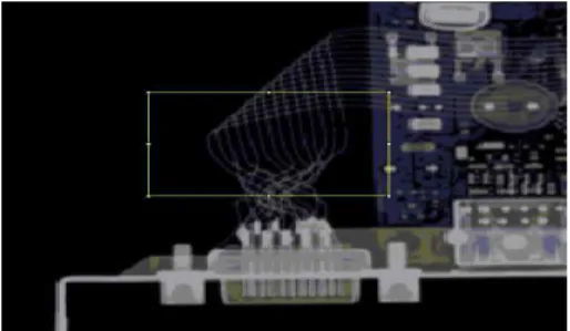
An interested region in the whole board image was selected for inspection. Fig. 9 shows the interested region selected.
Fig. 9. Determine of ROI
Fig. 10 shows the disconnection of the specimen which was found by thinning the selected interested region.
Fig. 10. Edge of Disconnected Circuit
IV. Results and Discussion
Fig. 10 shows the image which obtained edge by applying the Sobel algorithm. The highlight box in Fig. 10 indicates a disconnection. Compared with Fig. 9, which is the image before the application of the algorithm, the processed image is more readable and highlights the disconnection better.
Original image After image processing image Fig. 11. Compare original image and after
image processing image
With respect to the imaging conditions, if low voltage radiation employs, more X-ray radiation is absorbed due to the weak penetration power, showing the synthetic resin part better.
On the other hand, under high voltage radiation condition, the X-ray radiation better penetrates the synthetic resin part which is seen more transparent than the metal part. The radiation voltage may be determined according to the material of the suspected specimen region. The image of this study was obtained under the condition of the high voltage radiation, and thus the synthetic resin region was seen more transparent than the metal region[9]. If imaging is carried out at an inappropriate voltage, the pixels of the synthetic resin region may not be clearly seen, and the readability may not be sufficiently high even after the imaging processing.
In that case, the radiation voltage may be lowered to obtain better results after the imaging process. Since the obtained images are dependent on the applied radiation voltage and current conditions, application of the same algorithm may either improve the image quality or cause loss of the quality to make the reading more difficult.
Therefore, applying appropriate imaging conditions is very important. Depending on the performance of the device used for image obtaining or the resolution of the imaging device, a disconnected region may be seen as if not disconnected, or a region that is not disconnected may be seen as if disconnected. Therefore, evaluation of devices may be necessary.
V. Conclusion
In this study, an image processing algorithm was developed to increase readability of the obtained images from a precise instrument such as a KTX train for non-destructive inspection.
The image processing algorithm was realized by using a Visual C++ development tool. The algorithm developed in this study allows to select an interested region in the acquired images when the interested region is suspected to cause a problem, and applies a thinning process based Sobel operators to the selected region.
The experimental results showed that the readability of defect parts that are not visible to naked eyes was increased through edge detector. Application of the algorithm developed in this study may help to accurately read non-destructive inspection images.
REFERENCES
[1] Zang Hee Cho, Joie P. Jones, Manbir Singh,
“Foundations of Medical Imaging,” John Wiley &
Sons, Inc. 1993.
[2]Alex Pappachen James, Belur V. Dasarathy, “Medical image fusion: A survey of the state of the art,”
Information Fusion, Vol. 19, No. 4, pp.4-19, 2014.
[3] Justin G. Schneeman, “Industrial X-ray Interpretation,”
Intex Publishing Company, 1968.
[4] Martin Hoheisel, “Review of medical imaging with emphasis on X-ray detectors,” Nuclear Instruments and Methods in Physics Research, Vol. 563, No. 1, pp.215-224, 2006.
[5] Alexander Flisch, Joachim Wirth, Robert Zanini, Michael Breitenstsin, Adrian Rudin, Florian Wendt, Franz Mnich, Roland Golz, “Industrial Computed Tomography in
Reverse Engineering Applications,”
DGZ-fP-Proceedings BB, Vol. 4, No. 7, pp.45-53, 1999.
[6] David Bernard, “X-ray tube selection criteria for BGA/CSP X-ray inspection,” The Proceedings of SMTA International Conference, 2002.
[7] Kwang Baek Kim, Jae Hyun Cho, "Detection of Flaws in Air Deck using Non-Destructive Testing", The Korean Institute of Information and Commucation Engineering,
Volume 15, Issue 9, 2011, pp.1865-1870
[8] Sang Bock Lee, Jun Haeng Lee, Sam Yul Lee, Nam Jin Kim, Gye Hwan Jin, "Digital Medical Image Processing", pp.218-221, 1990
[9] Kwon Su Chon, Seung Jun Seo, Jae Hong Lim,
"Inspection of electronic components using dual X-ray energy", Journal of the Korean Society of Radiology, Vol. 9, No. 5, pp. 300~306, 2015
Authors
SangBock Lee received the M.S. and Ph.D. degrees in Computer Science and Electronic Engineering from Chongju University, Korea, in 1993 and 2000, respectively. And he was received Ph.
D. degree in Medicine from Chungbuk National University, Korea, in 2008, respectively.
Dr. Lee joined the faculty of the Dept. of Radiology at Nambu University, Gwangju, Korea, in 2003. He is currently a Professor in the Dept. of Radiology, Nambu University. He is interested in medical imaging and medical decision, and AI(in medicine).