Comparison of the Cytoprotective Effects of Several Natural and Synthetic Compounds against Oxidative Stress in Human Retinal Pigment Epithelial Cells
Da Hye Kim1, Jeong-Hwan Kim2, Seh-Kwang Park2,3, Ji-Won Jeong4, Mi-Young Kim2, Soo-Wan Nam5, Hyesook Lee6,7 and Yung Hyun Choi6,7*
1Department of Smart Bio-Health, Dong-eui University, Busan 47340, Korea
2Research and Development Department, BGN CARE Co., Ltd., Busan 47195, Korea
3BGN Eye Clinic, Seoul 05551, Korea
4BGN Eye Clinic, Busan 47195, Korea
5Department of Biotechnology, Dong-eui University, Busan 47340, Korea
6Anti-Aging Research Center, Dong-eui University, Busan 47340, Korea
7Department of Biochemistry, Dong-eui University College of Korean Medicine, Busan 47227, Korea Received August 28, 2020 /Revised September 25, 2020 /Accepted September 28, 2020
Oxidative stress causes injury to and degeneration of retinal pigment epithelial (RPE) cells. It is in- volved in several retinal disorders and leads to vision loss. In the present study, we investigated the effect of 14 kinds of natural compounds and two kinds of synthetic compounds on oxidative stress-in- duced cellular damage in human PRE cell lines (ARPE-19). From among them, we selected five kinds of compounds, including auranofin, FK-509, hemistepsin A, honokiol, and spermidine, which have in- hibitory effects against hydrogen peroxide (H2O2)-mediated cytotoxicity. In addition, we found that four kinds of compounds (excluding auranofin) have protective effects on H2O2-induced mitochondrial dysfunction. Furthermore, the expression of phosphorylation of histone H2AX, a sensitive marker of DNA damage, was markedly up-regulated by H2O2, whereas it was notably down-regulated by FK- 506, honokiol, and spermidine treatment. Meanwhile, five kinds of candidate compounds had no effect on H2O2-induced intracellular reactive oxygen species (ROS) levels, suggesting that the five candidate compounds have protective effects on oxidative stress-induced cellular damage through the ROS-in- dependent pathway. Taken together, according to the results of H2O2-mediated cellular damage—
such as cytotoxicity, apoptosis, mitochondrial dysfunction, and DNA damage—spermidine and FK-506 are the natural and synthetic compounds with the most protective effects against oxidative stress in RPE. Although further studies on the identification of the mechanism responsible are required, the results of the present study suggest the possibility of using spermidine and FK-506 to suppress the risk of retinal disorders.
Key words : Apoptosis, DNA damage, mitochondrial dysfunction, oxidative stress, retinal pigment epithelial cells
*Corresponding author
*Tel : +82-51-890-3319, Fax : +82-51-893-3333
*E-mail : [email protected]
This is an Open-Access article distributed under the terms of the Creative Commons Attribution Non-Commercial License (http://creativecommons.org/licenses/by-nc/3.0) which permits unrestricted non-commercial use, distribution, and reproduction in any medium, provided the original work is properly cited.
서 론
망막 색소상피 세포(retinal pigment epithelial cells, RPE cells)는 광 수용체와 맥락막 모세 혈관 층 사이의 계면에 위치 한 단층 세포로, 빛 수용, 물질 수송, 시각회로 조절, 식세포 작용, 면역 조절 등의 생화학적 기능을 수행하며, 정상 시력을 유지하는데 핵심적인 역할을 한다[15, 31]. 수많은 임상 및 실험 결과에 따르면, RPE의 구조적 결함 및 기능 손상을 초래하는
요인으로 산화적 스트레스의 중요성이 대두되고 있다[24, 25, 30]. 산화적 스트레스에 의해 유도된 반응성 산소종(reactive oxygen species, ROS)은 산소 대사의 부산물에서 나오는 자유 라디칼, 과산화수소 및 산소 이온을 포함하며 다양한 세포에 서 세포 손상 및 세포 자멸사를 유도한다고 알려져 있다[4, 10].
특히, RPE 세포와 같이 대사율이 높은 세포의 경우 산화적 손상에 더욱 취약하며, 지속적인 고농도 산화적 스트레스에 대한 노출은 RPE의 미토콘드리아 DNA 손상을 유발함으로 써, 미토콘드리아의 기능을 파괴할 뿐만 아니라 궁극적으로 RPE 세포의 세포사멸(apoptosis)을 초래한다[3, 24]. 망막 손상 으로 인한 시력 장애는 실명의 가장 중요한 원인 중 하나이며, 산화적 스트레스는 노인성 황반변성(age-related macular de- generation, AMD), 당뇨성 망막병증(diabetic retinopathy, DR) 및 색소성 망막염(retinitis pigmentosa, RP)과 같은 망막 질환의 원인으로 여겨지고 있다[12, 17, 22]. 따라서, 산화적
스트레스로부터 RPE 세포를 보호하거나 세포 손상을 지연시 키는 것이 망막질환의 예방 및 치료를 위한 주요 표적 접근법 이 될 수 있다.
식물, 동물 및 미생물에서 유래한 천연 화합물은 매우 중요 한 약물 스크리닝 자원으로 알려져 있으며, 약초에서 직접 추 출하거나 천연 화합물에서 유래한 다양한 시판 약물들이 임상 적으로 사용되고 있다[28]. 더욱이 19세기 분석 및 구조 화학의 발전을 통해 천연물로부터 다양한 화합물을 분리, 정제하고, 그 구조를 결정함으로써, 인체에 적용가능한 분자 표적 정보 를 제공하게 되었다[11, 21]. 더불어 20세기에 이르러 승인된 대부분의 약물이 천연물 또는 그로부터 파생된 유사체라는 점에서 천연물의 활용 가능성과 천연물이 일부 인간 표적에 대한 최적의 리간드 역할을 할 수 있음이 규명되었다[27]. 이러 한 관점에서, 망막질환의 치료 및 관련 문제의 해결을 위한 해결책으로 천연물 유래 화합물을 후보물질로 개발하는 것에 대한 관심이 증가하고 있다[34]. 최근, 천연 화합물이 AMD, DR 및 RP와 같은 망막질환에 미치는 영향을 뒷받침하는 결과 들이 보고되고 있다. Curcumin, caffeic acid phenethyl ester 및 sulforaphane과 같은 일부 화합물이 망막세포와 동물 모델 에서 항산화 및 항염증 효과를 통해 망막질환을 개선시킬 수 있음이 입증되었다[9, 32]. 또한 Kim 등[13]은 resveratrol이 nitric oxide-관련 기전을 조절함으로써 망막질환을 개선시킬 수 있음을 보고하였으며, Park 등[23]은 diphlorethohydrox- ycarmalol이 산화적 스트레스에 의한 RPE 세포의 DNA 손상 과 세포사멸을 억제시킬 수 있음을 규명하였다. 보다 최근에 는 glutathione이 비타민 C의 항산화능을 높임으로써 RPE 세 포를 보호할 수 있으며[29], astaxhantin이 항산화효소의 활성 을 통해 산화적 스트레스에 대한 망막 광수용체 세포 손상을 억제할 수 있음이 밝혀졌다[14]. 그러나 지금까지 규명된 천연 물의 망막질환 예방 및 개선 효능은 대부분 이들 물질의 항산 화능에 기인한 것으로, 다양한 천연 화합물에 대한 종합적인 효능 비교 연구는 미흡한 실정이다.
따라서, 본 연구에서는 천연물에서 유래한 14종의 유효 단 일 성분(coptisine chloride, cordycepin, diallyl trisulfide (DATS), fucoidan, genistein, glutathione, hemistepsin A, honokiol, morin, nargenicin A1, quercetin, schisandrin A, spermidine 및 sulforaphane)과 합성 화합물 2종(auranofin 및 FK-506)의 망막질환 개선 가능성을 평가하기위해, 사람유래 망막 색소상피 세포(ARPE-19)를 대상으로 산화적 스트레스 조건 하에서 유발된 세포 손상에 대한 보호 효능을 평가하고 자 하였다.
재료 및 방법
세포 배양
본 연구에서 사용된 사람 RPE 세포인 ARPE-19 세포는
American Type Culture Collection (Manassas, VA, USA)에 서 구입하였다. 이들 세포는 4-(2-hydroxyethyl)-1-piperazi- neethanesulfonic acid가 포함된 DMEM/F12 배지(1:1, Thermo Fisher Scientific, Carlsbad, CA, USA)에 10% fetal bovine se- rum (FBS; Thermo Fisher Scientific), 1% penicillin/strepto- mycin (Thermo Fisher Scientific)를 첨가하여 37℃, 5% CO2
하에서 배양하였다.
시험물질의 준비
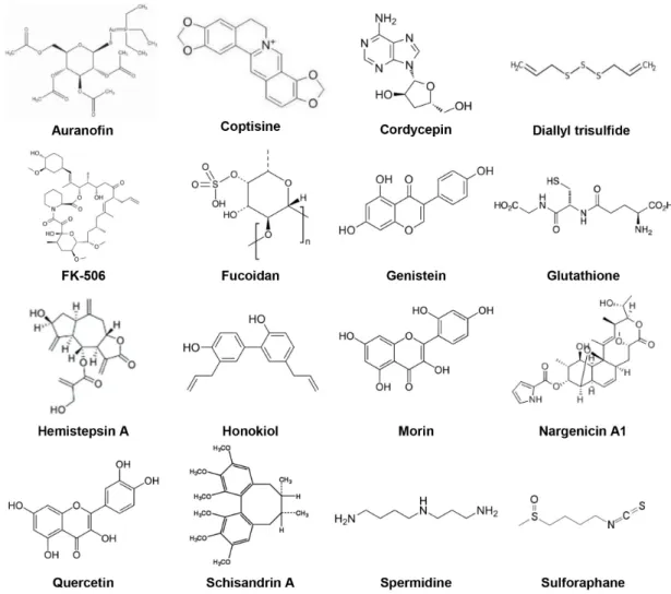
Auranofin, coptisine, cordycepin, DATS, fucoidan, genis- tein, glutathione, hemistepsin A, honokiol, morin, nargenicin A1, quercetin, schisandrin A, spermidine, sulforaphane 및 FK-506은 Sigma Aldrich Chemical Co. (St. Louis, MO, USA) 에서 구입하였으며, nargenicin A1은 Abcam Inc. (Cambridge, UK)에서 구입하여 실험에 사용하였다(Fig. 1). 모든 물질은 di- methyl sulfoxide (DMSO; Sigma Aldrich Chemical Co.)에 300 μM로 stock solution을 만들어 실험에 따라 적절하게 희 석, 혼합하여 사용하였다.
세포 생존율 평가
후보물질에 대한 세포 독성을 평가하기 위하여 3-(4,5-Dim- ethylthiazol–2-yl)-2,5-diphenyltetra-zolium bromide (MTT;
Invitrogen, Carlsbad, CA, USA) assay를 실시하였다. 우선, 약물 자체의 세포 독성을 확인하기 위해 6 well plate에 ARPE- 19 세포를 분주하여 90%의 confluence에 도달하면, 농도 별 후 보 물질을 처리하여 24시간 배양하였다. 각 well에 0.5 mg/ml MTT 용액을 넣은 후 37℃, 5% CO2 incubator에서 1시간 30분 배양하였다. 배지를 제거한 후, DMSO를 넣어 생성된 for- mazan을 모두 녹인 다음 microplate reader (Molecular Devi- ces, Sunnyvale, CA, USA)를 이용하여 540 nm에서 흡광도 값을 측정하였다. 각 물질에 대한 세포 독성은 대조군 값을 기준으로 백분율로 환산하여 나타내었다. 한편, 과산화수소 (hydrogen peroxide, H2O2; Junsei Chemical Co., Ltd., Tokyo, Japan) 처리에 따른 산화적인 스트레스에 대한 후보 물질의 세포 보호 효과를 확인하기 위하여 H2O2를 단독 또는 후보 물질을 3시간 전 처리 후 H2O2를 추가 처리 24시간 후, 0.5 mg/ml MTT 시약을 처리하여 생성된 formazan을 microplate reader를 이용하여 540 nm에서 흡광도 값을 측정하였다. 세포 보호 효과는 각각의 평균 흡광도 값을 구하여 대조군의 평균 흡광도 값에 대한 백분율로 나타내었다.
ROS의 생성 측정
H2O2 및 후보 물질에 의한 ARPE-19 세포에서의 ROS 생성 변화를 측정하기 위해 2’,7’-dichlorodihydrofluorescein diac- etate (DCF-DA; Invitrogen) 염색법을 이용하였다. H2O2에 의 해 생성된 ROS에 대한 후보 물질의 효능을 확인하기 위해
Fig. 1. Structure of 16 kinds of candidate compounds.
후보물질을 3시간 전 처리한 후 H2O2를 30분간 추가 배양하였 다. 준비된 세포들을 phosphate buffered saline (PBS)로 수세 후 10 μM의 DCF-DA를 37℃, 5% CO2 incubator에서 20분간 반응시킨 후, flow cytometer (BD Accuri C6 flow cytometer, BD Biosciences, San Jose, CA, USA)를 이용하여 ROS 생성 변화 여부를 평가하였다.
세포사멸의 정량적 분석
세포사멸 정도의 정량적 분석을 실시하기 위해 Annexin V Apoptosis Detection Kit (BD Biosciences)를 이용하여 flow cytometer로 분석하였다. ARPE-19 세포에 후보 물질을 3시간 전 처리한 후 H2O2를 처리하고 24시간 배양한 다음, 0.05%
trypsin-ethylenediaminetetraacetic acid를 처리하여 세포를 부유시킨 후, 2,000 rpm, 5분간 원심분리하였다. 상층액을 제 거한 뒤 binding buffer를 100 μl 넣은 후 annexin V-fluo- rescein isothiocyanate (FITC)와 propidium iodide (PI)를 처 리하여 암실에서 반응시켰다. 15분 후 binding buffer 200 μl을 추가하여 flow cytometer를 이용하여 세포사멸 유도 빈도를
측정하였다.
Mitochondrial membrane potential (MMP, Δψm)의 측정 후보 물질에 의한 MMP 변화를 측정하기 위해 ARPE-19 세포에 후보 물질을 3시간 동안 전처리하고 H2O2를 24시간 동안 추가 처리한 후, 10 μM 5,5', 6,6'-tetrachloro-1,1',3,3'-tet- raethyl-imidacarbocyanine iodide (JC-1, Sigma-Aldrich Chem- ical Co.)을 처리하여 20분간 염색하였다. 반응이 끝난 후 상층 액을 제거하고, PBS를 첨가하여 세포를 부유시킨 다음 flow cytometer를 이용하여 MMP 변화를 정량적으로 측정하였다.
미토콘드리아 활성 측정
후보 물질에 의한 미토콘드리아의 활성 변화를 측정하기 위 해 ARPE-19 세포에 후보 물질을 3시간 동안 전처리하고 H2O2
를 24시간 동안 추가 처리한 후, 50 nM MitoTracker® Red (Thermo Fisher Scientific, Bothell, WA, USA)을 처리하여 20 분간 염색하였다. PBS로 2-3회 세척한 다음 형광현미경(EVOS FS Auto, Thermo Fisher Scientific)을 이용하여 분석하였다.
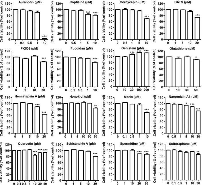
Fig. 2. Effect of 16 kinds of candidate compounds on the cell viability in ARPE-19 cells. ARPE-19 cells were treated with the indicated concentration of candidate compounds for 24 hr. And then cell viability was measured by an MTT assay. Data are expressed as the mean ± SD (n=6). The statistical analyses were conducted using analysis of variance (ANOVA-Tukey's post-hoc test) between groups. *p<0.05, **p<0.01 and ***p<0.001 when compared to control.
면역 형광 검사
후보 물질을 3시간 동안 전처리하고 H2O2를 24시간 동안 추가 처리한 ARPE-19 세포에 100% 메탄올을 넣어 -20℃, 10분 동안 고정시킨 후 PBS로 3번 세척하였다. Blocking buffer [5%
bovine serum albumin (BSA) in PBS-T (0.1% Triton X)]로 1시간 상온에서 반응시키고 2.5% BSA로 희석한 γH2AX anti- body (Santa Cruz Biotechnology, Inc., Santa Cruz, CA, USA) 를 첨가하여 4℃에서 over night 시킨 다음 PBS-T로 2회 10분 간 세척하였다. 그 후 4',6'-diamidino-2-phenylindole (DAPI;
Sigma-Aldrich Chemical Co.)를 처리하여 암실에서 20분 동안 반응시키고 PBS로 3회 세척한 다음 형광현미경을 이용하여
γH2AX의 발현 정도를 분석하였다.
통계 처리
GraphPad Prism® version 5.0 (Graphpad Inc., San Diego, CA, USA) 의 one-way ANOVA를 사용하여 통계분석을 실시 하였으며, Tukey's test로 사후 검정하여 p<0.05 값을 유의한 값으로 보고 통계 처리하였다.
결과 및 고찰
ARPE-19 세포에서 16종 후보 물질의 세포독성 확인
ARPE-19 세포에 대한 16종 후보물질의 세포 독성을 평가하 였다. Fig. 2의 결과에서 알 수 있듯이, auranofin, coptisine, fucoidan, morin, nargenicin A1 및 quercetin은 처리 농도1 μM까지는 세포독성을 나타내지 않았으나 그 이상의 농도에 서 유의적인 세포 생존율 감소를 보였다. 한편, DATS, FK-506, schisandrin A와 spermidine은 10 μM 농도까지 ARPE-19 세 포에 독성을 보이지 않았으며, 10 μM의 cordycepin과 hemi- stepsin A 처리는 APRE-19 세포에서 각각 62% 및 88%의 세포 생존율 감소를 나타내었다. Genistein은 200 μM 농도까지 세 포독성을 보이지 않았으나 30 μM 이상의 농도에서 세포 증식 을 유도하는 경향성을 나타내었다. Glutathione과 honokiol의 경우, 50 μM 농도에서 경미한 세포 생존률 저하를 보였으며, sulforaphane은 30 μM 농도에서 유의적인 세포 독성을 유발 하였다.
H2O2에 의한 ARPE-19 세포 독성에 대한 16종 후보물질 의 세포 보호 효과
상기 결과를 바탕으로, ARPE-19세포에 독성을 나타내지 않 는 농도 범위에서 16종의 후보물질을 대상으로 H2O2에 의해 유발된 세포 손상을 개선시키는 약물을 선별하였다. 이를 위 하여 ARPE-19 세포에서 H2O2의 처리에 의해 유발되는 산화 적 스트레스의 조건은 선행 연구에 준하여 300 μM로 설정하 였다[7, 18]. 300 μM의 H2O2에 24시간 동안 노출된 ARPE-19 세포의 생존율은 약 65%를 나타냈으며, 16종의 후보물질이 H2O2에 의해 유발된 이러한 세포독성을 개선시키는지 여부를 확인하였다. Fig. 3의 MTT assay 결과에 의하면, coptisine, cordycepin, DATS, fucoidan, glutathione, morin, nargenicin A1, quercetin, schisandrin A와 sulforaphane은 모든 농도에 서 H2O2에 의한 세포독성을 개선시키지 못하였다. 그러나 auranofin은 0.5 μM 이상의 농도에서 H2O2에 의해 유발된 세 포독성을 74% 수준으로 유의하게 개선시켰다. Hemistepsin A는 5 μM 전처리 조건 하에서 74% 수준까지 세포 생존율을 증가시켰으며, honokiol은 5~30 μM 범위에서 농도 의존적인 세포생존율 개선을 나타내었다. Spermidine과 FK-506의 경 우, 10 μM 처리시 H2O2-처리 세포 대비 증가된 세포 생존율을 나타내었다. 한편, genistein은 100 μM 이상의 고농도에서 H2O2에 의해 유발된 세포독성을 유의적으로 개선시켰다. 이 상의 결과를 바탕으로 16종의 후보물질 중 ARPE-19세포에 독성을 나타내지 않으며 동시에 H2O2에 의해 유발된 세포독 성을 개선시키는 약물을 선별하고 유효 농도를 설정하였다.
즉, 1 μM auranofin, 10 μM FK-506, 5 μM hemistepsin A, 30 μM honokiol과 10 μM spermidine을 바탕으로, 망막 색소상피 세포의 산화적 스트레스에 의한 세포 손상에 대한 방어 가능 성을 평가하기 위해 이후 실험을 수행하였다.
H2O2에 의한 ARPE-19 세포 내 ROS 생성에 미치는 후 보물질 5종의 효능
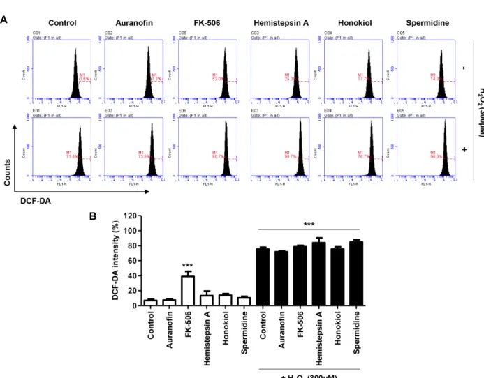
16종의 후보 물질 중 ARPE-19 세포에 대한 독성을 보이지 않으며, H2O2에 의해 유도된 세포 손상에 대한 유의미한 보호 효과를 나타낸 5종의 후보물질을 대상으로 ROS 생성능을 평 가하였다. 1 μM auranofin, 5 μM hemistepsin A, 30 μM hono- kiol, 10 μM spermidine 처리는 ARPE-19 세포의 ROS 생성에 영향을 미치지 않았으며, H2O2에 의해 유도된 세포 내 ROS 생성을 개선시키지 않았다(Fig. 4). 한편 FK-506은 10 μM 농도 에서 ARPE-19 세포 내 ROS의 생성을 유도하였으나, H2O2 처 리 세포에서 유의적인 ROS 증감을 나타내지 않음을 확인하였 다. 따라서, 5종의 후보물질은 산화적 스트레스에 의해 매개된 망막 색소상피 세포의 손상에 대해 ROS 비의존적 기전을 통 해 세포 보호효과를 나타낼 것으로 기대된다.
H2O2에 의한 ARPE-19 세포의 세포사멸에 대한 후보물 질 5종의 효능
세포사멸은 자가포식 세포사, 괴사와 함께 다양한 프로그램 화된 세포사 패턴의 마지막 단계로, H2O2에 노출된 ARPE-19 세포에서 세포사가 유도된다는 것은 잘 알려져 있다[6, 8]. 따 라서, 선정된 5종의 후보물질을 대상으로 H2O2에 의해 유도된 세포사멸에 대한 보호 효능을 평가하기 위해 annexin V-FITC/
PI 염색을 통한 flow cytometry 분석을 실시하였다. Fig. 5에 나타낸 바와 같이, 300 μM의 H2O2가 단독 처리된 ARPE-19 세포에서는 약 23%의 세포사멸을 유도되었으며, auranofin을 제외한 4종의 후보물질 전처리에 의해 유의적인 세포사멸의 감소가 나타났다. 한편, 5종의 후보물질 단독 처리 시 spermi- dine을 제외한 4종의 후보물질에서 ARPE-19 세포의 경미한 세포사멸의 증가를 보였다. 이러한 결과는 5종의 후보물질 중 산화적 스트레스에 의해 유발된 세포사멸에 대해 auranofin을 제외한 FK-506, hemistepsin A, honokiol과 spermidine의 유 의미한 효능을 보여주며, 특히 spermidine에 의한 세포사멸 보호효과가 가장 우수하였음을 제시한다.
H2O2에 의한 ARPE-19 세포의 미토콘드리아 기능 손상 에 대한 후보물질 5종의 효능
많은 연구들을 통해 H2O2로 인한 산화적 스트레스가 미토 콘드리아의 기능 장애를 유발하여 망막 색소상피 세포 손상을 초래할 수 있음이 입증되었다[5, 16, 33]. 또한 미토콘드리아 기능장애는 AMD를 포함한 다양한 노화관련 질병의 병태생 리학과 연루되어 있으며, 특히 미토콘드리아 결손으로 인한 내인성 세포사멸 경로의 활성화가 망막 질환의 주요 병인임이 밝혀져 있다[2, 20]. 따라서, 산화적 스트레스에 의한 망막 색소 상피 세포의 미토콘드리아 기능 손상에 대한 후보 물질 5종의 효능을 확인하기 위해 미토콘드리아 막전위(MMP)와 미토콘 드리아 활성을 평가하였다. 먼저, 미토콘드리아 막전위의 손
Fig. 3. Effect of 16 kinds of candidate compounds on the cell viability in H2O2-stimulated ARPE-19 cells. ARPE-19 cells were pre-treated with the indicated concentration of candidate compounds for 3 hr, and then treated with 300 μM H2O2 for 12 hr. Subsequently cell viability was measured by an MTT assay. Data are expressed as the mean ± SD (n=6). The statistical analyses were conducted using analysis of variance (ANOVA-Tukey's post-hoc test) between groups. ***p<0.001 when compared to control.
#p<0.05, ##p<0.01 and ###p<0.001 when compared to H2O2-treated cells.
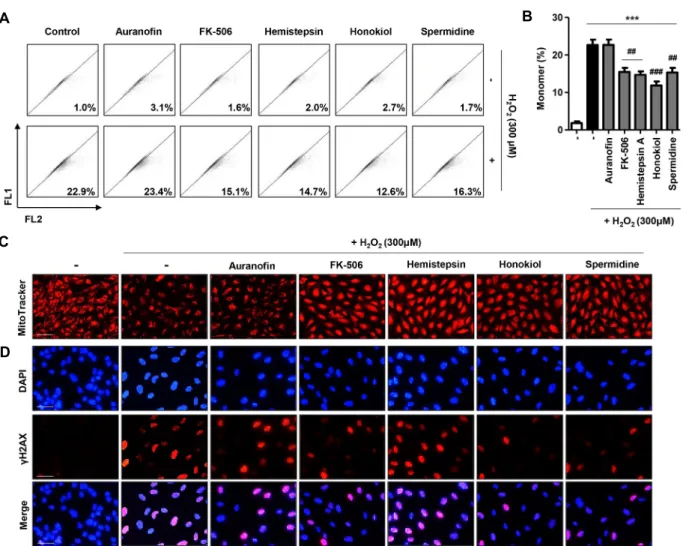
실 여부를 JC-1 염색을 통한 flow cytometry 분석으로 확인하 였다. JC-1은 미토콘드리아에 축적되는 catonic carbocyanine 감광체로, 높은 MMP에서 적색 형광의 응집체를 형성하는 반 면, 낮은 MMP에서는 녹색 단량체를 형성하는 특징을 가진다 [26]. 본 연구에서 정상세포의 경우, MMP의 손실 지표인 JC-1 단량체 비율이 1% 내외로 존재함을 통해 미토콘드리아 막이 정상적인 기능을 하고 있음이 입증되었다(Fig. 6A, Fig. 6B).
그러나 후보물질 5종 모두 단독 처리 시, ARPE-19 세포의
MMP에 아무런 영향을 미치지 않았다. 이에 반해, H2O2가 처 리된 ARPE-19 세포에서는 JC-1 단량체 비율이 약 23% 정도 증가하여 산화적 스트레스에 의해 망막 색소상피 세포의 MMP가 감소함을 확인할 수 있었다. 그리고 1 μM auranofin, 10 μM FK- 506, 5 μM hemistepsin A, 30 μM honokiol과 10 μM spermidine을 전처리한 세포의 MMP 변화를 확인한 결 과, auranofin을 제외한 4종의 후보물질 처리 세포에서 MMP 개선 효능이 관찰되었다(Fig. 6A, Fig. 6B). 이러한 결과를 바탕
A
B
Fig. 4. Effect of 5 kinds of candidate compounds on the intracellular ROS production in H2O2-stimualted ARPE-19 cells. ARPE-19 cells were pre-treated with candidate compounds for 3 hr, and then treated with 300 μM H2O2 for 30 min. The cells were incubated at 37℃ in the dark for 20 min with a culture medium containing 10 μM DCF-DA to monitor ROS production.
(A) The degree of ROS production was measured with a flow cytometer. (B) Quantitative data of DCF-DA intensity of 5 kinds of candidate compounds-treated cells with or without 300 μM H2O2. Data are expressed as the mean ± SD (n=3).
The statistical analyses were conducted using analysis of variance (ANOVA-Tukey's post-hoc test) between groups. ***p<0.001 when compared to control.
으로 MitoTracker® Red dye를 이용한 세포의 미토콘드리아 활성을 관찰하였다. Fig. 6C에서 알 수 있듯이, 정상세포에서 관찰된 미토콘드리아는 MitoTracker® Red의 높은 발현을 보 인데 반하여 H2O2가 처리된 세포의 경우 MitoTracker® Red 세포의 비율이 현저한 감소되었다. 이러한 결과를 통해 H2O2
처리에 의해 증가된 산화적 스트레스는 망막 색소상피 세포의 미토콘드리아 활성을 감소시킴을 알 수 있다. 그러나 FK-506, hemistepsin A, honokiol, 및 spermidine의 전처리는 H2O2에 의해 감소된 MitoTracker® Red 세포의 빈도 증가를 현저히 개선시켰다. 반면, auranofin은 미토콘드리아 활성에는 특이 적인 개선 효능을 보이지 않았다.
H2O2에 의한 ARPE-19 세포의 DNA 손상에 미치는 후보 물질 5종의 효능
세포 손상에 대한 초기 반응으로, DNA 이중 나선의 절단에 의해 히스톤 변이체 H2AX의 Ser-139 잔기의 인산화가 일어나 며, 이를 통해 γH2AX가 형성된다. 이러한 인산화된 히스톤 변이체는 DNA 손상에 대한 특징적이며, 민감한 바이오 마커 로 널리 사용되고 있다[19]. 뿐만 아니라 γH2AX가 DNA 손상 과 함께 염색질 리모델링 과정에서도 중요할 수 있음이 보고 되고 있으며, 세포사멸 과정에서 caspase-비의존적 세포자멸 사 유도에도 관여한다는 것이 입증되었다[1]. 따라서, 산화적 스트레스에 의해 매개된 ARPE-19 세포의 DNA 손상여부를 γH2AX 발현을 통해 관찰한 결과, 300 μM의 H2O2는 ARPE-19 세포의 핵 내 γH2AX의 발현을 현저히 증가시킴을 확인하였 다. 이러한 DNA 손상 marker인 γH2AX의 발현 증가는 FK-506, honokiol 및 spermidine의 전처리에 의해 두드러진 감소 경향을 보인데 반해, auranofin과 hemistepsin A에서는
A
B
Fig. 5. Effect of 5 kinds of candidate compounds on H2O2-induced apoptosis in ARPE-19 cells. The cells were treated with candidate compounds for 3 hr, and then incubated with or without 300 μM H2O2 for 24 hr. (A) Flow cytometry analysis was performed by annexin V and PI staining. Representative histograms are presented. (B) The percentages of apoptotic cells were determined by expressing the numbers of annexin V+ cells as percentages of all cells. Data are expressed as the mean ± SD (n=3).
The statistical analyses were conducted using analysis of variance (ANOVA-Tukey's post-hoc test) between groups. *p<0.05,
**p<0.01 and ***p<0.001 when compared to control. ###p<0.001 when compared to H2O2-treated cells.
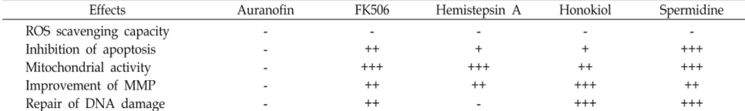
Table 1. Summary of the effect of candidates on H2O2-induced cellular damages in ARPE-19 cells
Effects Auranofin FK506 Hemistepsin A Honokiol Spermidine
ROS scavenging capacity Inhibition of apoptosis Mitochondrial activity Improvement of MMP Repair of DNA damage
- - - - -
- ++
+++
++
++
- + +++
++
-
- + ++
+++
+++
- +++
+++
++
+++
The effects of candidate compounds on cellular dysfunctions in H2O2-stimulated ARPE-19 cells are indicated as -, +, ++ and +++.
No effect expressed as -; + indicated significantly different at p<0.05 compared with H2O2-stimulated ARPE-19 cells and significantly different at p<0.001 compared with control; ++ indicated significantly different at p<0.01 compared with H2O2-stimulated ARPE-19 cells and significantly different at p<0.01 compared with control; +++ indicated significantly different at p<0.001 compared with H2O2-stimulated ARPE-19 cells and significantly different at p<0.05 compared with control.
유의한 효과를 나타내지 않았다(Fig. 6C).
이상의 결과를 요약하면, 16종의 후보물질 중 ARPE-19 세 포에 독성을 나타내지 않으면서 H2O2에 의해 유발된 세포독 성을 개선시키는 약물로 auranofin, FK-506, hemistepsin A,
honokiol 및 spermidine을 선정하였다. 이들 약물 중 산화적 스트레스에 의한 세포사멸, 미토콘드리아 기능 손상 및 핵 손 상에 대한 보호 효과를 평가하여 최종적으로 spermidine이 산화적 스트레스에 의한 망막 색소상피 세포의 보호 효능이
A B
C
D
Fig. 6. Effect of 5 kinds of candidate compounds on mitochondrial and DNA damages in H2O2-stimulated ARPE-19 cells. The cells were pretreated with candidate compounds for 3 hr, and then incubated with or without 300 μM H2O2 for 24 hr. (A) The cells were stained with JC-1 dye, and were then analyzed by a flow cytometer to evaluate the changes in MMP. Representative photographs are presented. (B) The percentages of monomeric cells were determined by expressing the numbers of JC-1 green as percentages of all cells. (C) Cells were stained with 50 nM MitoTracker® Red (scale bar; 75 μm). (D) Cell were stained with γH2AX antibody and DAPI was used to counterstain the nuclei (blue), then the cells were visualized using a fluorescence microscope. Representative fluorescence microscopy images, including a merged image, are shown (γH2AX;
red, DAPI; blue, scale bar; 25 μm).
가장 우수한 천연물 유래 단일 물질임을 규명하였으며, 동시 에 합성물질로는 FK-506이 망막 세포 보호 효능이 우수한 것 으로 나타났다(Table 1). 본 연구 결과를 통해 이들 물질이 망 막질환에 대한 위험을 감소시킬 가능성이 있음을 제시하였으 나, 이후 후속 연구를 통해 spermidine 및 FK-506의 망막 색소 상피 세포 손상에 대한 방어 기전 연구가 수반되어야 할 것으 로 사료된다.
감사의 글
본 연구는 BGN밝은눈안과병원과 (주)비지엔케어의 연구 비 지원으로 수행되었다.
The Conflict of Interest Statement
The authors declare that they have no conflicts of interest with the contents of this article.
References
1. Baritaud, M., Cabon, L., Delavallée, L., Galán-Malo, P., Gilles, M. E., Brunelle-Navas, M. N. and Susin, S. A. 2012. AIF- mediated caspase-independent necroptosis requires ATM and DNA-PK-induced histone H2AX Ser139 phosphoryla- tion. Cell Death Dis. 3, e390.
2. Barot, M., Gokulgandhi, M. R. and Mitra, A. K. 2011. Mito- chondrial dysfunction in retinal diseases. Curr. Eye Res. 36,
1069-1077.
3. Cai, J., Wu, M., Nelson, K. C., Sternberg Jr, P. and Jones, D. P. 1999. Oxidant-induced apoptosis in cultured human retinal pigment epithelial cells. Invest. Ophthalmol. Vis. Sci. 40, 959-966.
4. Dröge, W. 2020. Free radicals in the physiological control of cell function. Physiol. Rev. 82, 47-95.
5. Du, W., An, Y., He, X., Zhang, D. and He, W. 2018. Protection of kaempferol on oxidative stress-induced retinal pigment epithelial cell damage. Oxid. Med. Cell Longev. 2018, 1610751.
6. Han, D., Wu, X., Liu, L., Shu, W. and Huang, Z. 2018. Sodium tanshinone IIA sulfonate protects ARPE-19 cells against oxi- dative stress by inhibiting autophagy and apoptosis. Sci.
Rep. 8, 15137.
7. Hao, Y., Liu, J., Wang, Z., Yu, L. L. and Wang, J. 2019. Pice- atannol protects human retinal pigment epithelial cells against hydrogen peroxide induced oxidative stress and apoptosis through modulating PI3K/Akt signaling pathway.
Nutrients 11, E1515.
8. He, C. and Klionsky, D. J. 2009. Regulation mechanisms and signaling pathways of autophagy. Annu. Rev. Genet. 43, 67-93.
9. Head, K. A. 1999. Natural therapies for ocular disorders, part one: diseases of the retina. Altern. Med. Rev. 4, 342-359.
10. Jakubczyk, K., Dec, K., Kałduńska, J., Kawczuga, D., Koch- man, J. and Janda, K. 2020. Reactive oxygen species-sources, functions, oxidative damage. Pol. Merkur. Lekarski. 48, 124-127.
11. Ji, H. F., Li, X. J. and Zhang, H. Y. 2009. Natural products and drug discovery. Can thousands of years of ancient med- ical knowledge lead us to new and powerful drug combina- tions in the fight against cancer and dementia? EMBO Rep.
10, 194-200.
12. Kaarniranta, K., Pawlowska, E., Szczepanska, J., Jablkowska, A. and Blasiak, J. 2019. Role of mitochondrial DNA damage in ROS-mediated pathogenesis of age-related macular de- generation (AMD). Int. J. Mol. Sci. 20, 2374.
13. Kim, W. T. and Suh, E. S. 2010. Retinal protective effects of resveratrol via modulation of nitric oxide synthase on oxygen-induced retinopathy. Kor. J. Ophthalmol. 24, 108-118.
14. Lai, T. T., Yang, C. M. and Yang, C. H. 2020. Astaxanthin protects retinal photoreceptor cells against high glucose-in- duced oxidative stress by induction of antioxidant enzymes via the PI3K/Akt/Nrf2 Pathway. Antioxidants (Basel) 9, E729.
15. Lakkaraju, A., Umapathy, A., Tan, L. X., Daniele, L., Philp, N. J., Boesze-Battaglia, K. and Williams, D. S. 2020. The cell biology of the retinal pigment epithelium. Prog. Retin. Eye Res. 2020, 100846.
16. Li, S., Gaur, U., Chong, C. M., Lin, S., Fang, J., Zeng, Z., Wang, H. and Zheng, W. 2018. Berberine protects human retinal pigment epithelial cells from hydrogen peroxide-in- duced oxidative damage through activation of AMPK. Int.
J. Mol. Sci. 19, 1736.
17. Liang, F. Q. and Godley, B. F. 2003. Oxidative stress-induced mitochondrial DNA damage in human retinal pigment epi- thelial cells: a possible mechanism for RPE aging and age-re- lated macular degeneration. Exp. Eye Res. 76, 397-403.
18. Liu, Y., Ren, Y., Wang, X., Liu, X., Xu, Y. and He, Y. 2019.
Down regulation of UCP2 expression in retinal pigment epi- thelium cells under oxidative stress: an in vitro study. Int.
J. Ophthalmol. 12, 1089-1094.
19. Mah, L., El-Osta, A. and Karagiannis, T. 2010. γH2AX: a sen- sitive molecular marker of DNA damage and repair. Leuke- mia 24, 679-686.
20. Musat, O., Ochinciuc, U., Gutu, T., Cristescu, T. and Coman, C. 2012. Pathophysiology and treatment of ARMD. Oftalmo- logia 56, 45-50.
21. Newman, D. J., Cragg, G. M. and Snader, K. M. 2003.
Natural products as sources of new drugs over the period 1981-2002. J. Nat. Prod. 66, 1022-1037.
22. Nishimura, Y., Hara, H., Kondo, M., Hong, S. and Matsugi, T. 2017. Oxidative stress in retinal diseases. Oxid. Med. Cell Longev. 2017, 4076518.
23. Park, C., Lee, H., Hong, S. H., Kim, J. H., Park, S. K., Jeong, J. W., Kim, G. Y., Hyun, J. W., Yun, S. J., Kim, B. W., Kim, W. J. and Choi, Y. H. 2019. Protective effect of diphlor- ethohydroxycarmalol against oxidative stress-induced DNA damage and apoptosis in retinal pigment epithelial cells.
Cutan. Ocul. Toxicol. 38, 298-308.
24. Plafker, S. M., O'Mealey, G. B. and Szweda, L. I. 2012. Me- chanisms for countering oxidative stress and damage in reti- nal pigment epithelium. Int. Rev. Cell. Mol. Biol. 298, 135-177.
25. Ruan, Y., Jiang, S., Musayeva, A. and Gericke, A. 2020. Oxi- dative stress and vascular dysfunction in the retina: Ther- apeutic strategies. Antioxidants (Basel) 9, E761.
26. Salvioli, S., Ardizzoni, A., Franceschi, C. and Cossarizza, A.
1997. JC-1, but not DiOC6(3) or rhodamine 123, is a reliable fluorescent probe to assess delta psi changes in intact cells:
implications for studies on mitochondrial functionality dur- ing apoptosis. FEBS Lett. 411, 77-82.
27. Schmidt, B. M., Ribnicky, D. M., Lipsky, P. E. and Raskin, I. 2007. Revisiting the ancient concept of botanical therapeu- tics. Nat. Chem. Biol. 3, 360-366.
28. Sulaiman, R. S., Basavarajappa, H. D. and Corson, T. W.
2014. Natural product inhibitors of ocular angiogenesis. Exp.
Eye Res. 129, 161-171.
29. Tram, N. K., McLean, R. M. and Swindle-Reilly, K. E. 2020.
Glutathione improves the antioxidant activity of vitamin C in human lens and retinal epithelial cells: Implications for vitreous substitutes. Curr. Eye Res. 2020, 1-12.
30. Upadhyay, M., Milliner, C., Bell, B. A. and Bonilha, V. L.
2020. Oxidative stress in the retina and retinal pigment epi- thelium (RPE): Role of aging, and DJ-1. Redox Biol. 2020, 101623.
31. Vinores, S. A., Derevjanik, N. L., Ozaki, H., Okamoto, N.
and Campochiaro, P. A. 1999. Cellular mechanisms of blood- retinal barrier dysfunction in macular edema. Doc. Ophthal- mol. 97, 217-228.
32. Yu, M., Anderson, R. E. and Mandal, N. A. 2012. Natural compounds in retinal diseases. In: Stratton R., Hauswirth W. and Gardner, T. (eds) Studies on retinal and choroidal disorders. oxidative stress in applied basic research and clin- ical practice. Humana Press. 437-456.
33. Zhao, H., Wang, R., Ye, M. and Zhang, L. 2019. Genipin
초록:인간 망막 색소상피 세포에서 산화적 스트레스에 대한 천연 및 합성 화합물들의 세포 보호 효과 비교
김다혜1․김정환2․박세광2,3․정지원4․김미영2․남수완5․이혜숙6,7․최영현6,7*
(1동의대학교 스마트 바이오헬스학과, 2㈜비지엔케어 연구개발전담부서, 3BGN밝은눈안과의원, 4BGN밝은눈안과
병원, 5동의대학교 의생명공학과, 6동의대학교 항노화연구소, 7동의대학교 한의과대학 생화학교실)
산화적 스트레스에 의한 망막 색소상피 세포의 손상 및 퇴화는 시력 소실을 포함한 다양한 망막질환의 원인으 로 알려져 있다. 본 연구에서는 천연물 유래 14종 단일 성분과 합성 화합물 2종을 대상으로 망막질환 개선 가능성 을 평가하기 위해, 사람 유래 망막 색소상피 세포를 대상으로 산화적 스트레스에 의한 세포 손상에 대한 보호 효능을 평가하였다. 그 결과, 16종의 후보물질 중 H2O2에 의해 유발된 세포독성을 개선시키는 효능을 보인 5종 (auranofin, FK-509, hemistepsin A, honokiol 및 spermidine)을 선별하였다. 이를 기반으로 H2O2에 의한 미토콘드 리아 기능 손상 및 활성 저하 억제능을 평가하였으며, auranofin을 제외한 4종에서 유의한 세포 보호 효능이 관찰 되었다. 더불어 H2O2에 의해 유도된 DNA 손상에 미치는 영향을 조사한 결과, FK-506, honokiol과 spermidine이 DNA 손상을 개선시켰음을 확인하였다. 그러나 5종의 후보물질이 활성 산소종의 생성에는 뚜렷한 억제 효능을 나타내지 않았으며, 이는 5종의 후보 물질이 ROS-비의존적 기전을 통해 세포 보호효과를 나타낼 것으로 사료된 다. 이상의 결과를 바탕으로, 조사 대상 후보물질 중에서 spermidine이 산화적 스트레스로부터 망막 색소상피 세 포의 보호 효능이 가장 우수한 천연물 유래 단일 물질임을 규명하였으며, 동시에 합성물질로는 FK-506이 망막 세포 보호 효능이 우수한 것으로 나타났다. 비록 작용 기전에 대한 추가 연구가 필요하겠지만, 본 연구 결과는 spermidine과 FK-506이 산화적 스트레스에 의한 망막질환의 위험을 억제할 가능성이 있음을 시사한다.
protects against H2O2-induced oxidative damage in retinal pigment epithelial cells by promoting Nrf2 signaling. Int.
J. Mol. Med. 43, 936-944.
34. Zhou, F. 2018. The potentials of natural compounds in treat- ing human retinal diseases. J. Blood Disord. Transfus. 9, 22.