• 검색 결과가 없습니다.

The Effects of Direction Changes on the Muscular Activity of the Lower Extremities During Seated Reaching Exercises

N/A
N/A
Protected

Academic year: 2021

Share "The Effects of Direction Changes on the Muscular Activity of the Lower Extremities During Seated Reaching Exercises"

Copied!
8
0
0

로드 중.... (전체 텍스트 보기)

전체 글

(1)

| Abstract |

Purpose: Although multi-directional reaching exercises are commonly used clinically, the effects of specific movement directions on the muscle systems of the lower extremities have not been explored. We therefore investigated lower extremity muscle activity during reaching exercises with different sagittal and horizontal plane movements.

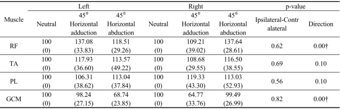
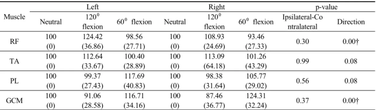
Methods: The surface electromyography responses of the bilateral rectus femoris, tibialis anterior, peroneus longus, and gastrocnemius muscles were measured during reaching exercises in three directions in the horizontal plane (neutral, 45°horizontal shoulder adduction, and 45°abduction) and three directions in the sagittal plane (neutral, 120° flexion, and 60° flexion). A total of 20 healthy, physically active participants completed six sets of reaching exercises. Two-way repeated ANOVA was performed:

body side (ipsilateral and contralateral) was set as the intra-subject factor and direction of reach as the inter-subject factor.

Results: Reaching at 45° horizontal shoulder adduction significantly increased the activity of the contralateral rectus femoris and gastrocnemius muscles, while 45° horizontal shoulder abduction activated the ipsilateral rectus femoris and gastrocnemius muscles. The rectus femoris activity was significantly higher with reaching at a 120° shoulder flexion compared to the other conditions. The gastrocnemius activity decreased significantly as the shoulder elevation angle increased from 60° to 120°.

Conclusion: Our results suggest that multi-directional reaching stimulates the lower extremity muscles depending on the movement direction. The muscles acting on two different joints responded to the changes in reaching direction, whereas the muscles acting on one joint were not activated with changes in reaching direction.

Key Words: Electromyography, Lower extremity, Reaching test

†Corresponding Author : Se-Yeon Park ([email protected]) PNF and Movement, 2019; 17(2): 207-214

https://doi.org/10.21598/JKPNFA.2019.17.2.207

Print ISSN: 2508-6227 Online ISSN: 2508-6472

Original Article Open Access

1)

The Effects of Direction Changes on the Muscular Activity of the Lower Extremities During Seated Reaching Exercises

Jwa-Jun Kim⋅Dae-Kyung Kim 1 ⋅Jae-Yong Kim 2 ⋅Jae-Wook Shin 2 ⋅Se-Yeon Park 3†

Department of Physical Therapy, Choonhae College of Health Sciences

1 Korea Proprioceptive Neuromuscular Facilitation Association Busan Branch

2 Rehabilitation Center, Busan Medical Center

3 Department of Physical Therapy, Kaya University

Received: May 19, 2019 / Revised: May 30, 2019 / Accepted: May 30, 2019

ⓒ 2019 Journal of Korea Proprioceptive Neuromuscular Facilitation Association

This is an Open Access article distributed under the terms of the Creative Commons Attribution Non-Commercial License

(http://creativecommons.org/licenses/by-nc/3.0) which permits unrestricted non-commercial use, distribution, and reproduction

in any medium, provided the original work is properly cited.

(2)

ԺԸՀ ք ՘ՖՎԨթնլԨՕշվխյխնռԨ՞շմԶԨԹԿԴԨՖշԶԨԺ

Ⅰ. Introduction

Sitting balance is the ability to maintain a sitting position with minimal sway during static or dynamic activities (Cabanas-Valdés et al., 2013; Dean et al., 2007).

This capacity can be assessed in a quiet state of sitting or during specific functional activities such as reaching for an object or putting on clothing (Crosbie et al., 1995;

Dean et al., 1999). Maintaining a balanced sitting position is a fundamental component of daily movement (Cabanas-Valdés et al., 2013), especially in patients who have experienced a cerebrovascular accident. Moreover, regaining sitting balance is considered a major factor affecting functional outcomes after cerebrovascular accidents (Dean et al., 2007). The ability to maintain balance is determined by one’s ability to integrate the somatosensory, visual, and vestibular systems. Previous studies have suggested that several exercises, including multi-directional reaching, sitting on an unstable surface, and raising one leg while in a sitting position, can enhance balance ability (Cabanas-Valdés et al., 2013). However, further studies are necessary for identifying specific exercises that improve specific components of balance abilities (Abuin-Porras et al., 2018; Bank et al., 2016).

The act of reaching is a basic human upper extremity skill that requires coordination of the lower and upper extremities (Dean et al., 1999). Due to this characteristic, reaching tasks have been used to evaluate upper extremity function and individual balance capacity (Katz-Leurer et al., 2009). Multi-directional reaching is a form of training that emphasizes balance-training effects (Crosbie et al., 1995). Although this exercise is commonly used clinically, few reports have explored the effect of adjusting the reaching direction on muscular systems of the lower extremities (Crosbie et al., 1995; Dean et al., 1999). Dean et al. (1999) suggested that reaching more than 140%

of the arm’s length activates lower extremity muscles,

but the specific effect of varying the reaching direction has not been explored. With reference to the horizontal plane of the human body, reaching to the contralateral side may activate contralateral limb muscles, and reaching to the ipsilateral side may activate ipsilateral limb muscles.

In the sagittal plane, reaching to a relatively higher position may activate the knee extensor muscles, and reaching to a relatively lower position may activate para-ankle muscles. However, this hypothesis has not yet been confirmed.

In this report, we investigated activities of the lower extremity muscles during various reaching exercises, which were divided into three conditions defined by the sagittal and horizontal planes. By comparing reaching conditions, we explored whether various directions of reaching activated specific lower extremity muscles.

Ⅱ. Materials and Method

1. Participants

Twenty asymptomatic males were recruited from a local university using convenience sampling. The subjects had a mean±SD age of 21.94±2.24 (range 20–28) years.

Subjects had no history of musculoskeletal disease and

neurological problem, or pain in the past 3 months. For

preventing potential influence of fatty tissue on measuring

surface electromyography (sEMG) activity, participants

with a body mass index (BMI) of 25 or higher were

excluded. The height and weight of the final sample

populations were 175.07±5.26cm and 66.60±8.39kg,

respectively. The average BMI was 21.69±2.23. All

participants were right hand dominant, and the reaching

exercise was also performed with right hand.

(3)

՜հխԨՍծծխիռջԨշծԨՌձպխիռձշնԨՋհթնկխջԨշնԨռհխԨՕսջիսմթպԨՉիռձվձռցԨշծԨռհխԨՔշտխպԨՍրռպխյձռձխջԨՌսպձնկԨ՛խթռխլԨغ ք ԺԸՁ

2. Design

The study employed a 3×2 factorial design with repeated measures on each factor. The first factor: sagittal elevation angle; had three levels: 60, 90, and 120°of glenohumeral flexion; horizontal abduction angle, had three levels: 45⁰ adduction, neutral, 45⁰ abduction. The second factor was set as intra subject factor as ipsilateral-contralateral lower extremity muscles. Subjects were tested on three conditions of reaching in each sagittal and horizontal plane, and the order was randomized.

3. Instrumentation

Electromyography (EMG) signals were acquired from the lower extremity muscles: rectus femoris (RF), tibialis anterior (TA), peroneus longus (PL), gastrocnemius (GCM). The muscular activities were collected by using surface EMG system (Myosystem TM DTS, Noraxon Inc., USA). The EMG signal was recorded by a computer and sampled at 1024 Hz, adjusted bandpass filter from 20Hz to 500Hz, and analyzed through the product provided software(MyoResearch XP master edition 1.06, Noraxon Inc., USA).

Surface electromyography (EMG) electrodes were placed over four muscles on the right and left side of the body. The electrodes were placed in a bipolar arrangement over the muscles in the recommended locations (Cram et al., 1998). The reference electrode was placed over the medial malleorus. For preparing electrode placement, the skin was shaved, lightly abraded with sandpaper and cleaned with ethyl alcohol.

4. Procedures

After 5 minutes of practice and rest time for acclimation to the reaching exercise variations, each subject performed

three different reaching exercise in horizontal plane and three different reaching exercise in sagittal plane. The reaching distance was set as maximal distance that each subject could reach. Commonly, subject’s lower body is fixed to bench with strap, not to take off the gluteal area from bench.

Subjects sat on a height-adjustable bench with erect lumbar posture. Subjects were asked to sat upright on a height-adjustable bench, looking straight ahead and the investigator corrected the position such that the alignment of spinal line was in a neutral position. The neutral reaching exercise, subjects asked to flex their dominant right arm to 90⁰ with elbow extension. After fixing the laser pointer to dorsal middle finger of a subject, one researcher stood one meter from a subject and hold a white paper which was drawn a circle reflecting light from the laser pointer. Subject asked to reach their arm, not to lift off the buttock, without light’getting out of the circle. For the variation of reaching in sagittal plane, 120⁰ and 60⁰ of shoulder flexion was determined and the same procedure was repeated. For the variation of reaching exercise condition on horizontal plane, 45⁰ horizontal adduction from neutral condition and 45⁰ horizontal abduction from neutral condition was determined and same procedure was repeated. For normalizing the sEMG data during reaching variations, Muscle activities collecting from the neutral reaching was set as reference voluntary contraction (RVC). The normalized values of left and right RF, TA, PL, and GCM are presented as %RVC. The averaged two values from each condition including normalized EMG were used for statistical analysis.

5. Statistical analy ysis

The root mean square of sEMG data during the test was obtained with a widow length of 0.125s and averaged.

The data are expressed as the %MVIC relative to

(4)

ԺԹԸ ք ՘ՖՎԨթնլԨՕշվխյխնռԨ՞շմԶԨԹԿԴԨՖշԶԨԺ

normalized data. PASW Statistics (version 18.0; SPSS, Chicago, IL, USA) was used to determine the significance of differences in %RVC values between test conditions.

The two way repeated measure of analysis was performed to determine the effects of reaching directions on the EMG data. For identifying the disparity between left and right side of muscle, ipsilateral and contralateral side was set as an intra-subject factor. When significant correlation was observed, subsequent analysis of the one-way repeated ANOVA was performed on each side of ipsilateral and contralateral muscle. In all analyses, p<0.05 was taken to indicate statistical significance.

Ⅲ. Results

1. Muscle activities of the lower extremity during the reaching exercise with horizontal shoulder adduction-abduction variation

The RF and GCM activity showed significant differences in factor of reaching direction conditions, which was different to ipsilateral and contralateral side of muscle (p<0.05) (Table 1). Subsequent analysis revealed

that reaching with 45⁰ horizontal shoulder adduction condition of reaching activated contralateral side of the RF activity, compared to the neutral and 45⁰ horizontal shoulder abduction conditions of reaching. In a ipsilateral side of RF, the reaching with the 45⁰ horizontal abduction condition of reaching showed higher right side of RF activity, compared with the neutral and 45⁰ horizontal adduction conditions of reaching (Fig. 1). The contralateral side of the GCM activity was significantly lower with the reaching with the 45⁰ horizontal shoulder abduction condition of reaching, compared with other conditions (p<0.05). On the other hand, the ipsilateral side of the GCM activity was significantly lower with the 45⁰ horizontal shoulder adduction condition, compared with other conditions (p<0.05) (Fig. 1). There were no significant differences in TA and PL activities (p>0.05).

2. Muscle activities of the lower extremity during the reaching exercise with sagittal shoulder flexion variation

There were significant differences in RF and GCM activity with a factor of reaching direction conditions, but not with a factor of the ipsilateral and contralateral

Muscle

Left Right p-value

Neutral

45⁰ Horizontal

adduction

45⁰ Horizontal

abduction

Neutral

45⁰ Horizontal

adduction

45⁰ Horizontal

abduction

Ipsilateral-Contr

alateral Direction

RF 100

(0)

137.08 (33.83)

118.51 (29.26)

100 (0)

109.21 (39.02)

137.64

(28.61) 0.62 0.00†

TA 100

(0)

117.93 (36.60)

113.57 (49.22)

100 (0)

108.68 (29.55)

116.50

(38.55) 0.69 0.10

PL 100

(0)

106.31 (38.62)

113.04 (37.84)

100 (0)

119.33 (43.30)

113.03

(52.93) 0.56 0.10

GCM 100

(0)

98.24 (27.15)

68.74 (23.85)

100 (0)

64.77 (33.76)

99.49

(26.99) 0.82 0.00†

NOTE. Each value represents the mean (SD, Standard deviation).

The values with different superscripts (†) in the column are significantly different in a factor of frontal plane movement (p<0.05) by Bonferroni’s measure.

Table 1. Comparison in %RVC values of the lower extremity muscles which depends on directions of the forward

reaching movement in horizontal plane (n=20, unit: %RVC)

(5)

՜հխԨՍծծխիռջԨշծԨՌձպխիռձշնԨՋհթնկխջԨշնԨռհխԨՕսջիսմթպԨՉիռձվձռցԨշծԨռհխԨՔշտխպԨՍրռպխյձռձխջԨՌսպձնկԨ՛խթռխլԨغ ք ԺԹԹ

Muscle

Left Right p-value

Neutral 120⁰

flexion 60⁰ flexion Neutral 120⁰

flexion 60⁰ flexion Ipsilateral-Co

ntralateral Direction

RF 100

(0)

124.42 (36.86)

98.56 (27.71)

100 (0)

108.93 (24.69)

93.46

(27.33) 0.30 0.00†

TA 100

(0)

112.64 (33.67)

100.40 (28.89)

100 (0)

113.09 (64.18)

101.26

(43.29) 0.99 0.08

PL 100

(0)

99.37 (27.43)

117.69 (40.83)

100 (0)

98.38 (31.64)

105.77

(29.02) 0.56 0.08

GCM 100

(0)

91.06 (28.58)

116.71 (34.16)

100 (0)

87.46 (36.77)

124.31

(32.24) 0.37 0.00†

NOTE. Each value represents the mean(SD, Standard deviation).

The values with different superscripts (†) in the column are significantly different in a factor of sagiital plane movement (p<0.05) by Bonferroni’s measure.

Table 2. Comparison in %RVC values of the lower extremity muscles which depends on directions of the forward

reaching movement in sagittal plane (n=20, unit: %RVC)

Fig. 1. Comparison of muscle activities of the rectus femoris and gastrocnemius following factors of reaching direction and that ipsilateral-contralateral side.

* significant difference between conditions.

(6)

ԺԹԺ ք ՘ՖՎԨթնլԨՕշվխյխնռԨ՞շմԶԨԹԿԴԨՖշԶԨԺ

side of muslce(p<0.05) (Table 2). The reaching with 120⁰ shoulder flexion condition of reaching significantly elevated the both sides of the RF activity, compared to the neutral and 60⁰ sagittal shoulder flexion conditions of reaching (p<0.05) (Fig. 1). Both sides of the GCM activities were significalty higher with the reaching with the 60⁰ sagittal shoulder flexion condition of reaching, compared with other conditions (p<0.05) (Fig. 1).

Ⅳ. Discussion

In this study, we investigated muscular activities of the lower extremity during reaching exercises performed in different directions in the sagittal and horizontal planes.

Clinical literature and previous studies have suggested that multi-directional reaching exercises are effective for transferring body weight (Barton JE., 2016; Kusoffsky et al., 2001). Moreover, reaching distance is a predictor of balance ability in standing and sitting positions (Katz-Leurer et al., 2009). Although a reaching exercise program is commonly prescribed and used for patients with ankle instability, the relationship of muscular activities of the lower extremities and reaching direction has not been evaluated. Similar to previous studies that proposed exercise variations (Dean et al., 1999), our study provides an approach to selectively activate lower extremity muscles through reaching exercises.

We showed that reaching direction had different effects on two-joint muscles such as the rectus femoris and gastrocnemius than did single-joint muscles such as the tibialis anterior and peroneus longus muscles. The reaching movement is one of many functional movements that require multi-joint cooperation (De Ridder et al., 2013). Although seated reaching requires least amount of upper body movement, muscle responses induced by weight transfer resulting from reaching are thought to involve only multi-joint linked muscles.

Regarding activity of the rectus femoris against reaching variations in the horizontal plane, the 45⁰ adducted condition primarily increased activity in the contralateral rectus femoris, and the 45⁰ abducted condition primarily activated the ipsilateral rectus femoris.

This result reflects rectus femoris activation in the direction of weight transfer. A previous finding also reported that ipsilateral horizontal reaching with adduction or abduction could indirectly activate the rectus femoris (Dean et al., 1999).

In the sagittal plane, 120⁰ reaching activated the bilateral rectus femoris to a greater degree than did the other sagittal reaching conditions. Similarly, during the pre-liftoff phase of the sit-to-stand movement, an effort to maintain the light of a laser-pointer within a circle requires eccentric control of the trunk extensors during 120⁰ reaching conditions (Doorenbosch et al., 1994). As strengthening the rectus femoris is essential to performing the sit-to-stand movement (Yoshiko et al., 2017), upward reaching could be used in training for this movement.

On the other hand, the gastrocnemius was strongly activated with downward reaching, such as in the 60⁰ sagittal direction. Although we did not collect data on the center of pressure under the buttocks, this activation be caused by excessive anterior transfer of the center of mass under the reaching condition. Among ankle strategies, plantar flexion could be used against the external perturbation in the anterior direction (Hwang et al., 2009).

This study has several limitations. The first was the

lack of EMG information for other lower extremity

muscles, such as the hamstring, which was not measured

because we could not eliminate contact between the bench

and electrode. Second, further studies exploring the

diagonal direction of reaching may be required because

the present study includes only sagittal and horizontal

reaching.

(7)

՜հխԨՍծծխիռջԨշծԨՌձպխիռձշնԨՋհթնկխջԨշնԨռհխԨՕսջիսմթպԨՉիռձվձռցԨշծԨռհխԨՔշտխպԨՍրռպխյձռձխջԨՌսպձնկԨ՛խթռխլԨغ ք ԺԹԻ

Ⅴ. Conclusion

Our results suggest that multi-directional reaching stimulates lower extremity muscles depending on the direction of movement. Muscles acting on two different joints responded to changes in the direction of reach; the rectus femoris was activated in the direction of weight transfer, and the gastrocnemius with downward reaching.

Acknowledgements

This study supported by Korea Proprioceptive Neuromuscular Facilitation Association Busan Branch.

References

Abuin-Porras V, Villafañe JH, Jiménez-Antona C, et al.

Relationship between attention and balance: a dual-task condition study in children. Journal of Exercises Rehabilitation. 2018;14(3):349-55.

Bank J, Charles K, Morgan P. What is the effect of additional physiotherapy on sitting balance following stroke compared to standard physiotherapy treatment: a systematic review. Topics in Stroke Rehabilitation.

2016;23(1):15-25.

Barton JE, Graci V, Hafer-Macko C, et al. Dynamic balanced reach: a temporal and spectral analysis across increasing performance demands. Journal of Biomechanical Engineering. 2016;138(12):10-15.

Cabanas-Valdés R, Cuchi GU, Bagur-Calafat C. Trunk training exercises approaches for improving trunk performance and functional sitting balance in patients with stroke: a systematic review. NeuroRehabilitation.

2013;33(4):575-92.

Cram JR, Kasman GS, Holtz J. Introduction to surface

electromyography. Gaithersburg. Aspen. 1998.

Crosbie J, Shepherd R, Squire T. Postural and voluntary movement during reaching in sitting: the role of the lower limbs. Journal of Human Movement Studies.

1995;28:103–26.

Dean CM, Channon EF, Hall JM. Sitting training early after stroke improves sitting ability and quality and carries over to standing up but not to walking: a randomised trial. The Austrian Journal of Physiotherapy.

2007;53(2):97-102.

Dean CM, Shepherd RB, Adams RD. Sitting balance II: reach direction and thigh support affect the contribution of the lower limbs when reaching beyond arm’s length in sitting. Gait & Posture. 1999;10(2):147-53.

De Ridder EM, Van Oosterwijck JO, Vleeming A, et al. Posterior muscle chain activity during various extension exercises: an observational study. BMC Musculoskeletal Disorders. 2013;14:204.

Doorenbosch CA, Harlaar J, Roebroeck ME, et al. Two strategies of transferring from sit-to-stand; the activation of monoarticular and biarticular muscles. Journal of Biomechanics. 1994;27(11):1299-307.

Hwang S, Tae K, Sohn R, et al. The balance recovery mechanisms against unexpected forward perturbation. Annals of Biomedical Engineering. 2009;37(8):1629-37.

Hsu WL, Yang YR, Hong CT, et al. Ankle muscle activation during functional reach in hemiparetic and healthy subjects. American Journal of Physiscal Medicine

& Rehabilitation. 2005;84(10):749-55.

Katz-Leurer M, Fisher I, Neeb M, et al. Reliability and validity of the modified functional reach test at the sub-acute stage post-stroke. Disability and Rehabilitation.

2009;31(3):243-8.

Kusoffsky A, Apel I, Hirschfeld H. Reaching-lifting-placing

task during standing after stroke: coordination among

ground forces, ankle muscle activity, and hand

movement. Archives of Physiscal Medicine and

(8)

ԺԹԼ ք ՘ՖՎԨթնլԨՕշվխյխնռԨ՞շմԶԨԹԿԴԨՖշԶԨԺ

Rehabilitation. 2001;82(5):650-60.

Yoshiko A, Kaji T, Sugiyama H, et al. Effect of 12-month resistance and endurance training on quality, quantity,

and function of skeletal muscle in older adults requiring long-term care. Experimental Gerontology. 2017;

98:230-237.

수치

Table 1. Comparison in %RVC values of the lower extremity muscles which depends on directions of the forward
Table 2. Comparison in %RVC values of the lower extremity muscles which depends on directions of the forward

참조

관련 문서

근활성도는 Table 1에 나타난 바와 같다.. RF: rectus femoris, VM: vastus medialis, VL: vastus lateralis, MH: medial hamstring, GM: medial head of gastrocnemius, TA: tibialis

Before and after water exercise, the lower limbs pennation angle change significantly increased in rectus femoris and tibialis anterior.. But, there wasn’t

The muscle activity of the elector spinae, rectus abdominis, gluteus maximus, gluteus medius, vastus medialis, vastus lateralis, tibialis anterior, and soleus muscles was

Conclusion: The biceps femoris, gastrocnemius, and tibialis anterior muscles showed greater muscle activation in the closed kinematic chain position when compared to the