Effects of Aquatic and Ground Obstacle Training on Balance and Muscle Activity in Patients With Chronic Stroke
Hyun-chul Hwang, MSc, PT, So-hee Kim, MSc, PT, Tae-ho Kim, PhD, PT
Dept. of Physical Therapy, Daegu University, Republic of KoreaAbstract 1)
Background: Obstacle training affects lower limb muscle activity, balance, reducing the risk of falls, and making gait more stable.
Objects: This study aimed to investigate the effects of aquatic and ground obstacle training on balance and muscle activity in patients with chronic stroke.
Methods: The study subjects included 30 patients with stroke, who were divided into aquatic (n
1= 15) and ground (n
2=15) groups. Groups underwent obstacle training three times per week, 30 min per session, for six weeks that went as follows: walking over sites with the paralyzed leg, stepping onto and down from a box step, and walking over obstacles with the non-paralyzed leg.
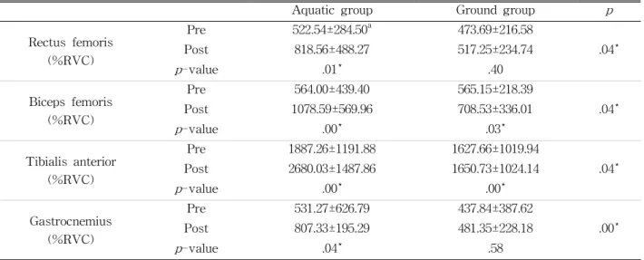
Results: The experimental results were obtained by comparing muscle activity. Activity of the rectus femoris, biceps femoris, tibialis anterior, and gastrocnemius were significantly increased in the aquatic group (p<.05). Activity of the biceps femoris and tibialis anterior were significantly increased in the ground group (p<.05); however, the rectus femoris and gastrocnemius were not significantly different. In the comparison of maximal distance regarding the limits of stability, it was significantly increased on the non-affected side, affected side, and anterior and posterior distance in the aquatic group (p<.05). It was significantly increased in the non-affected side and anterior and posterior distance the ground group (p<.05); however, maximal distance on the affected side distance was not significantly different.
Conclusion: Gait training with aquatic and ground obstacles is effective for improving balance and gait ability of patients with stroke. However, it was more effective for the aquatic group than for the ground group.
Key Words: Aquatic therapy; Balance; Muscle activity; Stroke.
Introduction
Stroke severely restricts bodily movement owing to impairment in sensory and motor functions (Duncan et al, 2002). Particularly, compromised ca- pacity to support the body and instability in weight bearing (Ikai et al, 2003) have an enormous impact on gait function and may cause secondary injuries owing to falls (Orlin et al, 2000).
A fall is when part of the body, other than a foot sole, touches the ground unintentionally. Falls reduce mobility, cause injuries, and generate fear of falls in strong patients (Teasell et al, 2002). Reportedly, 55%
of patients with stroke experience one or more falls within a year of stroke onset, and 42% of these 55%
patients experience multiple falls during that time (Ashburn et al, 2008). Accordingly, a plethora of ex- ercise programs are conducted to prevent falls in pa- tients with stroke. However, as research suggests that patients with stroke frequently fall owing to ob- stacles regularly encountered in everyday life, such as thresholds, doorsteps, curbstones, and bumpy ground, these aspects need to be studied (Austin et al, 1999).
Patients with stroke employ strategies to walk over
obstacles that differ from normal, such as increasing
vertical distance between the obstacle and the leading
Corresponding author: So-hee Kim [email protected]
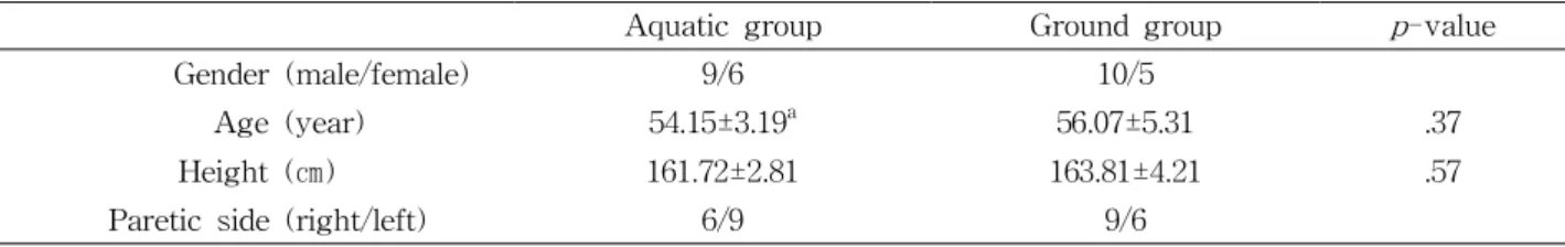
Aquatic group Ground group p -value
Gender (male/female) 9/6 10/5
Age (year) 54.15±3.19
a56.07±5.31 .37
Height (㎝) 161.72±2.81 163.81±4.21 .57
Paretic side (right/left) 6/9 9/6
a
mean±standard deviation
Table 1. General characteristics of the subjects (N=30)
leg to prevent a fall, reducing walking speed to pro- vide a stable support to posture, and positioning the leg as close as possible to an obstacle (Chen et al, 1991). These factors suggest the need for ongoing gait training with environmental obstacles resembling the community setting. Accordingly, such gait train- ing is usually provided on the ground (Miller et al, 2008), although patients with stroke tend to shift the center of the body to the non-paralyzed side and do not actively engage in gait training owing to fear of a fall (Melzer et al, 2008).
However, patients with stroke can be more active in gait training in water (Eich et al, 2004). This is because aquatic training provides a safer exercise environment by reducing the fear of falls and risk of injury (Hauer et al, 2002). Aquatic training increases stability because buoyancy offsets gravity, and the physiologic effects of lower heart rate and deeper breathing stabilize heart rate based on water depth (Noh et al, 2008). Because of its benefits, the aquatic environment has been incorporated into gait training, as demonstrated by the incorporation of aquatic ther- apy in programs to strengthen cardiovascular, neuro- logic, and skeletal muscular systems, including swimming exercise, aquarobics, training with floating devices, and walking over obstacles in water, in many countries such as the United States and Japan (Eyvaz et al, 2018).
Despite the numerous benefits of aquatic therapy, research has been limited to the Bad Ragaz Ring method, Halliwick, and Watsu interventions, and no studies have been published about gait training with obstacles in water. To propose a clinically effective intervention, this study investigated the effects of
gait training with obstacles in water and on the ground on muscle activity and balance in the lower limbs in patients with chronic stroke.
Methods
Subjects
The study was conducted after the approval of the Institutional Review Board of Daegu University (approval number: 104061-201501-HR-022-02).
The study included 30 patients with hemiplegia and stroke diagnosed by computed tomography (CT) or magnetic resonance imaging (MRI), who had been hospitalized for >6 months and who provided volun- tary consent to participate. Stroke onset had to be ≥ 6 months earlier to minimize the potential for spon- taneous recovery, and patients had to have no fear of water and no aquatic training experience.
Inclusion criteria comprised unilateral brain injury identified by CT or MRI; ability to walk for ≥10 m with or without a walker; stiffness in the paralyzed lower limb of G2 or lower scores on the Modified Ashworth Scale; absence of orthopedic disease in both lower limbs; no restricted range of motion; and a score of ≥24 on the Mini Mental State Examination (Korean version), indicating ability to understand and comply with researchers’ instructions. General charac- teristics of subjects are presented in Table 1.
Instruments
EMG equipment (TeleMyo DTS system, Noraxon
Inc. USA) was used to analyze the surface muscle ac-
tivity in four muscles: paralyzed rectus femoris, biceps
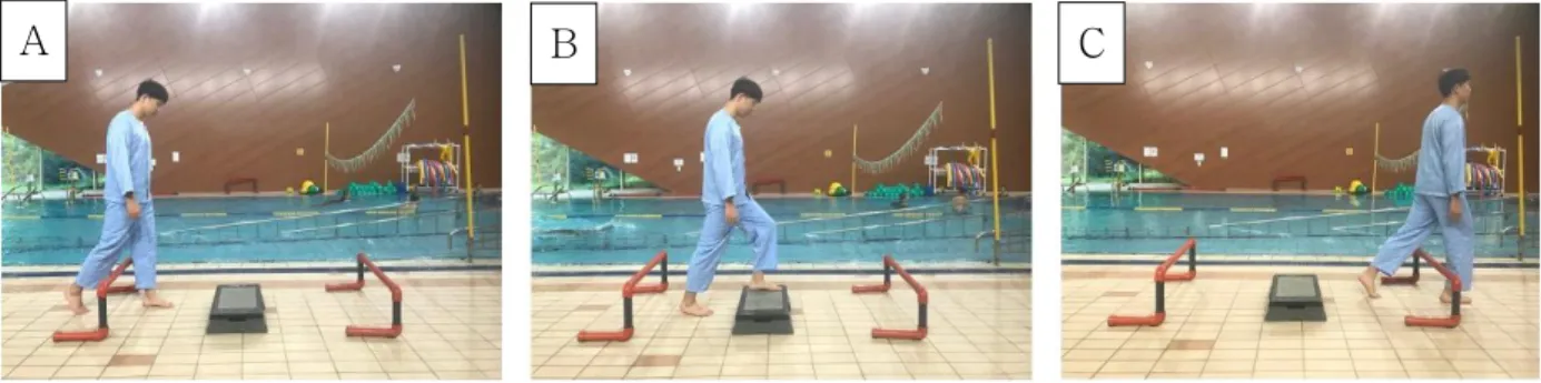
A B C
Figure 1. Obstacle training program (A: task 1, B: task 2, C: task 3).
femoris, tibialis anterior, and gastrocnemius. Electrode positions were the half point of a line connecting the anterior superior iliac spine and the superior patella (for the rectus femoris); the half point of a line con- necting the ischial tuberosity and lateral epicondyle of the tibia (for the biceps femoris); two phalanges below the tibial tuberosity (for the tibialis anterior); and the third-point of a line connecting the fibular head and the heel (for the gastrocnemius). The standardization method was percent reference voluntary contraction (%RVC) (Stegeman et al, 2000). Measurement of the gait cycle was conducted by attaching a foot switch to the heel bone and the first metatarsus to activate the pressure sensor during walking.
Balance was measured using digital balance tester (BIO Rescue, RM Engineering Inc. France). This de- vice measures the total area of moment of the center of pressure as the subject makes the maximum shift of the center of pressure by bearing body weight in eight directions (east, west, south, north, northeast, northwest, southeast, and southwest) as instructed on the front monitor. On the monitor, subjects watched a video and demonstration about how to shift weight.
We measured the limit of stability in the standing position that allowed the maximum range of shift in the center of gravity while maintaining balance ac- cording to the direction displayed on the monitor.
Procedures
The 30 subjects were randomly assigned to aquat- ic and ground-training groups in equal numbers. All subjects received neurodevelopmental treatment five
times a week, while participating in a 6-week, 30-min obstacle training program either in water or on the ground three times a week. The obstacle pro- gram comprised three tasks developed by modifying the techniques of Means & O’Sullivan (Means et al, 2000). Water and ground training included identical tasks with obstacle courses as follows: (Figure 1).
Task 1: Walking over obstacles with the paralyzed leg
Subjects walked over obstacles with the paralyzed leg as the leading leg and the non-paralyzed leg as the swinging leg in a 10-min walk with obstacles placed at 1-m intervals.
Task 2: Stepping on and down a box step
Subjects stepped onto a box step with the non-paralyzed leg first and then with the paralyzed leg; they then stepped down with the non-paralyzed leg first, followed by the paralyzed leg. Next, the se- quence was reversed. Subjects continued stepping on and down for 10 min by switching the leading leg.
Task 3: Walking over obstacles with the non-par- alyzed leg
Subjects walked over obstacles with the non-para- lyzed leg as the leading leg and the paralyzed leg as the swinging leg in a 10-min walk with obstacles placed at 1-m intervals.
Statistical Analyses
Data analysis was performed using Statistical
Package for the Social Sciences (SPSS ver. 18.0 for
Windows) (IBMcorp., Armonk, NY, USA). Analyses
included a paired t-test to examine the pretest–
Aquatic group Ground group p Rectus femoris
(%RVC)
Pre 522.54±284.50
a473.69±216.58
Post 818.56±488.27 517.25±234.74 .04*
p -value .01* .40
Biceps femoris (%RVC)
Pre 564.00±439.40 565.15±218.39
Post 1078.59±569.96 708.53±336.01 .04*
p -value .00* .03*
Tibialis anterior (%RVC)
Pre 1887.26±1191.88 1627.66±1019.94
Post 2680.03±1487.86 1650.73±1024.14 .04*
p -value .00* .00*
Gastrocnemius (%RVC)
Pre 531.27±626.79 437.84±387.62
Post 807.33±195.29 481.35±228.18 .00*
p -value .04* .58
a
mean±standard deviation, *p<.05
Table 2. Comparison of muscle activation (N=30)
Aquatic group Ground group p
Non-Affected side (㎜)
Pre 2310.74±2028.51 2238.24±1321.91
.01*
Post 3980.66±1537.65 2730.59±754.39
p -value .00* .02*
Affected side (㎜)
Pre 1680.98±1521.85 1850.62±1447.41
.04*
Post 3559.31±1764.57 2463.50±875.51
p -value .00* .13
Forward (㎜)
Pre 2108.54±1215.44 2059.62±1126.65
.04*
Post 3881.22±1379.71 2901.38±1101.65
p -value .00* .00*
Backward (㎜)
Pre 2387.64±2445.27 2063.62±1175.85
.01*
Post 4273.39±1802.89 2750.25±1276.03
p -value .01* .0*
a