• 검색 결과가 없습니다.

The Effects of Water Exercise Program on Pennation Angle of the Lower Limb Muscle with Women in Their 20’s

N/A
N/A
Protected

Academic year: 2021

Share "The Effects of Water Exercise Program on Pennation Angle of the Lower Limb Muscle with Women in Their 20’s"

Copied!
5
0
0

로드 중.... (전체 텍스트 보기)

전체 글

(1)

Hwa-Young Cho, PT; Moon-Jung Kim, Researcher

1

; Se-Won Yoon, PT, PhD

Department of Physical Therapy, Graduate School of Seonam University;

1

Department of Physical Therapy, Kwang-Ju Women’s University

Purpose: This study was designed to investigate the effect of a water exercise program on the pennation angle of the lower-limb muscle in women in their 20s.

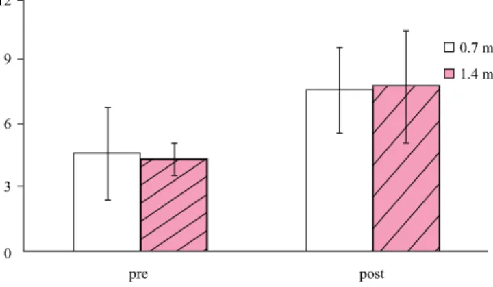
Methods: Ten female subjects were randomly divided into two groups, with 5 subjects exercising in water 0.7 m deep and 5 subjects exercising in water 1.4 m deep. They did the water exercising program for 40 minute per day, 3 days per week, for total 6 weeks. We measured the pennation angle of lower-limb muscle using ultrasonography. All measurements for each group were performed at pre-training and after 6 weeks of training.

Results: The pennation angle was compared before and after the water exercise period for each group, and statistically significant changes within each group in measurements of the rectus femoris and tibialis anterior (p<0.05). However, there was no significant difference in muscle architecture by water depth (p>0.05) between the two groups.

Conclusion: These results show that the pennation angle of the lower-limb muscle of women in their 20s changed after 6 weeks of participating in a water exercise program, but these changes were not dependent on the depth of the water in which the exercises were performed.

Keywords: Water exercise, Depth, Sonography, Pennation angle Received: May 11, 2010

Revised: June 10, 2010 Accepted: June 14, 2010

Corresponding author: Se-Won Yoon, [email protected]

the Lower Limb Muscle with Women in Their 20’s

The Journal Korean Society of Physical Therapy

I. Introduction

Without the danger of accident, water exercise improves muscle strength and endurance all of the same exercise as you can on the ground. Buoyancy and resistance is the only difference in kinetics.

1

Buoyancy and resistance will increase the movable range of joints, strengthen muscles and muscular endurance and could even improve cardiovascular endurance and balance.

2

Consequently it different water depth which loaded weight, it comes 10%, 25% and 50% of the weight at each neck, breast and lumbar level.

3

Effect of buoyancy is different with following water depth. The speed and volume of water moving in an action receives enough resistance so that the momentum for personal physical strength can be controlled.

4

Geometric arrangement of the fascicle within the muscle is the defined of the architecture of a skeletal muscle. Muscle

architecture is mainly characterized by the fascicle length, the pennation angle and muscle thickness. In pinnate muscles, fascicles are arranged obliquely with respect to the tendon, and this angulations (pennation angle) is altered by contractions.

5

Compared and figure out size, thickness, type of muscle fiber is very important to examine and analysis for function of skeletal muscle.

6,7

Low cost, non-invasive and easy access, and the possi- bility of a dynamic examination is advantages of sonography examinations. Since the early 1990s, sonography has been used to measure the changes in muscle thickness, muscle fiber penna- tion angle, muscle fascicle length, and muscle cross-sectional area during isometric and dynamic contractions.

8

Buoyancy and resistance is different depend on depth and will changes in muscles. So, it will be need to know about change following water depth for application of water exercise program.

Therefore, we tested the hypothesis that the water exercise

(2)

Table 1. Water exercise program

Program Practice method Application time

(minute) Total Warming-up The upper body and the lower body give a tension and relaxation

with each region by freely. 5

Water walking 40 exercise

Water walking I The knee spreads and walks like kicking the ball.

And water flow identifies. (1.4 m raising hands up) 10 Intermediate exercise I Sits and does a leg dumping, to release the muscle

and the ligament. 5

Water walking II Equality with water walking I 10

Intermediate exercise II Equality with intermediate exercise I 5 Arrangement motion Becomes likewise the progress with warming-up exercise and gives a tension

and relaxation with each region by freely. 5

Figure 1. Measurement of the pennation angle at tibialis anterior on longitudinal section.

program is useful for muscular architecture and the different of water depth affect lower limb muscle group. Our first objective was to investigate the water exercise program improve on the muscular pennation angle in lower limbs. And, the other was to examine the water exercise affects on the muscular pennation angle of the low limbs depend on depth.

II. Methods

1. Subject

We have studied the effects of water exercise program on the pennation angle of the lower limbs, June 18th∼July 31st 2009.

Subjects were ten female volunteers from G University student in Kwang-Ju. The subjects agreed to participate in the experi- ment after being explained the aims of the study. They are randomized in two groups which one is 0.7 m (about depth of the hip) (n=5, age=21.2±0.6, height=164.0±4.3, weight=55.1±

5.1) and other is 1.4 m (about depth of the chest) (n=5, age=

21.1±0.6, height=162.4±4.8, weight=56.3±7.4). Exclusion criteria involved contraindications for water exercise; heart disease, light sense illness, and mental illness.

9

2. Experimental process

A water exercise programs performed walking exercise in water at a depth of 0.7 m and 1.4 m during the experiment, the exercise program perform at the same time.

Water walking program was conducted 3 times a week for six

weeks, 0.7 m standard height of a pool of 7 × 15 m (width × height), and 1.4 m in height 13 × 25 m (width × height) was, water temperature is 29°, maintain the interior temperature was 30°. Table 1 shows the water exercise program.

3. Measurement

We used Ultrasound MyLab25 (Esaote, Italy) for gathering ultrasonic images to measure muscle pennation angle. This machine’s Frequency Range is 12 MHz. As well as, dynamic Range (13) and Density (High) are fixed price in all kind of test.

Then, we used Image Pro Plus 4.5 (Media Cybernetics, USA) program for analysis of gathering images and measured that pre and post experiment each.

Pennation angle means an angle made by fascicles and deep

aponeurosis.

10

In this study, we measured images what gathering

from left lower limbs muscle’s sagittal plane. We measured

(3)

Figure 2. Comparison of pennation angle at rectus femoris before and after water exercise program.

M±SD: Mean±Standard Deviation

*p<0.05

Figure 3. Comparison of pennation angle at tibialis anterior before and after water exercise program.

M±SD: Mean±Standard Deviation

*p<0.05

Figure 4. Comparison of pennation angle at gastrocne- mius before and after water exercise program.

M±SD: Mean±Standard Deviation

pennation angle by fascicles in deep aponeurosis (Figure 1).

For measurement of rectus femoris, we measured above 10 cm from patella base by sitting position keeping flexion 90°

knee. We measured tibialis anterior below 10 cm from patella base by sitting position keeping flexion 90° knee. And we measured 13 cm below from fossa posterior which between knee joint and condyle keeping prone position putting ankle joint on the edge of table for measurement of gastrocnemius. We are marked by oil-based pen to make sure measurement about same part before start the test.

4. Statistical analyses

SPSS 12.0 statistical program used for data analysis. The single- sample Kolmogorov-Smirnov test was conducted for find out normal distribution whether of general characteristics of subjects and each measurement items. The result has been recognized

normal distribution, general characteristics of subjects for the group differences between independent sample test were analyzed.

Take the repeated measure ANOVA for comparing to the pennation angle from pre and post experimentation and the significance level was assumed 0.05.

III. Results

1. Comparison of pennation angle at rectus femoris In 0.7 m, rectus femoris pennation angle was 3.21±0.61° and 5.52±0.29°at before and after water exercise. And rectus femoris pennation angle in 1.4 m was 3.54±1.99° and 5.41±4.06° at before and after water exercise. There was significantly increased (p<0.05). Rectus femoris pennation angle was increased, but the analysis showed that there was no significant difference by water depth (Figure 2).

2. Comparison of pennation angle at tibialis anterior Before water exercise tibialis anterior pennation angle in 0.7 m was 1.77±1.28° and 4.03±1.28° was after. In 1.4 m was 1.99±

0.74° and 5.31±2.62° at before and after water exercise. Tibialis anterior pennation angle were significantly increased in both group (p<0.05). Tibialis anterior more increase an average, but there was no significant difference between groups (Figure 3).

3. Comparison of pennation angle at gastrocnemius

Gastrocnemius pennation angle in 0.7 m was increased from

4.65±2.16° to 7.59±2.01° but there was no significant difference

by statistical after water exercise. Also, in 1.4m showed statisti-

(4)

cally increase from 4.36±1.97° to 7.77±1.23°, but there was no significant difference by statistical after water exercise. Gastro- cnemius pennation angle was increased, but there was no signi- ficant difference by water depth (p>0.05)(Figure 4).

IV. Discussion

On this study, we divided ten normal people into two groups, testing one with water exercise program by 3 times a week for 6 weeks. As the result, we found out there were significantly increased pennation angle in rectus femoris and tibialis anterior, but gastrocnemius was not significant difference. And, we found out there wasn’t any difference of pennation angle between each groups. This study shows that water exercise program was useful for improve of muscular architecture, but the following water depth was not affect on lower limb group.

Previous studies suggest that for measure muscle architectural parameters, such as pennation angle ultrasonography is a fea- sible method. Kawakami et al.

11

was found that in muscle contraction the change of pennation angle with the fascicle length, because in a given muscle volume, the decrease of fascicle length would increase the pennation angle. And Narici et al.

12

found with aging, pennation angle and fiber length are decrease.

Bates and Hanson

13

reported that increase of muscle fiber, and the size or volume of muscle with the increase of vessel density of muscle and protein mass. And it improves muscle strength. It shows that these various factors affect the increase of pennation angle and muscle density. This has mean increase of pennation angle was related with muscle strength. In our study, the result that increased of pennation angle may be considered improve muscle strength. Narici et al.

14

reported resistance training during for 16 weeks caused in muscle architecture and the pennation angle of triceps brachii muscle is significantly increased by 4.8°. Although above research, increased the pennation angle of lower limb by water exercise program is result from resistance of water. And buoyancy has role of resistance.

Park et al.

4

reported the activity of quadriceps femoris in- creases in the deeper but gastrocnemius decrease. But, in this study could not find that the water exercise program according to the depth of water affects the increase of pennation angle. We think there were lack of subjects and too short period of the study.

We could find that increase of pennation angle in water exercise program according to the passage of time. But, there were no difference of following. It is result from limitation of this study. The limitation is lack of subjects, and short-term exercise programs. And the water depth of pool was not suitable for each subject height. Therefore to support this study more we have to have many subjects, and develop the study of various two-way water exercise program and the combined water exercise with ground exercise. We need study to estimate and measure the effects of the long-term exercise programs. And more studies should be considered in all the realistic parts to progress as like cost-effectiveness and living standard with objectivity.

V. Conclusion

Before and after water exercise, the lower limbs pennation angle change significantly increased in rectus femoris and tibialis anterior. But, there wasn’t significant difference pennation angle increasing between two groups depends on water depth. We find out the muscle pennation angle is no any similar differences according to water depth. In this study investigate that the water exercise is feasible method for improve muscle architecture especially pennation angle.

There were many studies about water exercise that subject is various patient such as stroke,

15

cerebral palsy,

16

osteoarthritis,

17

rheumatoid arthritis.

18

But, there was no study about effect of water depth. We believe that knowledge of effect following water depth is useful for application on patient for suitable purpose.

Author Contributions Research design: Yoon SW

Acquisition of data: Cho HY, Kim MJ

Analysis and interpretation of data: Cho HY, Kim MJ Drafting of the manuscript: Cho HY, Kim MJ Research supervision: Yoon SW

References

1. Mitarai G, Mano T, Mori S et al. Electromyographic study on human standing posture in experimental hypographic state.

Annu Rep Res Inst Environ Med Nagoya Univ. 1972;19:1-9.

(5)

2. Kim ES. Effects of aqua exercise program on the improve- ment of static balance in child with mental retardation. J Kor Soc Phys Ther. 2004;16(1):208-46.

3. McNeal RL. Aquatic therapy for patients with rheumatic disease. Rheum Dis Clin North Am. 1990;16(4):915-29.

4. Park JW, Park DS, Park HS. The principle effect of aquatic exercise. DaeBul University bulletin. 2000;6(1):489-513.

5. Lee JW, Yoon SW. The effects of neuromuscular electrical stimulation on skeletal muscle architecture and qualitative pro- perties in vivo. International Journal of Contents. 2009;5(4):

35-9.

6. Seo SK.The analysis of ultrasonography on gastrocnemius medialis of patient with stroke. J Kor Soc Phys Ther. 2007;

19(2):11-9.

7. Jeong JG, Kim TY, Kim YN et al. Analysis of sEMG median frequency and ultrasound image echodensity of normal ske- letal muscle. J Kor Soc Phys Ther. 2006;18(1):83-94.

8. Lim EY, Kim IG, Kim TY et al. Correlation between MVIC and muscle architecture in the extensor carpi radialis longus muscle during maximum voluntary isometric contraction. J Kor Soc Phys Ther. 2007;19(6):23-30.

9. Rho KT, Choi JH, Kim HJ. Effects of the 12-weeks aquatic complex exercise on physical function and psychological function in the elderly. Journal of Physical Growth and Motor Development. 2005;13(4):63-73.

10. Lee JW. The effect of neural adaptation and change of

skeletal muscle architecture by electrical stimulation. Dong- shin University. Dissertation of Doctorate Degree. 2007.

11. KawakamiY, Ichinose Y, Fukunaga T. Architectural and functional features of human triceps surae muscles during contraction. J Appl Physiol. 1998;85(2):398-404.

12. NariciMV, Binzoni T, Hiltbrand E et al. In vivo human gastrocnemius architecture with changing joint angle at rest and during graded isometric contraction. J Physiol. 1996;

496(Pt1):287-97.

13. Bates A, Hanson N. Aquatic exercise therapy. Philadelphia, WB Saunders Company, 1996:1-28.

14. Narici MV, Maganaris CN, Reeves ND et al. Effect of aging on human muscle architecture. J Appl Physiol. 2003;95(6):

2229-34.

15. Song JM, Kim SM. The effect of aquatic exercise on the improvement of physical and pulmonary function after stroke.

J Kor Soc Phys Ther. 2009;21(2):15-22.

16. Kelly M, Darrah J. Aquatic exercise for children with cerebral palsy. Developmental medicine and child neurology. 2005;

47(12):838-42.

17. Cochrane T, Davey RC, Matthes Edwards SM. Randomised controlled trial of the cost-effectiveness of water-based the- rapy for lower limb osteoarthritis. Health Tech Assess. 2005;

9(31):iii-iv, ix-xi, 1-114.

18. Kim TY, Kim GY, Lambeck Johan. Hydrotherpay in rheuma-

toid arthritis. J Kor Soc Phys Ther. 2002;12(3):407-14.

수치

Figure 1. Measurement of the pennation angle at tibialis  anterior on longitudinal section.
Figure 3. Comparison of pennation angle at tibialis anterior  before and after water exercise program.

참조

관련 문서

The aquatic-training group showed a statistically significant increase in muscle activity in the paralyzed rectus femoris, biceps femoris, tibialis anterior, and

After wearing the thick insole standing position(0') had increased EMG activity of vastus lateralis muscle, the knee angle is rectus femoris, vatus

Muscle activity of rectus femoris and biceps femoris was statistically increased in both heel rocker shoes during gait cycle on treadmill.. The maximum peak

Conclusion : In conclusion, maximum muscle activation of rectus femoris is affected by hip angle, and the findings from this study may be helpful for physical therapists

The knee joint angle significantly increased and the muscle activity of tibialis anterior was significantly decreased after visual biofeedback training.. It was

Methods: The surface electromyography responses of the bilateral rectus femoris, tibialis anterior, peroneus longus, and gastrocnemius muscles were measured during

The muscle activity of the elector spinae, rectus abdominis, gluteus maximus, gluteus medius, vastus medialis, vastus lateralis, tibialis anterior, and soleus muscles was

The Effect of Lower Extremity Muscle Activity on Bridging Exercise According to the Knee Joint Angle.. Kyung-hwan Kim, P.T., MSc., Kyong-il Ki, P.T.,