레스베라트롤이 니페디핀의 약물동태에 미치는 영향
최병철·최준식*,#
건강보험 심사평가원, *조선대학교 약학대학
(Received March 4, 2010; Revised June 10, 2010; Accepted June 10, 2010)
Effects of Resveratrol on the Pharmacokinetics of Nifedipine in Rats
Byung-Chul Choi and Jun Shik Choi*,#
Health Insurance Review and Assessment Service
*College of Pharmacy, Chosun University, Gwangiu 501-759, Korea
Abstract — The aim of this study was to investigate the effect of resveratrol on the pharmacokinetics of nifedipine in rats.
The pharmacokinetic parameters of nifedipine were measured after the oral administration of nifenipine (6 mg/kg) in the presence or absence of resveratrol (0.5, 2.5 and 10 mg/kg, respectively). The effect of resveratrol on the P-glycoprotein (P- gp), CYP 3A4 activity was also evaluated. Resveratrol inhibited CYP3A4 enzyme activity in a concentration-dependent man- ner with 50% inhibition concentration (IC50) of 0.94
µM. In addition, resveratrol significantly enhanced the cellular accu-
mulation of rhodamine 123 in MCF-7/ADR cells overexpressing P-gp. Compared to the control groups, the presence of 2.5 mg/kg and 10 mg/kg of resveratrol significantly (p<0.05, p<0.01) increased the area under the plasma concentration- time curve (AUC) of nifedipine by 49~75%, and the peak concentration (Cmax) of nifedipine by 48~66%. The absolute bio- availability (AB%) of nifedipine was significantly (p<0.05) increased by 22.9-34.8% compared to the control (19.8%). The terminal half-life (T1/2) of nifedipine was significantly (p<0.05) increased compared to the control. While there was no sig- nificant change in the time to reach the peak plasma concentration (Tmax) of nifedipine in the presence of resveratrol. It might be suggested that resveratrol altered disposition of nifedipine by inhibition of both the CYP3A and P-glycoprotein efflux pump in the small intestine of rats. In conclusion, the presence of resveratrol significantly enhanced the oral bio- availability of nifedipine, suggesting that concurrent use of resveratrol or resveratrol-containing dietary supplenment with nifedipine should require close monitoring for potential drug interation.Keywords □ nifedipine, resveratrol, pharmacokinetics, CYP3A, p-glycoprotein, first-pass metabolism, rats
Resveratrol(레스베라트롤)은 피토알렉신으로 뽕나무, 땅콩, 포 도, 적포도주 등에 널리 분포되여 있으며 특히 적포도주는 일상 생활에서 널리 이용된다.1,2)또한 resveratrol의 항산화작용에 의 해 심혈관질질환 및 대사증후군 등을 예방한다.3,4) Resveratrol 은 간의 아포지단백과 지질의 합성을 조절하고5)혈소판 응집을 억제하며 혈소판과 중성구에서의 에이코사노이드 생산을 저해한 다.6)게다가 resveratrol은 암의 발생도 억제한다고 보고되었다.7,8) Resveratrol은 1상 대사효소인 CYP3A4 활성을 억제하므로 사람과 쥐의 간마이크로솜에서 CYP3A subfamily와 CYP1A subfamily를 억제한다.9,10) Resveratrol은 piceatannol으로 대사되
어 쥐에서 CYP1A1와 CYP1A2를 억제한다.11) Resveratrol은 KB-C2 세포에서 농도 의존적으로 다우노루비신의 축적을 증가 시키므로 P-gp와 관련이 있다고 보고되었다.12)그러나 이런 결 과들이 일괄성이 결여되어 resveratrol이 CYP3A subfamily 및 P-gp 활성 억제효과를 재평가 하였다.
Nifedipine(니페디핀)은 지용성 약물로서 경구투여시 장관막에 서 신속하게 흡수되며 전신순환에 도달하기 전에 초회통과효과 로 인해 생체이용률이 45~60%로 낮고 개체간 차이도 크다.13.14) 이 약물은 장관 막에서 흡수 후 소장 및 간에서 극성대사체로 대 사되어 신장으로 약 80% 정도 배설된다.13) Nifedipine은 칼슘채 널 차단제로서 협심증, 고혈압, 레이노증후군 등 많은 혈관성 질 환에 사용하고 있다.15,16) Nifedipine은 cytochome P-450(CYP3A4) 에 의해 대사된다.17,18) CYP3A4는 주로 간에 존재하지만 장관막 에도 존재한다.19,20)소장 장관막 세포에서 CYP3A4는 P-당단백
#본 논문에 관한 문의는 저자에게로 (전화) 062-230-6365 (팩스) 062-222-5414 (E-mail) [email protected]
종설
질과 함께 존재하여21) CYP3A4에 의해 대사되는 약물의 생체이 용률을 협동적으로 감소시킨다고 한다.22-25)즉 P-glycoprotein(당 단백질)은 약물을 장관 세포에서 장관 내로 배출(분비)시켜 흡수 를 저해시킬뿐만 아니라 CYP3A4와 접촉 시간을 연장시킴으로 써 약물의 대사를 증가시킨다. P-당단백질(P-gp)은 장관강막, 담 낭내막, 신장 및 혈액-뇌관문 등의 혈관 내 분비성세포에 존재하 며 약물의 유입을 차단하는 방어 작용이 있다.26,27) P-당단백질은 종양 세포막에도 다량 존재하며 파크리탁셀, 독소루비신, 도노마 이신, 빈크리스틴 및 빈플라스틴 등 많은 약물들의 세포내 유입 을 저해시킨다는 것이 보고되었다.28-30) Nifedipine은 베라파밀 및 딜티아젬 등 다른 칼슘채널 차단제와 같이 P-당단백질의 기질이 며 이를 억제하는 작용을 가지고 있다.
생체실험에서 nifedipine을 항산화제인 세친, 모린 및 나린 진과 병용경구투여시 nifedipine의 Cmax와 AUC 등은 유의성 있 게 증가하였다.31-33) Resveratrol과 딜티아젬 및 니칼르디핀과 병 용경구투여시 딜티아젬 및 니칼르디핀의 Cmax와 AUC 등은 유 의성 있게 증가하였다.34,35)
항산화제인 resveratrol과 nifedipine은 심혈관계 질환의 치료 와 예방을 위해 combination therapy로써 병용처방이 가능하다.
그러나 resveratrol과 nifedipine의 병용투여에 대한 보고된바가 없다. 그러므로 본 실험에서 resveratrol이 CYP3A subfamily 및 P-gp 활성 억제효과와, 더불어 resveratrol과 nifedipine을 병용경 구투여시 resveratrol이 고혈압치료제인 nifedipine의 약물동태에 미치는 영향을 연구하고자 하였다.
실험방법
시료, 시약 및 기기
Nifedipine, resveratrol과 내부표준물질인 nisoldipine은 Sigma-Aldrich사(St. Louis, MO, 미국)의 것을, 메탄올, tert- butylmethylether과 핵산은 Merck사(Darmstadt, 독일)의 HPLC 등급을, 인산 및 수산화나트륨은 Junshei사(Tokyo, 일본)의 것을 사용하였다.
기기로는 Shimadzu사의 HPLC CBM 10A series를, 질소기류 증발장치(dry thermo bath)는 Rikakikai Co., LTD(Tokyo, Japan), 교반기는 Scientific Industries, INC.(Bohemia, N.Y., USA), 원 심분리기는 Hitachi Co., Ltd.(Tokyo, Japan)것을 사용하였다.
실험동물
일정한 조건하에서 사육한 체중 270~300 mg의 Sprague- Dawle계 웅성 흰쥐를 1군을 6마리씩으로, 물은 자유로이 공급하 면서 24시간 절식시킨다. 우측 대퇴동맥에 폴리에칠렌관을 삽입 하여 혈액을 채취하였으며 혈액응고를 방지하기 위해 관내에 주 사용 생리식염수에 녹인 헤파린(75 IU/ml)을 주입하였다. 그리고
채취한 혈액량 만큼의 생리 식염수를 피하주사로 보충해 주었다.
약물투여
대조군(control)은 nifedipine 6 mg/kg을 경구투여하였다.
Resveratrol 병용투여군(coadministration)에서는 resveratrol 0.5, 2.5 및 10 mg/kg을 각각 nifedipine 6 mg/kg과 증류수에 용해시 켜 경구투여하였다. 정맥주사투여군(I.V.)은 nifedipine 1.5 mg/kg 을 우측대퇴정맥으로 1분에 투여하였다.
혈액채취는 경구투여군에서 약물투여 후 0.25, 0.5, 1, 2, 3, 4, 8, 12 및 24시간에서, 그리고 정맥 투여군은 약물투여 후 0, 0.1, 0.25, 0.5, 1, 2, 3, 4, 8, 12 및 24시간에서 각각 혈액 0.4 ml 채 취하여 130,000 rpm에서 5분간 원심분리한 후 혈장 0.2 ml을 취 해 분석 시까지 -40oC에서 냉동 보관하였다.
혈장 중 nifedipine 분석
혈장 중 nifedipine 농도는 Grundy 등36)의 방법을 수정하여 측 정하였다. 혈장 0.2 ml에 nisoldipine(0.005 mg/ml) 0.02 ml, 1 M NaOH 용액 0.2 ml와 tert-butylmethylether : 헥산(75 : 25, v/v) 1.2 ml를 가하고 vortex로 2분 추출하였다. 이것을 10분간 방치 한 후 130,000 rpm에서 10분간 원심분리를 한 다음 유기용매층 1.0 ml를 다른 시험관으로 옮긴 후 질소가스 기류 하에 30oC에 서 날려 보냈다. 잔사에 0.01 N 염산 0.2 ml를 넣고 교반기에서 2분간 용해시킨 후 0.05 ml를 HPLC에 주입하였다.
컬럼은 Symmetry®C18(4.6×150 mm, particle size 5µm, Waters)을, 이동상은 메탄올 : 삼차증류수 : 아세트산 : 트리에틸아민 을 600 : 400 : 4 : 0.8(v/v/v/v) 비율로 섞고 초음파로 기포를 제거한 후 사용하였으며, 유속은 1.5 ml/min으로 350 nm에서 측정하였다.
혈장 중 니페디핀 및 내부표준물질의 크로마토그램은 양호하게 분 리되었고, 공혈장의 크로마토그램은 측정에 영향을 주지 않았다.
검량곡선은 nifedipine이 혈장에서 10, 20, 50, 100, 200, 500 및 1,000 ng/ml에 해당하는 양을 각 시험관에 0.05 ml씩 넣어 정 상 흰쥐에서 채취한 혈장 0.2 ml를 가하여 교반기로 2분 교반한 후 위의 측정방법과 동일한 방법으로 정량하여 검량선을 작성하 였다. 이렇게 작성한 검량선은 y=0.0052x+0.005(r=0.9993)으 로 좋은 직선성을 나타내었다.
CYP inhibition assay
인체의 CYP3A4 효소 활성의 inhibition assay는 전에 보고된 방법인37)CYP inhibition assay kit를 사용한 multiwell plate에서 시행하였다. 간단히 설명하자면 인간의 CYP 효소는 baculovirus 에 감염된 곤충세포를 통해 얻었다. CYP 기질은 potassium phosphate buffer에서 1 pmol의 CYP3A4 효소와 NADPH 생산 체계(1.3 µM NADP, 3.54 mM glucose 6-phosphate, 04 U/ml glucose 6-phosphate dehydrogenase와 3.3 mM MgCl2)의 반응
혼합물을 첨가한 것과 첨가하지 않을 것을 배양하였다. 반응은 stop solution을 첨가한 후 45분 후 종결되었다. 대사체의 농도 는 여기파장(excitation wavelength) 409 nm 형광파장(emission wavelength) 530 nm로 설정하여 형광광도계(spectrofluorometer) 로 측정하였다. Positive control(1 µM ketoconazole)은 같은 plate에서 시행되었고 99% 저해를 나타냈다. 모든 실험은 동일 하게 수행하였고 결과는 저해 퍼센트지로 표현하였다.
Rhodamine-123 retention assay
MCF-7/ADR cell을 105의 seeding 농도로 24-well plates에 seed한다. 80%의 융합지점에서 FBS-free DMEM을 18시간 동 안 배양한다. 배지를 Hanks' balanced salt solution으로 바꾸고 cell을 37도에서 30분간 배양한다. Cell에 20 µM의 rhodamine- 123을 첨가하여 90분 동안 배양한 후 배지를 완전히 제거한다.
cell은 ice-cold phosphate buffer(pH 7.0)으로 3번 씻는다. cell lysis 후 lysate된 cell 안의 rhodamine-123은 여기파장(excitation wavelength 480 nm 형광파장(emission wavelength) 540 nm으 로 측정한다. 형광값은 각각 sample의 총단백질 함량으로 정규 화(normalized)하고 control 값에 대한 비율로 표시한다.
Nifedipine 약물동태학적 분석
모델 비의존적인 Lagrange 방법을 이용한 LAGRAN computer program38)을 사용하여 nifedipine의 약물동태학적 파라미터를 구 하였다. 무한대까지의 혈장중농도곡선하면적(area under the plasma concentration-time curve; AUC)은 사다리꼴공식에 의해 통상의 방법에 따라 계산하였다. 최고혈중농도(Cmax)와 최고혈중 농도 도달시간(Tmax)은 실험동물 각 개체의 실제 측정치에서 구 했으며, 반감기(T1/2)는 0.693/Kel로 구했다.
그리고 절대생체이용률(A.B.)은 정맥투여군의 AUC에 대한 병 용투여군 AUC의 백분율로 구했으며, 상대생체이용률(R.B.)은 대 조군의 AUC에 대한 병용투여군 AUC의 백분율로 구했다. 즉 정 맥투여한 nifedipine과 경구투여한 nifedipine을 비교한 절대적 생 체이용률 계산법은 다음과 같다.
경구투여한 nifedipine 상대적 생체이용률의 계산법은 다음과 같다.
통계 처리
각 실험군의 약물동태학적 파라미터의 유의성 검정은 ANOVA 를 적용하여 대조군과 비교해 p<0.05일 때 유의성 있는 것으로
처리하였다.
결과 및 고찰
Inhibitory effect of apigenin on CYP3A4
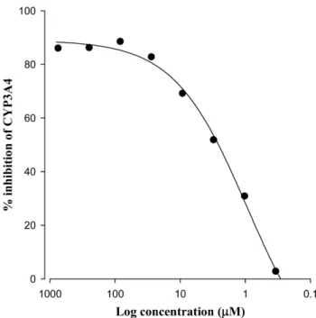
CYP3A4 활동에 대한 resveratrol의 저해 효과는 Fig. 1에서 볼수 있다. Resveratrol은 CYP3A4 효소의 활성을 저해하고 CYP3A4에 대한 resveratrol의 50% 저해 농도값(IC50)은 0.94 µM 이었다.
Rhodamine-123 retention assay
Fig. 2에서 보여지듯 MCF-7/ADR cell에 p-gp가 overexpressing 되면 P-gp가 부족한 MCF-7 cell에 비해 Rhodamine-123의 축적 이 감소함을 알수 있다. Resveratrol을 병용사용할때 농도 의존 적 방법으로 Rhodamine-123의 cellular uptake가 증가하였고 100µM에서 통계확적으로 유의성(p<0.01)을 보였다. 이번 결과 는 resveratrol이 P-gp의 활동을 유의성있게 저해함을 암시한다.
Resveratrol이 nifedipine의 약동학 변수에 미치는 영향 Nifedipine 대조군과 병용투여군의 평균혈장농도 추이는 Fig.
3과 같았으며, 약물동태학적 파라미터는 각각 Table I과 같았다.
혈장중농도곡선하면적(AUC)은 대조군에서 3058±714 ng/ml·
hr, resveratrol 병용투여군(2.5 및 10 mg/kg)에서 각각 4547±
1109 ng/ml·hr 및 5365±1312 ng/ml·hr로 유의성(p<0.05,
Absolute bioavailability A B%( , )=AUCoral AUCIV
--- IV dose Pral dose ---
× ×100
Relative bioavailability R B%( , )=AUCcombined AUCcontral --- 100×
Fig. 1 − Inhibitory effect of resveratrol on CYP3A4 activity. All experiments were performed in duplicate, and results are expressed as the percent of inhibition (IC
50
; 0.94µM).p<0.01)있게 증가하였다. 최고혈중농도(Cmax)는 대조군에서 248±
60.1 ng/ml, resveratrol 병용투여군(2.5 및 10 mg/kg)에서 각각 367±77.6 ng/ml 및 412±84.3 ng/ml로 유의성(p<0.05, p<0.01) 있게 증가하였으며, t1/2는 대조군에 비해 병용투여군에서 유의성 있게 연장되었다. Nifedipine의 절대생체이용률(AB%)은 대조군 에서 19.8±4.2%, resveratrol 병용투여군(2.5 및 10 mg/kg)에서 각각 29.5±6.3% 및 34.8±7.0%로 유의성(p<0.05, p<0.01)있게 증가하였다. Nifedipine의 상대생체이용률(RB%)은 대조군에 비 해 resveratrol 병용투여군에서는 1.15~1.75배로 현저히 증가하 였다.
이러한 결과는 P-당단백질과 CYP3A4이 소장막의 내피세포에
같이 존재하고 있으므로32)많은 약물들의 흡수를 저해하며 흡수 전 대사(first-pass effect)를 받는데 협동작용을 한다고 보고된 것 같이,33) resveratrol은 CYP3A4와 P-당단백질 억제약물로서 nifedipine과 병용투여시 증가된 생체이용률은 resveratrol이 흰 쥐의 장관막 세포에서 P-당단백질의 유출작용을 억제하며, CYP3A 로 인한 대사를 감소시킨 것으로 사료된다.
본 실험 결과는 모린, 나린진 및 세친과 병용투여시 흰쥐에서 nifedipine의 생체이용률을 증가시켰다는 보고와 일치하였다.19-21) 또한 Hong22)등의 연구결과인 resveratrol과 딜티아젬을 병용투 Fig. 2 − Rhodamine-123 retention. After incubation of MCF-7/ADR
cells with 20µM R-123 for 90 min. Data represents means±SEM of 4 separate samples (significant versus the control MCF-7 cells, **p<0.01). Verapamil (100 µM) was used as a positive control.
Fig. 3 − Mean plasma concentration of nifedipine after oral admin- istration of nifedipine (6 mg/kg) coadministered with resveratrol and after intravenous administration of nifedipine (1.5 mg/kg) in rats. (●) Control (nifedipine, 6 mg/kg); (○) Coadministered with resveratrol, 0.5 mg/kg; (▼) Coadmin- istered with resveratrol, 2.5 mg/kg; (△), Coadministered with resveratrol, 10 mg/kg; (■) I.V. (nifedipine 1.5 mg/kg).
Table I − Mean (±S.D.) Pharmacokinetic parameters of nifedipine after oral administration of nifedipine (6 mg/kg) coadministered with resveratrol in rats (Mean±SD, n=6)
Parameters Nifedipine
control
Resveratrol coadministration I.V.
1.5 mg/kg
0.5 mg/kg 2.5 mg/kg 10 mg/kg
AUC (ng/ml·h) 3058±714 3529±772
04547±1109* 005365±1312**
3856±902C
max
(ng/ml)0.248±58.8 0.303±62.1 0.367±77.6* 00.412±84.3**
T
max
(h)00.8±0.3 00.8±0.3
0.46±0.10 0.46±0.10t
1/2
(h) 11.6±2.8 12.5±3.0 14.0±3.3* 14.7±3.6*08.7±2.3
AB (%) 19.8±4.2 22.9±4.5 29.5±6.3*
034.8±7.0**
100RB (%) 100 115 148 175
*p<0.05 compared to control.
AUC: area under the plasma concentration-time curve from 0 h to infinity.
C
max
: peak plasma concentration; Tmax
: time to reach peak concentration; t1/2
: terminal half-life; RB (%): relative bioavailability, compared AUCcombined
to AUCcontrol
; AB (%): absolute bioavailability.여시 딜티아젬의 생체이용률을 증가시켰다는 보고와도 일치하였 다 또한 Choi23)등의 연구결과인 resveratrol과 니카르디핀을 병 용투여시 니카르디핀의 AUC와 Cmax을 증가시켰다는 보고와도 일치하였다.
소장에서 P-당단백질과 CYP3A4의 억제제인 퀄세친 및 모린 이 베라파밀과 및 딜티아젬의 생체이용률을 증가시켰다.30-31)또 한 본 실험 결과는 Nabekura 등28)의 연구결과와 일치하다. 즉, KB-C2 세포에서 resveratrol은 다우노루비신(P-당백질의 기질) 의 세포내 축척을 농도의존적으로 증가시켰다는 보고와 일치하였다.
항고혈압제인 nifedipine의 생체이용효율증가 즉 효과증가는 resveratrol이 소장 및 간장에서 cytochrome P450(CYP) 3A4를 억제하고 소장에서 P-당단백질의 유출작용(P-gp efflux trans- porter)을 억제하여 나타난 것으로 사료된다. 동물 모델에서 이 런 결과가 인체실험에서 확인된다면, nifedipine을 resveratrol 과 병용투여시 nifedipine의 약물동태학적 상호작용을 고려하여 임 상에서 용량을 조절하는 것이 바람직하다고 사료된다.
결 론
Nifedipine 6 mg/kg을 경구투여한 대조군과 resveratrol(0.5, 2.5 및 10 mg/kg)을 nifedipine과 병용투여한군에서 nifedipine의 생체이용률 및 약물동태 파라미터의 변화와 resveratrol이 CYP3A4 효소의 활성억제 및 P-gp의 활성억제효과를 검토한 결 과는 다음과 같다.
1. Resveratrol은 CYP3A4 효소의 활성을 저해하고 CYP3A4 에 대한 resveratrol의 50% 저해 농도값(IC50)은 0.94 µM이었다.
2. Resveratrol을 병용사용할때 농도 의존적으로 Rhodamine- 123의 cellular uptake가 증가시킨 결과는 resveratrol이 P-gp의 활성을 유의성 있게 저해 하였다.
3. Nifedipine혈장중농도곡선하면적(AUC) 및 최고혈중농도 (Cmax)는 대조군에 비해 resveratrol 병용투여군(2.5 및 10 mg/kg) 에서 유의성(p<0.05, p<0.01)있게 증가하였다.
4. 반감기(t1/2)는 대조군에 비해 resveratrol 병용투여군에서 유 의성 있게 연장되엇다.
5. Nifedipine의 절대적 생체이용률(AB%)은 대조군에서 비해 resveratrol 병용투여군(2.5 및 10 mg/kg)에서 유의성(p<0.05, p<0.01)있게 증가하였다.
6. Nifedipine의 상대적 생체이용률(RB%)은 대조군에 비해 resveratrol 병용투여군에서는 1.15~1.75배로 현저히 증가하였다.
Nifedipine의 생체이용효율증가 즉 효과증가는 resveratrol이 소장 및 간장에서 cytochrome P450(CYP) 3A4를 억제하고 소 장에서 P-당단백질의 유출작용(P-gp efflux transporter)을 억제 하여 나타난 것으로 사료된다.
본 연구에 의하면 resveratrol이 nifedipine의 생체이용률을 증
가시킬 가능성이 있으므로 임상에서 nifedipine을 resveratrol 또 는 resveratrol을 함유하는 식품과 병용하여 사용할 경우 nifedipine의 용량을 조절하는 것이 바람직하다고 사료된다.
참고문헌
1) Soleas, G. J., Diamandis, E. P. and Golberg, D. M. : Resveratrol: a molecule whose time has come? and gone?
Clin. Biochem. 30, 91 (1997a).
2) Soleas, G. J., Diamandis, E. P. and Golberg, D. M. : Wine as a biological fluid: history, production, and role in disease prevention. J. Clin. Lab. Anal. 11, 287 (1997b).
3) Constant, J. : Alcohol, ischemic heart disease, and the French paradox. Coronary Artery. Dis. 8, 645 (1997).
4) Fauconneau, B., Waffo-Teguo, P., Huguet, F., et al. : Comparative study of radical scavenger and antioxidant properties of phenolic compounds from vitis vinifera cell cultures using in vitro tests. Life Sci. 61, 2103 (1997).
5) Frankel, E. N., Waterhouse, A. L. and Kinsella, J. E. : Inhibition of human LDL oxidation by resveratrol. Lancet. 341, 1103 (1993).
6) Bertelli, A. A. E., Giovanni, L., Stradi, R., et al. : Kinetics of trans- and cis-resveratrol (3,4',5-trihydroxystilbene) after red wine oral administration in rats. Int. J. Clin Pharmacol Res. 26, 77 (1996).
7) Jang, M., Cai, L., Udeani, G. O., et al. : Cancer chemopreventive activity of resveratrol, a natural product derived from grapes.
Science 275, 218 (1997).
8) Guengerich, F. P. : Roles of cytochrome P-450 enzymes in chemical carcinogenesis and cancer chemotherapy. Cancer Res.
48, 2946 (1988).
9) Chan, W. K. and Delucchi, A. B. : Resveratrol, a red wine constituent, is a mechanism-based inactivator of cytochrome P450 3A4. Life Sci. 67, 3103 (2000).
10) Piver, B., Berthou, F., Dreano, Y., et al. : Inhibition of CYP3A, CYP1A and CYP2E1 activities by resveratrol and other non volatile red wine components. Toxicol. Lett. 125, 83 (2001).
11) Chang, T. K. H., Chen, J. and Yu, C. T. : In vitro inhibition of rat CYP1A1 and CYP1A2 by piceatannol, a hydroxylated metabolite of resveratrol. Drug Metab. Lett. 1, 13 (2007).
12) Nabekura, T., Kamiyama, S. and Kitagawa, S. : Effects of dietary chemopreventive phytochemicals on P-glycoprotein function. Biochem. Biophys. Res. Commun. 327, 866 (2005).
13) Walter, D. G., Gruchy, B. S., Renwick, A. G., et al. : The first- pass metabolism of nifedipine in man. Br. J. Clin. Pharmacol.
18, 951 (1984).
14) Raemsch, K. and Sommer, J. C. : Pharmacokinetics and metabolism of nifedipine. Hypertension. 5, 18 (1983).
15) Henry, P. D. : Comparative pharmacology of calcium
antagonists: nifedipine, verapamil and diltiazem. Am. J.
Cardiol. 46, 1047 (1980).
16) Sorkin, E. M., Clissold, S. P. and Brogden, R. N. : Nifedipine.
A review of its pharmacodynamic and pharmacokinetic properties, and therapeutic efficacy, in ischaemic heart disease, hypertension and related cardiovascular disorders. Drugs. 30, 182 (1985).
17) Guengerich, F. P., Brian, W. R. and Iwasaki, M. : Oxidation of dihydropyridine calcium channel blockers and analogues by human liver cytochrome P-450 3A4. J. Med. Chem. 4, 1838 (1991).
18) Iribarne, C., Dréano, L. G., Bardou, J. F., et al. : Interaction of methadone with substrates of human hepatic cytochrome P450 3A4. Toxicology 117, 13 (1997).
19) Watkins, P. B., Wrighton, S. A., Schuetz, E. G., et al. : Identification of glucocorticoid-inducible cytochromes P-450 in the intestinal mucosa of rats and man. J Clin Invest. 80, 1029 (1987).
20) Kolars, J. C., Schmiedlin-Ren, P., Dobbins, 3rd W. O., et al. : Heterogeneity of cytochrome P450IIIA expression in rat gut epithelia. Gastroenterology 102, 1186 (1992).
21) Gottesman, M. M. and Pastan, I. : Biochemistry of multidrug resistance mediated by the multidrug transporter. Annu. Rev.
Biochem. 62, 385 (1993).
22) Gan, S. L., Moseley, M. A., Khosla, B., et al. : CYP3A-Like cytochrome P450-mediated metabolism and polarized efflux of cyclosporin A in Caco-2 cells: interaction between the two biochemical barriers to intestinal transport. Drug Metab.
Dispos. 24, 344 (1996).
23) Watkins, P. B. : The barrier function of CYP3A4 and P- glycoprotein in the small bowel. Adv. Drug Deliv. Rev. 27, 161 (1996).
24) Wacher, V. H., Silverman, J. A., Zhang, Y., et al. : Role of P- glycoprotein and cytochrome P450 3A in limiting oral absorption of peptides and peptidomimetics. J. Pharm. Sci. 87, 1322 (1998).
25) Ito, K., Kusuhara, H. and Sugiyama, Y. : Effects of intestinal CYP3A4 and P-glycoprotein on oral drug absorption theoretical approach. Pharm. Res. 16, 225 (1999).
26) Chaudhary, P. M. and Robinson, I. B. : Expression and activity of P-glycoprotein, a multidrug efflux pump, in human
hematopoietic stem cells. Cell. 66, 85 (1991).
27) Van Asperen, J., Van Tellingen, O., Sparreboom, A., et al. : Enhanced oral bilavailability of diltiazem in mice treated with the p-glycoprotein blocker. Br. J. Cancer. 76, 1181 (1997).
28) Biedler, J. L. and Riehm, H. : Cellular resistance to actinomycin D in Chinese hamster cells in vitro: cross- resistance, radioautographic, and cytogenetic studies. Cancer Res. 30, 1174 (1970).
29) Ford, J. M. : Modulators of multidrug resistance preclinical studies. Hematol. Oncol. Clin. N. Am. 9, 337 (1995).
30) Fruehauf, J. P. and Manetta, A. : Use of extreme drug resistance assay to evaluate mechanisms of resistance in ovarian cancer: Taxol resistance and MDR1 expression.
Contrib. Gynecol. Obstet. 19, 39 (1994).
31) Lee, J. K. and Choi, J. S. : Effect of morim on the pharmacokinetics of nifedipine in rats. Yakhak Hoeji 51, 169 (2007).
32) Na, J. H. and Choi, J. S. : Effect of naringin on the pharmacokinetics of nifedipine in rats. J. Kor. Pharm. Sci. 35, 101 (2005).
33) Han, H. K., Lee, I. K. and Choi, J. S. : Pharmacokinetic interaction between nifedipine and quercetin in rabbits. J. Kor.
Pharm. Sci. 34, 283 (2004).
34) Hong, S. P., Choi, D. H. and Choi, J. S. : Effects of resveratrol on the pharmacokinetics of diltiazem and its major metabolite, desacetyldiltiazem, in rats. Cardiovascular. 26, 269 (2008).
35) Choi, J. S., Choi, B. C. and Kang, K. W. : Effect of resveratrol on the pharmacokinetics of oral and intravenous nicardipine in rats: possible role of P-glycoprotein inhibition by resveratrol.
Pharmazie. 64, 49 (2009).
36) Grundy, J. S., Kherani, R. and Foster, R. T. : Sensitive high- performance liquid chromatographic assay for nifedipine in human plasma utilizing ultraviolet detection. J. Chromatogr B.
Biomed. Appl. 654, 146 (1994).
37) Crespi, C. L., Miller, V. P. and Penman, B. W. : Microtiter plate assays for inhibition of human, drug-metabolizing cytochromes P450. Anal. Biochem. 248, 188 (1997).
38) Rocci, M. L. and Jusko, W. J. : LAGRAN program for area and moments in pharmacokinetic analysis. Computer Programs in Biomedicine. 16, 203 (1983).