215
암로디핀이 레파그리니드의 약물동태에 미치는 영향
최동현·최준식*
조선대학교 의과대학, 조선대학교 약학대학
(2011년 6월 22일 접수·2011년 8월 2일 수정·2011년 8월 30일 승인)
Effects of Amlodipine on the pharmacokinetics of Repaglinide
Dong-Hyun Choi and Jun-Shik Choi*
College of Pharmacy, College of Medicine, Chosun University, Gwangju 501-759, Republic of Korea (Received June 22, 2011·Revised August 2, 2011·Accepted August 30, 2011)
암로디핀과 레파그리니드의 병용은 당뇨병의 합병증으로인한 고혈압 유발 시 병용 처방될 수 있다. 암로디핀과 레파 그리니드의 약동학적 상호작용 연구를 위하여 암로디핀 (0.1 및 0.4 mg/kg) 과 레파그리니드를 흰 쥐에 경구 (0.5 mg/kg) 및 정맥 (0.2 mg/kg) 투여하여 연구를 실시하였다. 암로디핀이 cytochrome P450 (CYP) 3A4 활성과 P-glycoprotein (P-gp)의 활성에 미치는 영향도 평가하였다. 암로디핀의 CYP3A4의 50% 효소활성억제는 9.1 µM 이 었다. 암로디핀은 P-gp의 활성에는 영향을 미치지 않았다. 암로디핀 (0.4 mg/kg)은 레파그리니드의 혈장곡선하면적 (AUC)과 최고혈장농도 (Cmax)를 40.2% 와 22.2% 각각 유의성 (p < 0.05)있게 증가시켰다. 따라서, 레파그리니드의 상대적생체이용률 (RB)은 암로디핀과 병용투여 시 1.18-1.40 배 증가되었으며, 또한 레파그리니드의 절대적생체이용률 (AB)은 대조군과 비교하여 41.0% 유의성 있게 증가되었다. 경구 투여 시와는 대조적으로, 암로디핀은 정맥 내로 투 여된 레파그리니드에서는 약동학적 파라미터에 어떤 영향도 미치지 않았다. 따라서 암로디핀이 레파그리니드의 생체이 용률을 증가시킨 것은 신장배설 감소 또는 P-gp 활성억제 보다는 암로디핀이 소장 또는 간장에서 CYP3A4을 억제 시켰기 때문으로 사료된다. 암로디핀과 레파그리니드의 병용투여 시 레파그리니드의 용량을 조절하는 것이 안전하다고 사료된다.
□ Key words - Repaglinide, Pharmacokinetics, Amlodipine, CYP3A4, Rat
Amlodipine, a dihydropyridine calcium channel blocker, causes coronary and peripheral vasodilatation by blocking the influx of extracellular calcium across cell membranes. Amlodipine is arterioselective and effective for the treatment of hypertension, myocardial ischemia, and vasospasm in surgical patients.1) Amlo- dipine is rapidly absorbed and is extensively metabo- lized in the liver while it shows linear dose-related pharmacokinetic characteristics and, at steady-state, there are relatively small fluctuations in plasma concen- trations across a dosage interval.2) Although structurally related to other dihydropyridine derivatives, amlodipine displays significantly different pharmacokinetic charac-
teristics.3) Amlodipine is a substrate of cytochrome P450 (CYP) 3A subfamily, specifically CYP3A4.4-5) In addition, amlodipine is also a P-glycoprotein (P-gp) substrate.6-7)
Repaglinide is a new carboxymethyl benzoic acid derivative, also known as 2-ethoxy-4-[2-[[3- methyl-1- [2-(1-piperidinyl) phenyl] butyl] amino]-2-exoethyl]
benzoic acid.8) It is a novel post prandial glucose regu- lator for the treatment of type 2 diabetes mellitus.9-10) It reduces fasting glucose concentrations in patients with type 2 diabetes mellitus.8) It helps to control blood glu- cose by increasing the amount of insulin released by the pancreas.11) Repaglinide stimulates the release of insu- lin from the pancreatic β-cell by binding to and closing of ATP-dependent potassium channels. This depolarizes the plasma membrane, leading to the opening of volt- age-dependent calcium channels. Influx of calcium ions, which increases intracellular Ca2+, triggers exocy-
Correspondence to : Jun-Shik Choi
College of Medicine, Chosun University, Gwangju 501-759, Republic of Korea
Tel: +82-62-230-6365, Fax: +82-62-222-5414 E-mail : [email protected]
tosis of insulin.12)
Repaglinide is rapidly absorbed from the gastrointes- tinal tract after oral administration. It differs from other antidiabetic agents in its structure, binding profile, duration of action and mode of excretion.13) Repaglin- ide is primarily metabolized via oxidative biotransfor- mation involving the hepatic microsomal cytochrome P450 system, particularly the CYP3A4 isoform.14) The metabolic pathway of repaglinide indicates two major sites for the principal biotransformation of repaglinide:
the piperidine ring and the aromatic carboxylic acid group.15) Repaglinide has affinity for P-gp and it can significantly contribute to potential drug-drug interac- tions with other P-gp substrates or inhibitors.16) Kajosaari et al.17) reported that co-administration of repaglinide with the known P-gp inhibitor cyclosporine A could significantly increase the plasma concentra- tions of repaglinide in humans.
There are few reports about CYP enzyme and P-gp activity for amlodipine. Therefore, we evaluated the inhi- bition of CYP enzyme activity and P-gp activity by amlo- dipine using CYP inhibition assays and rhodamine-123 retention assays in P-gp-overexpressing MCF-7/ADR cells.
Clinically repaglinide and amlodipine can be pre- scribed for prevention or treatment of cardiovascular disease as complications of diabetes. However, pharma- cokinetic interaction between repaglinide and amlo- dipine has not been reported. Therefore, the present study aims to investigate the effect of amlodipine on CYP3A4 activity, P-gp activity and the pharmacokinet- ics of repaglinide after oral and intravenous administra- tion in rats.
MATERIALS AND METHODS
Materials
Repaglinide, amlodipine and indomethacin (internal standard) were purchased from the Sigma-Aldrich Co.
(St. Louis, MO, USA). HPLC-grade methanol and ace- tonitrile were acquired from Merck Co. (Darmstadt, Germany). All other chemicals for this study were of
reagent grade and were used without further purifica- tion. Apparatus used in this study included an HPLC equipped with a Waters 1515 isocratic HPLC Pump, a Waters 717 plus auto sampler and a Waters 2487 scan- ning UV detector (Waters Co., Milford, MA, USA), an HPLC column temperature controller (Phenomenex Inc., CA, USA), a Bransonic Ultrasonic Cleaner (Bran- son Ultrasonic Co., Danbury, CT, USA), a vortex-mixer (Scientific Industries Co., NY, USA), and a high-speed microcentrifuge (Hitachi Co., Tokyo, Japan).
Animal experiments
Male Sprague-Dawley rats (weighing 270-300 g) were purchased from the Dae Han Laboratory Animal Research Co. (Choongbuk, Korea) and were given access to a commercial rat chow diet (No. 322-7-1, Superfeed Co., Gangwon, Korea) and tap water. The animals were housed, two per cage, and maintained at 22±2oC and 50-60% relative humidity under a 12:12 h light-dark cycle. The experiments were initiated after acclimation under these conditions for at least 1 week.
The Animal Care Committee of Chosun University (Gwangju, Korea) approved the design and the conduct of this study. The rats were fasted for at least 24 h prior to the experiments and each animal was anaesthetized lightly with ether. The left femoral artery and vein were cannulated using polyethylene tubing (SP45, i.d.
0.58 mm, o.d. 0.96 mm; Natsume Seisakusho Co. LTD., Tokyo, Japan) for blood sampling and i.v. injection, respectively.
Oral and intravenous administration of repaglinide
The rats were divided into six groups (n=6, each): an oral control group (0.5 mg/kg of repaglinide dissolved in distilled water, 1.0 ml/kg) without or with 0.1 and 0.4 mg/kg of amlodipine (mixed in distilled water, 3.0 ml/kg), and an i.v. control group (0.2 mg/kg of repa- glinide, dissolved in 0.9% NaCl solution, 1.5 ml/kg) with- out or with 0.1 and 0.4 mg/kg of amlodipine (mixed in distilled water, 3.0 ml/kg). Oral repaglinide was adminis- tered intragastrically using a feeding tube, and amlo- dipine was administered in the same manner 30 minprior to the oral administration of repaglinide. Repa- glinide for i.v. administration was injected through the femoral vein within 0.5 min. A 0.4-ml aliquot of blood was collected into heparinized tubes from the femoral artery at 0.25, 0.5, 0.75, 1, 2, 4, 6, and 10 h after repa- glinide oral administration and at 0 (to serve as con- trol), 0.1, 0.25, 0.5, 1, 2, 6, and 10 h after repaglinide i.v. administration. The blood samples were centrifuged at 13,000 rpm for 3 min, and the plasma samples were stored at –40oC until HPLC analysis.
HPLC assay
Plasma concentration of repaglinide was determined by HPLC as reported by Ruzilawati et al.12) with a slight modification. Briefly, a 50-µl aliquot of 1 µg/ml indomethacin, as an internal standard, and a 0.2-ml ali- quot of 0.1 M potassium dihydrogen orthophosphate (KH2PO4, pH 5.9) were mixed with a 0.2-ml aliquot of the plasma sample. After the mixture was vortexed, 1 ml of ethylacetate, 50µl of isoamylalcohol and 35 µl of 1.0 M NaOH were added. The resulting mixture was then vortex-mixed for 10 min and centrifuged at 9,000 rpm for 10 min. After centrifugation, the ethylacetate phase was transferred into a clean test tube and evapo- rated under a gentle stream of nitrogen gas at 45_C.
The dried extract was reconstituted with 150µl of mobile phase, vortex-mixed and transferred to a clean autosampler vial. A 70-ml aliquot of the supernatant was injected into the HPLC system. Chromatographic separation was achieved using a µBondapak C18 col- umn (3.9300 mm i.d., 10µm, Waters Co.) attached to a µBondapak C18 guard column (3.9×20 mm i.d., 10 µm, Waters Co.). The mobile phase consisted of acetoni- trile:0.01 M ammonium formate (pH 2.7, adjusted with phosphoric acid) (56:44, v/v). The flow-rate of the mobile phase was maintained at 1.0 ml/min. Chroma- tography was performed at 30 and regulated by an HPLC column temperature controller. The UV detector was operated at a wavelength of 244 nm. Repaglinide and indomethacin were eluted with retention times at 7.4 and 8.4 min, respectively. The lower limit of quanti- fication for repaglinide in rat plasma was 20 ng/ml. The
intra- and inter-day coefficients of variation of repaglin- ide were below 12.1 and 11.5%, respectively.
CYP inhibition assay
The assays of inhibition of human CYP3A4 enzyme activity were performed in a multiwell plate using CYP inhibition assay kit (GENTEST, Woburn, MA) as described previously.18) Briefly, human CYP enzymes were obtained from baculovirus-infected insect cells.
CYP substrate (7-BFC) was incubated with or without test compounds in the enzyme/substrate buffer consist- ing of 1 pmol of P450 enzyme and a NADPH generat- ing system (1.3 mM NADP, 3.54 mM glucose 6- phosphate, 0.4 U/ml glucose 6-phosphate dehydroge- nase and 3.3 mM MgCl2) in a potassium phosphate buffer (pH 7.4). Reactions were terminated by adding stop solution after 45-min incubation. Metabolite con- centrations were measured by spectrofluorometer (Molecular Devices, Sunnyvale, CA) at an excitation wavelength of 409 nm and an emission wavelength of 530 nm. Positive control (1µM ketoconazole) was run on the same plate and produced 99% inhibition. All experiments were performed in duplicate, and results are expressed as the percent of inhibition.
Rhodamine-123 retention assay
The procedures used for the Rho-123 retention assay were similar to a previously reported method.19) MCF- 7/ADR cells were seeded in 24-well plates and preincu- bated with amlodipine for 30 min. At 80% confluence, the cells were incubated in FBS-free DMEM for 18 h.
The culture medium was changed to Hanks’ balanced salt solution and the cells were incubated at 37oC for 30 min. After incubation of the cells with 20µM rhodamine- 123 in the presence or absence of amlodipine (1, 3 and 10µM) for 90 min, the medium was completely removed. The cells were then washed three times with ice-cold phosphate buffer (pH 7.0) and lysed in EBC lysis buffer. Rhodamine-123 fluorescence in the cell lysates was measured using excitation and emission wavelengths of 480 and 540 nm, respectively. Fluores- cence values were normalized to the total protein con-
tent of each sample and are presented as the ratio to control.
Pharmacokinetic analysis
The plasma concentration data were analyzed by the non-compartmental method using WinNonlin software version 4.1 (Pharsight Co., Mountain View, CA, USA).
The elimination rate constant (Kel) was calculated by log-linear regression of repaglinide concentration dur- ing the elimination phase. The terminal half-life (t1/2) was calculated by 0.693/Kel. The peak plasma concen- tration (Cmax) and time to reach peak plasma concentra- tion (Tmax) of repaglinide in plasma were obtained by visual inspection of the data from the concentration–
time curve. The area under the plasma concentration- time curve from 0 h to the time (AUC0–t) of last mea- sured concentration (Clast) was calculated by the linear trapezoidal rule. The AUC from zero to infinite (AUC0–∞) was obtained by the addition of AUC0–t and the extrapo- lated area determined by Clast/Kel. Total body clearance (CL/F) was calculated by:
The absolute bioavailability (AB) was calculated by:
While the relative bioavailability (RB) was calculated by:
Statistical analysis
Statistical analysis was conducted using one-way ANOVA followed by a posteriori testing with the Dun- nett correction. Differences were considered to be sig- nificant at p < 0.05. All mean values are presented with their standard deviation (Mean±SD).
RESULTS
Inhibition of CYP3A4
The inhibitory effect of amlodipine on CYP3A4 activ- ity is shown in Fig. 1. Amlodipine inhibited CYP3A4 activity in a concentration-dependent manner. Amlo- dipine inhibited CYP3A4 activity with an IC50 value of 9.1µM.
Rhodamine-123 retention assay
The effect of amlodipine on the cellular accumulation of rhodamine-123 in MCF-7 and MCF-7/ADR cells is shown in Fig. 2. Accumulation of rhodamine-123 was reduced in MCF-7/ADR cells overexpressing P-gp compared to that in MCF-7 cells lacking P-gp. The rel- CL F⁄ Dose
AUC---
=
A.B. AUCoral AUCi.v.
--- Dosei.v.
Doseoral ---
× ×100
=
R.B. AUCwith amlodipine
AUCcontrol
--- 100×
=
Fig. 1. Inhibitory effects of ketoconazole (A) and amlodipine (B) on CYP3A4 activity. All experiments were performed in duplicate, and results are expressed as the percent of inhibition.
ative cellular uptake of rhodamine-123 was comparable at the concentration range of 1-10µM amlodipine. This result suggests that amlodipine could not inhibit P-gp activity.
Effect of amlodipine on the pharmacokinetics of oral
repaglinide
Mean arterial plasma concentration-time profiles of repaglinide following oral administration of repaglinide (0.5 mg/kg) to rats without or with amlodipine (0.1 and 0.4 mg/kg) are shown in Fig. 3; the corresponding phar- macokinetic parameters are shown in Table 1. Amlodipine significantly altered the pharmacokinetic parameters of repaglinide. Compared to the control group (given oral repaglinide alone), amlodipine significantly increased the area under the plasma concentration-time curve (AUC0–∞, p < 0.05 at 0.4 mg/kg) and the peak plasma concentration (Cmax, p < 0.05 at 0.4 mg/kg) of repaglinide by 40.2%
and 22.2%, respectively. Amlodipine increased the absolute bioavailability (AB) of repaglinide by 41%
(0.4 mg/kg, p<0.05) compared to the oral control group, and the relative bioavailability (RB) of repaglin- ide by 1.18- to 1.40-fold. There were no significant dif- ferences in the time to reach peak plasma concentration (Tmax) and terminal half-life (t1/2) of repaglinide between the presence and absence of amlodipine.
Effect of amlodipine on the pharmacokinetics of intravenous repaglinide
Mean plasma concentration-time profiles of repaglin- ide after the intravenous administration of repaglinide (0.2 mg/kg) to rats without or with amlodipine (0.1 and Fig. 2. Rhodamine-123 retention. MCF-7/ADR cells were
pre-incubated with amlodipine for 30 min and then MCF-7/
ADR cells were treated with 20µM R-123 for 90 min. Data represents mean±SD of 6 separate samples (significant versus the control MCF-7 cells).
Fig. 3. Mean plasma concentration–time profiles of repaglinide following an oral (0.5 mg/kg) administration of repaglinide to rats with or without amlodipine (Mean±SD, n=6). (●) Control (repaglinide 0.5 mg/kg, oral), (○) with 0.1 mg/kg amlodipine, (▼) with 0.4 mg/kg amlodipine.
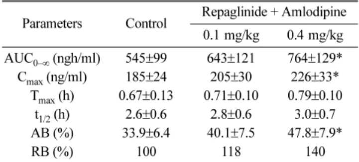
Table 1. Pharmacokinetic parameters of repaglinide after the oral administration of repaglinide (0.5 mg/kg) to rats without or with amlodipine (0.1 and 0.4 mg/kg) (Mean±SD, n=6)
Parameters Control Repaglinide + Amlodipine 0.1 mg/kg 0.4 mg/kg AUC0–∞ (ngh/ml) 545±99 643±121 764±129*
Cmax (ng/ml) 185±24 205±30 226±33*
Tmax (h) 0.67±0.13 0.71±0.10 0.79±0.10 t1/2 (h) 2.6±0.6 2.8±0.6 3.0±0.7 AB (%) 33.9±6.4 40.1±7.5 47.8±7.9*
RB (%) 100 118 140
*p < 0.05 significant difference compared to the control (given oral repaglinide alone).
AUC0–∞: area under the plasma concentration-time curve from 0 h to infinity; Cmax: peak plasma concentration; Tmax: time to reach peak plasma concentration; t1/2: terminal half-life; AB(%): absolute bioavailability; RB(%): relative bioavailability.
0.4 mg/kg) are shown in Fig. 4, while the correlated pharmacokinetic parameters are shown in Table 2. The AUC of repaglinide was increased, but was not statisti- cally significant compared to that in the control. The t1/2 of repaglinide was also prolonged, but this increase was not significant. The pharmacokinetics of intravenous repaglinide was not affected by the concurrent use of amlodipine in contrast to those of oral repaglinide.
Accordingly, the enhanced oral bioavailability in the
presence of amlodipine, while there was no significant change in the pharmacokinetics of intravenous repaglin- ide, may be mainly due to inhibition of the CYP3A- mediated metabolism of repaglinide in the small intes- tine and/or in the liver by amlodipine rather than renal elimination of repaglinide.
DISCUSSION
Type 2 diabetes mellitus is a common health problem associated with cardiovascular disease, with an increas- ing incidence worldwide.20) In clinical, repaglinide and amlodipine could be prescribed for prevention or treat- ment of cardiovascular disease as complications of dia- betes. Considering that the drugs used in combination therapy often share the same metabolic pathways or cellular transport pathways, there is a high potential for pharmacokinetic drug interactions between repaglinide and amlodipine. Consequently, this study was to inves- tigate the effect of amlodipine on CYP3A4 activity, P- gp activity and the pharmacokinetics of repaglinide after oral and intravenous administration in rats.
In vitro and in vivo studies have assessed the potential for repaglinide to interact with other drugs in clinical use. One of the principal cytochrome P450 isoforms involved in repaglinide metabolism in human liver microsomes is CYP3A4. CYP3A4 is the most abundant isoform of cytochrome P450 in the human liver21) and is responsible for the metabolism of many different drugs.22) Levels of CYP3A4 vary substantially between individuals; these variations may be related to induction of the isoform by various drugs and may partially account for the observed inter-individual variations in repaglinide metabolism.23) As CYP3A9 expressed in rat corresponds to the ortholog of CYP3A4 in human,24) and rat CYP3A2 is similar to human CYP3A4.25-26) Human 3A4 and rat 3A1 have 73% of protein homol- ogy.27) Rats were selected as an animal model in this study to evaluate the potential pharmacokinetic interac- tions mediated by CYP3A4, although there should be some difference in enzyme activity between rat and human.28)
Fig. 4 Mean plasma concentration–time profiles of repaglinide following an intravenous (0.2 mg/kg) administration of repaglinide to rats with or without amlodipine (Mean±SD, n=6). (●) Control (repaglinide 0.2 mg/kg, i.v.), (○) with 0.1 mg/kg amlodipine, (▼) with 0.4 mg/kg amlodipine.
Table 2. Pharmacokinetic parameters of repaglinide after the intravenous administration of repaglinide (0.2 mg/kg) to rats without or with amlodipine (0.1 and 0.4 mg/kg) (Mean±SD, n=6)
Parameters Control Repaglinide + Amlodipine 0.1 mg/kg 0.4 mg/kg AUC0–∞ (ngh/ml) 643±118 686±122 752±144
t1/2 (h) 2.6±0.5 2.7±0.6 2.8±0.6 CLt (ml/min) 5.1±1.0 4.9±0.9 4.4±0.9
RB (%) 100 107 117
AUC0–∞: area under the plasma concentration-time curve from 0 h to infinity; t1/2: terminal half-life; CLt: total body clearance; RB(%):
relative bioavailability.
Considering that repaglinide is a substrate of both CYP3A4 and P-gp,14,16) the effects of amlodipine on the CYP3A4 activity and the cell-based P-gp activity were evaluated.
CYPs enzymes make a considerable contribution to the first-pass metabolism and oral bioavailability of many drugs. Moreover, induction or inhibition of CYPs may be responsible for significant drug and drug interactions.29-30) Modulators of P-gp can enhance or limit the permeability of a number of therapeutic agents that are considered sub- strates of this efflux pump protein.6) Therefore amlodipine, a inhibitor against both CYP3A4, should have a great impact on the bioavailability of many drugs where CYP3A4 metabolism, is the major barrier to the systemic bioavailability of its substrates.31-32)
As shown in Fig. 1, amlodipine inhibited CYP3A4 activ- ity with an IC50 value of 9.1µM. However, the relative cel- lular uptake of rhodamine-123 was comparable (Fig. 2). It suggested that amlodipine could not inhibit P-gp activity.
The inhibitory effect of amlodipine against CYP3A-medi- ated metabolism was confirmed by the employment of recombinant CYP enzymes. Poor solubility and first-pass metabolism in the liver and epithelial cells of the small intestine result in low bioavailability of repaglinide (56%).16) CYP3A4 inhibitors might alter the bioavailabil- ity and pharmacokinetics of repaglinide.
In this study, amlodipine significantly increased AUC0–∞
by 40.2% and the RB of repaglinide by 1.18- to 1.40-fold.
Because amlodipine considerably increased Cmax of repa- glinide, it seems that amlodipine inhibited the CYP3A4- mediated biotransformation of repaglinide mainly during first-pass metabolism. CYP3A4 is present in consider- able quantities in the small-intestine mucosa,33-34) and the intestine CYP3A4 has been shown to play a major role in drug interactions with CYP3A4 inhibitors. These results are consistent with a report by Niemi et al.35) showing that clarithromycin, a CYP3A4 inhibitor, signifi- cantly increased the AUC0–∞ and Cmax of repaglinide.
Niemi et al.36) reported that gemfibrozil significantly increased the AUC0–∞ of repaglinide 8.1-fold. This result is also consistent with our study.
After intravenous administration of repaglinide with
amlodipine, the AUC0–∞ of repaglinide was increased, but was not significant. The CLt values of repaglinide tend to decrease, but was not statistic significant. This suggests that the effects of intravenous amlodipine on the inhibition of hepatic metabolism of repaglinide via CYP3A4 were almost negligible. In contrast to those of oral repaglinide, the pharmacokinetics of intravenous repaglinide was not affected by the concurrent use of amlodipine. Accordingly, while there was no significant change in the pharmacokinetics of intravenous repaglin- ide, the enhanced oral bioavailability may be mainly due to inhibition of the CYP3A-mediated metabolism of repaglinide in the small intestine and/or in the liver by amlodipine rather than P-gp and/or renal elimination of repaglinide. Although there are potential adverse effects, this interaction may provide a therapeutic bene- fit whereby it enhances bioavailability, allowing a reduction in the dose administered. Since the present study has raised the awareness of potential drug interac- tions by concomitant use of amlodipine with repaglin- ide, the clinical significance of this finding needs to be further evaluated in clinical studies.
CONCLUSION
In conclusion, amlodipine significantly enhanced the oral bioavailability of repaglinide. The enhanced bio- availability of repaglinide may be attributable to the inhibition of the CYP3A4-mediated metabolism in the small intestine and/or in the liver rather than both to inhibition of P-gp in the small intestine and to reduction of renal elimination of repaglinide by amlodipine. The increase in the oral bioavailability of repaglinide should be taken into consideration of potential drug interac- tions when coadministering repaglinide and amlodipine.
REFERENCES
1. Kungys G, Naujoks H, Wanner C. Pharmacokinetics of amlodipine in hypertensive patients undergoing haemodialysis. Eur J Clin Pharmacol 2003; 59: 291-295.
2. Abernethy DR. Pharmacokinetics and Pharmacodynamics of amlodipine. Cardiology 1992; 80: 31-36.
3. Meredith PA, Elliott HL. Clinical pharmacokinetics of amlodipine. Clin Pharmacokinet 1992; 22: 22-31.
4. Nishio S, Watanabe H, Kosuge K, et al., Interaction between amlodipine and simvastatin in patients with hypercholesterolemia and hypertension. Hypertens Res 2005; 28: 223-227.
5. Kim KA, Park PW, Park JY. Effect of cytochrome P450 3A5*3 genotype on the stereoselective pharmacokinetics of amlodipine in healthy subjects. Chirality 2009; 21:
485-491.
6. Darvari R, Boroujerdi M. Concentration dependency of modulatory effect of amlodipine on P-glycoprotein efflux activity of doxorubicin - a comparison with tamoxifen. J Pharm Pharmacol 2004; 56: 985-991.
7. Harmsze AM, Robijns K, van Werkum JW, et al., The use of amlodipine, but not of P-glycoprotein inhibiting calcium channel blockers is associated with clopidogrel poor-response. Thromb Haemost 2010; 103: 920-925.
8. El-Houssieny BM, Wahman LF, Arafa NM. Bioavailability and biological activity of liquisolid compact formula of repaglinide and its effect on glucose tolerance in rabbits.
Biosci Trends 2010; 4: 17-24.
9. Marbury TM, Ruckle JL, Hatorp V, et al., Pharmacokinetic of repaglinide in subjects with renal impairment. Clin Pharmacol Ther 2000; 67: 7-15.
10. Hatorp V, Won-Chin H, Strange P. Repaglinide pharmacokinetic in healthy young adult and eldery subjects. Clin Ther 1999; 21: 702-710.
11. Gromada J, Dissing S, Kofod H, et al., Effects of the hypoglycaemic drugs repaglinide and glibenclamide on ATP-sensitive potassium-channels and cytosolic 113 calcium levels in beta TC3 cells and rat pancreatic beta cells. Diabetologia 1995; 38: 1025-1032.
12. Ruzilawati AB, Wahab MS, Imran A, et al., Method development and validation of repaglinide in human plasma by HPLC and its application in pharmacokinetic studies. J Pharm Biomed Anal 2007; 43: 1831-1835.
13. Culy JR, Jarvis B. Repaglinide: a review of its therapeutic use in type 2 diabetes mellitus. Drugs 2001;
61: 1625-1660.
14. Bidstrup TB. Björnsdottir, I., Sidelmann, U. G., et al., CYP2C8 and CYP3A4 are the principal enzymes involved in the human in vitro biotransformation of the insulin secretagogue repaglinide. Br J Clin Pharmacol 2003; 56: 305-314.
15. Bauer E, Beschke K, Ebner T, et al., Biotransformation of [14C] repaglinide in human, cynomolgus monkey, dog, rabbit, rat and mouse. Diabetologia 1997; 1: 326-332.
16. Chang C, Bahadduri PM, Polli JE, et al., Rapid identification of P-glycoprotein substrates and inhibitors.
Drug Metab Dispos 2006; 34: 1976-1984.
17. Kajosaari L, Niemi M, Neuvonen M, et al., Cyclosporine markedly raises the plasma concentrations of repaglinide.
Clin Pharmacol Ther 2005; 78: 388-399.
18. Crespi CL, Miller VP, Penman BW. Microtiter plate assays for inhibition of human, drug-metabolizing cytochromes P450. Anal Biochem 1997; 248: 188-190.
19. Han CY, Cho KB, Choi HS, et al., Role of FoxO1 activation in MDR1 expression in adriamycin-resistant breast cancer cells. Carcinogenesis 2008; 29: 1837-1844.
20. Gomes MB, Giannella-Neto D, Faria M, et al., Estimating cardiovascular risk in patients with type 2 diabetes: a national multicenter study in Brazil. Diabetol Metab Syndr 2009; 1: 22-28.
21. Gonzalez FJ. Cytochrome P450 in humans. In: Schenkman JB, Grein H, editors. Cytochrome P450: handbook of experimental pharmacology. 1993; Vol. 105: Berlin: Springer- Verlag.
22. Li AP, Kaminski DL, Rasmussen A. Substrates of human hepatic cytochrome P450 3A4. Toxicology 1995; 104: 1-8.
23. Wrighton SA, Stevens JC. The human hepatic cytochromes P450 involved in drug metabolism. Crit Rev Toxicol 1992; 22: 1-21.
24. Kelly PA, Wang H, Napoli KL, et al., Metabolism of cyclosporine by cytochromes P450 3A9 and 3A4. Eur. J.
Drug Metab Pharmacokinet 1999; 24: 321-328.
25. Bogaards JJ, Bertrand M, Jackson P, et al., Determining the best animal model for human cytochrome P450 activities: a comparison of mouse, rat, rabbit, dog, micropig, monkey and man. Xenobiotica 2000; 30: 1131-1152.
26. Guengerich FP, Martin MV, Beaune PH, et al., Characterization of rat and human liver microsomal cytochrome P-450 forms involved in nifedipine oxidation, a prototype for genetic polymorphism in oxidative drug metabolism. J Biol Chem 1986; 261: 5051-5060.
27. Lewis DFV. Cytochrome P450. Substrate specificity and metabolism. In: Cytochromes P450. Structure, Function, and Mechanism. Taylor & Francis: Bristol, 1996; 122- 123.
28. Cao X, Gibbs ST, Fang L, et al., Why is it challenging to predict intestinal drug absorption and oral bioavailability in human using rat model. Pharm Res 2006; 23: 1675-1686.
29. Cummins CL, Jacobsen W, Benet LZ. Unmasking the dynamic interplay between intestinal P-glycoprotein and CYP3A4. J Pharmacol Exp Ther 2002; 300: 1036-1045.
30. Benet LZ, Cummins CL, Wu CY. Transporter-enzyme interactions: implications for predicting drug–drug interac- tions from in vitro data. Curr Drug Metab 2003; 4: 393-398.
31. Saeki T, Ueda K, Tanigawara Y, et al., P-glycoprotein- mediated transcellular transport of MDR-reversing agents.
FEBS Lett 1993; 324: 99-102.
32. Wacher VJ, Salphati L, Benet LZ. Active secretion and enterocytic drug metabolism barriers to drug absorption.
Adv Drug Deliv Rev 2001; 46: 89-102.
33. Kivistö KT, Bookjans G, Formm MF, et al., Expression of CYP3A4, CYP3A5 and CYP3A7 in human duodenal
tissue. Br. J Clin Pharmacol 1996; 42: 387-389.
34. Zhang QY, Dunbar D, Ostrowska A, et al., Characterization of human small intestinal cytochromes P-450. Drug Metab Dispos 1999; 27: 804-809.
35. Niemi M, Neuvonen PJ, Kivistö KT. The cytochrome P450 3A4 inhibitor clarithromycin increases the plasma concentrations and effects of repaglinide. Clin Pharmacol
Ther 2001; 70: 58-65.
36. Niemi M, Backman JT, Neuvonen M, et al., Effects of gemfibrozil, itraconazole, and their combination on the pharmacokinetics and Pharmacodynamics of repaglinide:
potentially hazardous interaction between gemfibrozil and repaglinide. Diabetologia 2003; 46: 347-351.