담배연기 응축물과 Lipopolysaccharide의 투여로 유발된 COPD에 대한 백지 추출물의 억제효과
곽호근·임흥빈† 충북 충북대학교 특용식물학과
Inhibitory Effects of Angelicae Dahuricae Radix Extract on COPD induced by Cigarette Smoke Condensate and Lipopolysaccharide in Mice
Ho Geun Kwak and Heung Bin Lim†
Department of Industrial Plant Science & Technology, Chungbuk National University, Chungju 361-763, Korea.
ABSTRACT : This study was carried out to investigate the inhibitory effect of Angelicae Dahuricae Radix (ADR) extract on chronic obstructive pulmonary disease (COPD) induced by cigarette smoke condensate (CSC) and lipopolysaccharide (LPS) in mice. COPD was induced by intratracheal instillation of LPS and CSC 5 times for 12 days; this increased airway hyper- resoponsiveness (AHR) and inflammatory cells in bronchoalveolar lavage fluid (BALF). ADR extract was administered orally at a dose of 50 and 200 ㎎/㎏. The concentration of imperatorin, a major component of ADR and therefore used as a measure of quality control, was 0.098% ± 0.018%. Treatment of the mice with ADR extract (50 and 200 ㎎/㎏) alleviated AHR and reduced inflammatory cell counts. Treatment with cyclosporin A (CSA; 10 ㎎/㎏) also modulated AHR and reduced inflammatory cells effectively. Compared with CSA treatment, treatment with ADR (50 ㎎/㎏) extract reduced neutrophil and CD4
+/CD3
+cell counts by 22.67% and 44.92%, respectively. In addition, compared with CSA treatment, treatment ADR 200 ㎎/㎏ reduced neutrophils, CD4
+/CD3
+cells and CD8
+/CD3
+cells, by 32.10%, 83.17% and 82.11%, respectively. These results indicate that ADR extract may have an inhibitory effect on COPD induced by LPS and CSC in mice.
Key Words : COPD, Angelicae Dahuricae Radix, Lipopolysaccharide
INTRODUCTION
Chronic obstructive pulmonary disease (COPD) is a major cause of mortality worldwide and is expected to be the third commonest cause of death by 2020 (Petty, 2006; Martorana et al., 2006). It is associated with chronic inflammation of the lungs and airways, caused by environmental exposure to cigarette smoke and air pollution, and is characterized by restriction of airflow (Petty, 2002). The chronic inflammation is associated with increased numbers of neutrophils and macrophages at the site of inflammation, as well as inflammatory mediators, imbalance between proteases and protease inhibitors, and oxidative stress (MacNee, 2003). An increase in neutrophils in the bronchus is associated with development of COPD or reduction in pulmonary function and has been observed in bronchoalveolar lavage fluid (BALF) and sputum samples
obtained from COPD patients (Vlahos et al., 2006). The numbers of inflammatory cells are indicative of the severity of the disease.
Lymphocyte levels have also been found to increase in the lungs and airways of COPD patients, and the number of CD4
+and CD8
+cells is also associated with the severity of the disease (Daheshia, 2005).
The presence of lipopolysaccharide (LPS; a major constituent of gram negative bacterial cell walls) is implicated in the inflammatory response and has been found to play a part in chronic pulmonary inflammation (Tavares et al., 2004; Vernooy et al., 2002; Shen et al., 2009). Cigarette smoke (CS; regarded as the main cause of COPD) contains chemical compounds, free radicals, and oxidative molecules associated with the pathogenesis of COPD (Barnes 2000; Brusselle et al., 2006;
Chan et al., 2009). As a result, CS has been used for COPD modeling in other studies too (Martorana et al., 2006; Chan et
†
Corresponding author: (Phone) +82-43-261-2521 (E-mail) [email protected]
Received 2011 September 6 / 1st Revised 2011 October 14 / 2nd Revised 2011 October 19 / Accepted 2011 October 20
al., 2009; Zheng et al., 2009).
Angelicae Dahuricae Radix (ADR), the root of Angelica dahurica (Umbelliferae), has been used for treating the common cold, headache, toothache, ulcers, melena, etc. in oriental medicine (Lee et al., 2007). ADR contains effective components such as coumarins and terpenoid (Kim et al., 1998; Joo and Kang, 2005). Its some coumarins on ADR are reported to have anti-microbial, oxidative, and thrombotic activity (Lee et al., 2007; Kwon et al., 1997; Ryu et al., 2001; Lechner et al., 2004, Song et al., 2005). Meanwhile, inflammation is associated with the various diseases and other studies on anti-inflammatory activity have reported in medical plants (Shin et al., 1988; Son et al., 2007). In addition, ADR also have considered to play a anti- inflammatory effect in inflammatory response (Yoon et al., 2010;
Jin et al., 2011). Therefore, since COPD is an inflammatory disease of the lung and bronchus, ADR might have an inhibitory effect on it. However, no studies have reported an inhibitory effect of ADR on respiratory disease. Therefore, the present study investigated the inhibitory effect of ADR extracts on COPD induced by CSC and LPS in mice. The concentration of imperatorin, a major constituent of ADR, was used as a marker for quality control of the ADR.
MATERIALS AND METHODS
1. Materials and Regents
Angelicae Dahuricae Radix was harvested in Yeongju, KyungSang Buk Do, Korea and its quality was assessed by the Kyoungdong Herb Co. (Daejeon, Korea). CORESTA Monitoring Cigarette No. 6 (CM6) was purchased from ITG Reemtsma Tobacco Co. (Berlin, Germany). Imperatorin (from Angelica archangelica), diethyl pyrocarbonate (DEPC), chloroform, trichloroacetic acid, isopropanol, Tris-HCl, KCl, MgCl
2, ACK lysis solution, Dulbecco's modified Eagle's minimum essential medium (DMEM), Dulbecco's phosphate buffered saline (D- PBS), sulforhodamine B (SRB), 2-isopropanol, sodium dodecyl sulfate (SDS), PMA, ionomycin, FK506, and antibiotics were purchased from Sigma Co. (St Louis, USA). Fetal bovine serum (FBS) was purchased from Hyclone Co. (Logan, USA). Anti- mouse-CD3-PE (phycoerythrin), anti-mouse-CD4-FITC (fluorescein isothiocyanate), anti-mouse-CD8-FITC, anti-mouse-neutrophil mAb, and anti-mouse-IgG-FITC, were purchased from Phar- mingen Co. (Torreyana, USA). Cyclosporin A (CSA) was purchased from Choongwae Pharma Co. (Seoul, Korea). All other chemicals were of special reagent grade.
2. Instruments
High performance liquid chromatography (HPLC) 1100 series (Agilent Technologies, USA) equipped with a multi-wavelength detector (MWD; Agilent Technologies, USA) was used to determine the concentration of imperatorin in the ADR. A rotary evaporator (EYELA, Japan), hot water extractor (Daewoong Co., Korea), and rotary vacuum evaporator (Büchi B-480, Switzerland) were used for extraction. A FACSCalibur (BD, USA) was used for inflammatory cell counts. In addition, a freeze dryer (EYELA FDU-540, Japan), CO
2incubator (Forma scientific Co., USA), spectrophotometer (Shimazu, Japan), centrifuge (Sigma, USA), deep-freezer (Sanyo, Japan), homo- genizer (OMNI, USA), plate shaker (Lab-Line, USA), VarioMACS (Bergisch Gladbach, Germany), and real-time PCR system (Applied Biosystems 7500, USA) were used in this study.
3. Collection of cigarette smoke condensate and ADR extraction
CM6 (CORESTA approved Monitor No. 6) reference cigarette was conditioned by ISO conditioning at a temperature of 22 ± 2
℃, and relative humidity of 60% ± 5% for 48 hours or more.
The reference cigarettes were smoked under ISO conditions (puff volume, 35 mL; duration, 60 sec; interval, 2 sec) using an automatic smoking machine (Borgwaldt RM20, Germany).
Cigarette smoke condensate (CSC) was trapped in a 92 ㎜ Cambridge filter pad, and then extracted with methanol for 3 days at room temperature. After extraction, the solvent was evaporated under vacuum.
ADR (100 g) was extracted in boiling water (distilled water, 2 ℓ) for 3 hours. After filtration, the solvent was evaporated under vacuum and was then lyophilized by freeze drying. The freeze- dried crude extract was stored in the deep-freezer ( −84℃) until use. To determine the imperatorin concentration in ADR, 10 g of ADR was extracted with 50 ㎖ of methanol (100%) using sonication for 9 hours. After filtration, the crude extract was evaporated under vacuum.
4. Animals and COPD model
Female specific pathogen-free (SPF) BALB/c mice (age, 8- weeks; weight, 18~20 g) were purchased from Orient Bio Inc.
(Sungnam, Korea). The experimental animal protocol was
approved by the Institutional Animal Care and Use Committee of
Deajeon University. The mice were housed in a room maintained
at 22 ± 2℃, with relative humidity of 55 ± 15% and 12 h/12 h
light/dark cycle. They were given a standard laboratory diet
(Samyangfeed, Korea) of solid feed, and tap water was provided ad libitum. The animals were divided into 5 groups as follows: 1.
Normal group (n = 6) was treated with water; 2. Control group (n
= 6) with LPS and CSC; 3. LPS + CSC + cyclosporin A (CSA) group (n = 6) with LPS, CSC, and CSA; 4. LPS + CSC + ADR (50 ㎎/㎏) group (n = 6) with LPS, CSC, and ADR extracts (50 ㎎/㎏); and 5. LPS + CSC + ADR (200 ㎎/㎏) group (n = 6) with LPS, CSC, and ADR extracts (200 ㎎/㎏).
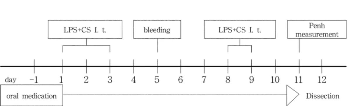
COPD was induced by intratracheal injection of 50 ㎕ of LPS (50 ㎍/㎖ in PBS) and CSC (10 ㎎/㎖ in PBS) for 5 days, after intraperitoneal administration of anesthetics. On the fifth day, blood was collected from the retro-orbital plexus and the neutrophil count measured by flow cytometry to select mice in which COPD had been successfully induced. Airway hyper- responsiveness (AHR) was measured by a methacholine challenge test on the eleventh day. Fig. 1 indicates the time schedule for the experimental procedure. Crude extracts of ADR (50 and 200 ㎎/㎏) and 10 ㎎/㎏ CSA (used as a positive control) were administered orally to the mice for 12 days. The next day, the mice were sacrificed.
5. Determination of imperatorin in ADR
Imperatorin is a major constituent of plants belonging to the species Umbelliferae and Rutaceae (Chen et al., 2007). In this study, the imperatorin content was measured for the purpose of quality control of the ADR extract used.
A stock solution of imperatorin (1 ㎎/㎖) was prepared in methanol by sonication at 30 ℃ for 30 min. The standard solutions for the calibration curve were prepared at 10 - 300
㎍/㎖ in methanol. The HPLC 1100 series, equipped with a degasser, an autosampler, a multi-wavelength detector, and a Zorbax Eclipse XDB-C18 column (250 ㎜ × 4.6 ㎜ I.D.; 5 ㎛ particle size; Agilent Technologies, USA) were used. The mobile phase of water:methanol (30 : 70) was delivered at a flow rate of 1 ㎖/min with an injection volume of 10 ㎕ and the wavelength
of the MWD set at UV 254 ㎚.
6. Measurement of AHR
Enhanced pause (Penh), a unitless measure of AHR, was estimated using a plethysmograph (BUXCO BioSystem XA, USA). Mice were placed in a whole-body chamber and baseline airflow measured for 30 min. Methacholine dissolved in PBS at concentrations of 12.5, 25, and 50 ㎎/㎖ was delivered via aerosol for 3 min. After nebulization, Penh was recorded by a flow transducer for 10 min and calculated as follows:
Penh = (Te − Tr)/Tr − PEP/PIP (Te, expiratory time; Tr, relaxa- tion time; PEP, peak expiratory pressure; PIP, peak inspiratory pressure)
7. Collection of BALF and counting of the total cells Eleven days after LPS and CSC administration, the trachea and airways of the anaesthetized mice were lavaged 3 times using a syringe (containing 1 ㎖ of 10% FBS/DMEM medium) to obtain BALF containing cells from the trachea and lungs. The BALF was treated with ammonium chloride (ACK) solution for 5 min at 37 ℃. BALF cells were washed with D-BPS, stained with 0.04% of trypan blue, and counted. Following bron- choalveolar lavage, the mice lungs were dissected and weighted.
8. Flow cytometry
After being counted, the BALF cells were stained using immunofluorescence and incubated with anti-mouse-CD3-PE (phycoerythrin), anti-mouse-CD4-FITC (fluorescein isothiocyanate), anti-mouse-CD8-FITC, anti-mouse-neutrophil mAb, and anti- mouse-IgG-FITC for 30 min at 4 ℃. After antibody reaction, all cells were washed 3 times with PBS. Cluster of differentiation antigen 4
+(CD 4
+)/CD3
+, CD8
+/CD3
+, and neutrophils were analyzed by a flow cytometer using Cell Quest software and the absolute numbers in each tissue were calculated from the number of total cells.
Fig. 1. The time schedule for COPD induction in mice. †I. t. : intratracheal injection.
9. Histological analysis
The dissected lung tissue was fixed in 10% formaldehyde solution for 24 hours, then rinsed in water for 8 hours, dehydrated, and embedded in paraffin using standard methods.
Tissue sections, 5 ㎛ thick, were stained with hematoxylin-eosin (H&E) and Masson-Trichrome (M-T), to aid examination by light microscopy.
10. Statistical analysis
Data are expressed as mean ± standard error of the mean
(SEM). Statistical analysis of comparisons between groups was performed using the Student T test. Significance was assessed as p < 0.05 or more.
RESULTS AND DISCUSSION
1. Determination of imperatorin in ADR
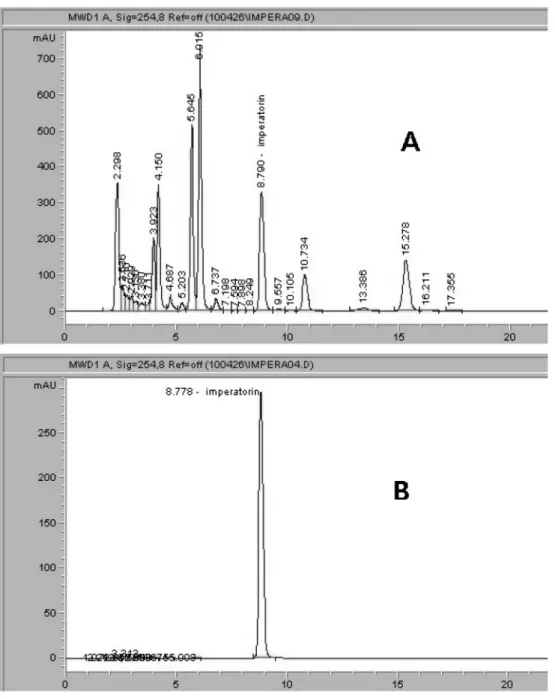
Fig. 2 shows chromatograms for the standard solution and
ADR extract. The calibration curve of a single compound
showed excellent linearity with a correlation coefficient (R²) of
Fig. 2. Chromatograms of ADR extract (A) and imperatorin (B).0.999 or more and the regression equation Y = 0.026X + 1.39 for imperatorin. The retention time for imperatorin was 8.773 ± 0.003 min. The yield of methanol extracts from ADR was 11.79 ± 0.63%, and the level of imperatorin in ADR was 0.098 ± 0.008%. Other research has reported the concentrations of imperatorin as 0.072 ± 0.008% and 0.068 ± 0.007% in Chinese Angelica dahurica Bench (Ketai et al., 2001). The concentration of imperatorin in ADR collected in China was 0.078 ± 0.035%, as measured by HPLC with electrospray tandem mass spectrometry (Zheng et al., 2010).
2. Effects of ADR on AHR
Changes in Penh value are associated with bronchoconstriction and therefore indicative of the severity of airflow obstruction (Hamelmann et al., 1997). In this study, the Penh level was determined using a whole-body plethysmograph to measure the response to a methacholine challenge test. The dose-response curves of AHR are shown in Fig. 3. Compared with the normal group, the LPS + CSC group showed significant increase in Penh level in response to methacholine doses of 25 ㎎/㎖ and 50 ㎎/㎖ (p < 0.01). However, the Penh level was significantly reduced in the LPS + CSC + ADR (200 ㎎/㎖) group compared with the LPS + CSC group at methacholine doses of 12.5, 25 and 50 ㎎/㎖ (p < 0.01). Compared with the ADR + CSC + CSA group, the LPS + CSC + ADR (50 ㎎/㎖) group was inhibited the Penh level of the LPS + CSC group by 65.87% and 70.74%
at the methacholine doses of 25 ㎎/㎖ and 50 ㎎/㎖. In addition, the LPS + CSC + ADR (200 ㎎/㎖) group had reduced Pehn levels of the LPS + CSC group by 91.44% and 96.76% at the methacholine doses of 25 ㎎/㎖ and 50 ㎎/㎖, when compared with the ADR + CSC + CSA (10 ㎎/㎖) group.
3. The effects of ADR on inflammatory cells in BALF To identify the inhibitory effect of ADR on COPD, inflammatory cells in the mouse BALF were counted using flow cytometry (Table 1). The number of neutrophils was significantly higher in the LPS + CSC group than in the normal group (p <
0.01). In contrast, the neutrophil count was significantly decreased in the LPS + CSC + ADR (50 and 200 ㎎/㎖) groups compared with the LPS + CSC group (p < 0.01). The number of neutrophils was reduced in the LPS + CSC + ADR (50 and 200
㎎/㎖) groups by 22.67% and 32.10%, respectively, compared with the LPS + CSC + CSA group.
CD3
+is known as an expression molecule of cell-surface in T cells. It forms a T-cell receptor (TCR) complex by association
with TCR and plays a crucial role in TCR signal transduction (Meuer et al., 1983; Yamaguchi et al., 2008). CD4
+and CD8
+are expressed on the surface of helper T and cytotoxic T cells and interact with a portion of the Class I and Class II MHC molecules to form antigen-presenting cells (Zamoyska, 1998).
CD4
+cells are involved in cell-mediated immunity, macrophage activation, maturation of B cells, and allergic inflammation, while CD8
+cells have been reported to be involved in cell- mediated immunity and antigen-specific cytotoxicity (Larosa and Orange, 2008). In the mice used in this study, the number of CD4
+/CD3
+and CD8
+/CD3
+cells significantly increased in the LPS + CSC group compared with the normal group (p < 0.01). In contrast, the number of CD4
+/CD3
+cells in the LPS + CSC + ADR (50 and 200 ㎎/㎖) groups was significantly lower than that in the LPS + CSC group (**: p < 0.05, ***: p < 0.01).
Furthermore, the number of CD8
+/CD3
+cells in the LPS +
Fig. 3. The effect of ADR extract on airway hyperresponsiveness.Mice were treated with LPS (50㎍/mL) and CSC (10㎎/mL) by intratracheal injection 5 times for 12 days. †Values are expressed as mean
± S.E (N = 5).
Statistical analysis of data was performed using Student's T-test. *: p < 0.01 compared with normal group. ***: p < 0.01 compared with control group. ‡Normal : Balb/c mice, LPS : Lipopolysaccharide (50㎍/㎖), CSC : Cigarette smoke condensate (10㎎/㎖), CSA : Cyclosporin A (10㎎/㎖), ADR : Angelicae Dahuricae Radix, MCH : Methacholine
CSC + ADR (200 ㎎/㎖) group was significantly reduced compared with the LPS + CSC group (p < 0.05). The number of CD4
+/CD3
+cells was reduced in the LPS + CSC + ADR (50 and 200 ㎎/㎖) groups by 44.92% and 83.17%, respectively, compared with the LPS + CSC + CSA group. Furthermore, the number of CD8
+/CD3
+cells was reduced in the LPS + CSC + ADR (50 and 200 ㎎/㎖) groups by 40.31% and 82.11% , respectively, compared with the LPS + CSC + CSA group.
4. Histological analysis
Infiltration of inflammatory cells in the tracheal and alveolar tissues was observed by H&E staining. Collagen deposition was assessed by M-T staining. The chronic inflammation in the bronchi caused remodeling of the bronchial wall as a result of destruction of the alveolar septa, and increase of collagen in the septa. These changes are seen in lung emphysema (Zheng et al., 2009). In this study, there was increased damage to the tracheal
and alveolar tissues, with infiltration of inflammatory cells and collagen deposition, in the LPS + CSC group compared with the normal group (Fig. 4A, B). On the other hand, the LPS + CSC + ADR (50 and 200 ㎎/㎖) and LPS + CSC + CSA groups showed reduced damage to the tracheal and alveolar tissues, as well as reduced infiltration of inflammatory cells and collagen deposition in the lung tissue compared with the LPS + CSC group (Fig. 4C, D, E).
In conclusion, the results of this study indicate that administration of LPS and CSC exacerbates AHR and induces accumulation of inflammatory cells in BALF, as well as histological changes. However, these responses are modulated by treatment with CSA, and this study has demonstrated that treatment with ADR extract produces similar results to those seen with CSA. However, CSA is rarely used in the treatment of respiratory diseases because of its side effect profile (Caramori et al., 2003; Barnes, 2008). In contrast, side effects associated with
Table 1. The number of inflammatory cells in BALF.Group Neutrophils (×103) CD4+/CD3+ cells (×103) CD8+/CD3+ cells (×103)
Normal 0.216 ± 0.061 0.25 ± 0.06 0.013 ± 0.003†
CSC + LPS (Control) 2.113 ± 0.278* 2.43 ± 0.04* 0.460 ± 0.128*
CSC + LPS + CSA 0.199 ± 0.002*** 0.84 ± 0.12*** 0.156 ± 0.011**
CSC + LPS + ADR (200 ㎎/㎏) 0.620 ± 0.070*** 1.01 ± 0.08*** 0.190 ± 0.026**
CSC + LPS + ADR (50 ㎎/㎏) 0.878 ± 0.067*** 1.87 ± 0.26** 0.387 ± 0.019 The number of inflammatory cells in bronchoalveolar lavage fluid (BALF) was counted using the fluorescence activated cell sorter (FACS). †Values are expressed as mean ± S.E (N = 6). Statistical analysis of data was performed using Student's T-test. *: p < 0.01 compared with normal group.
**: p < 0.05 and ***: p < 0.01 compared with control group. ‡Normal : Balb/c mice, LPS : Lipopolysaccharide (50 ㎍/㎖), CSC : Cigarette smoke condensate (10㎎/㎏), CSA : Cyclosporin A (10 ㎎/㎏), ADR : Angelicae Dahuricae Radix extract
Fig. 4. The histological analysis of lung tissue. †Normal : Balb/c mice, LPS : Lipopolysaccharide (50㎍/㎖), CSC : Cigarette smoke condensate (10㎎/㎖), CSA : Cyclosporin A (10 ㎎/㎏), ADR : Angelicae Dahuricae Radix extract.
ADR have yet to be reported. Therefore, ADR may be used as a natural plant-based therapy for COPD.
LITERATURE CITED
Barnes PJ. (2000). Chronic obstructive pulmonary disease. The New England Journal of Medicine. 343:269-281.
Barnes PJ. (2008). Drugs for airway disease. Medicine. 36:181- 190.
Brusselle GG, Bracke KR, Maes T, D'hulst AI, Moerloose KB, Joos GF and Pauwels RA. (2006). Murine models of COPD.
Pulmonary Pharmacology & Therapeutics. 19:155-165.
Caramori G and Adcock I. (2003). Pharmacology of airway inflammation in asthma and COPD. Pulmonary Pharmacology
& Therapeutics. 16:247-277.
Chan KH, Ho SP, Teung SC, So WHL, Cho CH, Koo MWL, Lam WK, Ip MSM, Man RYK and Mak JCW. (2009).
Chinese green tea ameliorates lung injury in cigarette smoke- exposed rats. Respiratory Medicine. 103:1746-1754.
Chen Y, Fan G, Zhang Q, Wu H and Wu Y. (2007). Fingerprint analysis of the fruits of Cnidium monnieri extract by high- performance liquid chromatography-diode array detection- electrospray ionization tandem mass spectrometry. Journal of Pharmaceutical and Biomedical Analysis. 43:926-936.
Daheshia M. (2005). Pathogenesis of chronic obstructive pulmonary disease (COPD). Clinical and Applied Immunology Reviews. 5:339-351.
Hamelmann E, Schwarze J, Takeda K, Oshiba A, Larsen GL, Irvin CG and Gelf EW. (1997). Noninvasive measurement of airway responsiveness in allergic mice using barometric plethysmography. American Journal of Respiratory and Critical Care Medicine. 156:766-775.
Jin HL, Lee BR, Lim KJ, Debneth T, Shin MH and Lim BO.
(2011). Anti-inflammatory effects of Prunus mume mixture in colitis induced by dextran sodium sulfate. Korean Journal of Medicinal Crop Science. 19:16-23.
Joo EY and Kang WJ. (2005). Analysis on the components of the Angelica dahurica root. Korean Journal of Food Preservation. 12:476-481.
Ketai W, Huitao L, Xingguo C, Yunkun Z and Zhide H.
(2001). Identification and determination of active components in Angelica dahurica Benth and its medicinal preparation by capillary electrophoresis. Talanta. 54:753-761.
Kim KS, Kim YH, Kang KD, Chung SH, Lee PS and Lee SC.
(1998). Essential oil content and composition of aromatic constituents in leaf of Saururus chinensis, Angelica dahurica and Cnidium officinale. Korean Journal of Medicinal Crop Science. 6:299-304.
Kwon YS, Kobayashi A, Kajiyama SI, Kawazu K, Kanzaki H and Kim CM. (1997). Antimicrobial constituents of Angelica dahurica roots. Phytochemistry. 44:887-889.
Larosa DF and Orange JS. (2008). Lymphocytes. Journal of Allergy and Clinical Immunology. 121:S364-S369.
Lechner D, Stavri M, Oluwatuyi M, Miranda RP and Gibbons S. (2004). The anti-staphylococcal activity of Angelica dahurica (Bai Zhi). Phytochemistry. 65:331-335.
Lee YS, Jang SM and Kim NW. (2007). Antioxidative activity and physiological function of the Angelica dahurica roots.
Journal of The Korean Society of Food Science and Nutrition.
36:20-26.
MacNee W. (2003). COPD: Causes and pathology. Medicine, 31:71-75.
Martorana PA, Cavarra E, Lucattelli M and Lungarella G.
(2006). Models for COPD involving cigarette smoke. Drug Discovery Today: Disease Models. 3:225-230.
Meuer SC, Fitzgerald KA, Hussey RE, Hodgdon JC, Schlossman SF and Reinherz EL. (1983). Clonotypic structures involved in antigen-specific human T cell function.
Journal of Experimental Medicine. 157:705-719.
Petty TL. (2002). COPD: Clinical phenotypes. Pulmonary Pharmacology & Therapeutics. 15:341-351.
Petty TL. (2006). The history of COPD. Interational Journal of COPD. 1:3-14.
Ryu SY, Kim JC, Kim YS, Kim HT, Kim SK, Choi GJ, Kim JS, Lee SW, Heor JH and Cho KY. (2001). Antifungal activities of coumarins isolated from Angelica gigas and Angelica dahurica aganist plant pathogenic fungi. The Korean Journal of Pesticide Science. 5:26-35.
Shen T, Kim DJ and Cho JY. (2009). Immunomodulatory effect of Pueraria lobata on the functional activation of macrophages by lipopolysaccharide treatment. Korean Journal of Medicinal Crop Science. 17:8-14.
Shin KH, Kim ON and Woo WS. (1988). Effects of the constituents of Angelicae Dahuricae Radix on hepatic drug metabolizing enzyme activity. Korean Journal of Pharma- cognosy. 19:19-27.
Son DW, Lee PJ, Lee JS, Lee SH, Choi SY, Lee JW and Kim SY. (2007). Neuroprotective effect of scopoletin from Angelica dahurica on oxygen and glucose deprivation-exposed rat organotypic hippocampal slice culture. Food Science and Biotechnology. 16:632-635.
Song DK, Kim JY, Li G, Lee KS, Seo CS, Yan JJ, Jung JS, Kim HJ, Chang HW and Son JK. (2005). Agents protecting against sepsis from the roots of Angelica dahurica. Biological and Pharmaceutical Bulletin. 28:380-382.
Tavares E, Ojeda ML, Maldonado R and Minano FJ. (2004).
Neutralization of macrophage inflammatory protein-2 blocks the fedrile response induced by lipopolysaccaride in rats. Journal of Thermal Biology. 29:413-421.
Vernooy JHJ, Dentener MA, van Suylen RJ, Buurman WA and Wouters EFM. (2002). Long-term intratracheal lipopolysaccharide exposure in mice results in chronic lung inflammation and persistent pathology. American Journal of Respiratory Cell and Molecular Biology. 26:152-159.
Vlahos R, Bozinovski S, gualano RC, Ernst M and Anderson GP. (2006). Modelling COPD in mice. Pulmonary Pharma- cology & Therapeutics. 19:12-17.
Yamaguchi H, Matsuo J, Sugimoto S, Utsumi M and Yamamoto Y. (2008). Inhibition of lymphocyte CD3 expression by Chlamydophila pneumoniae. Microbial Pathogenesis. 45:
290-296.
Yoon TS, Cheon MS, Kim SJ, Lee AY, Moon CB, Chun JM, Choo BK and Kim HK. (2010). Ecaluation of solvent
extraction on the anti-inflammatory efficacy of Glycyrrhiza uralensis. Korean Journal of Medicinal Crop Science. 18:28-33.
Zamoyska R. (1998). CD4 and CD8: Modulators of T-cell receptor recognition of antigen and of immune responses.
Current Opinion in Immunology. 10:82-87.
Zheng H. Liu Y, Huang T, Fang Z, Li G and He S. (2009).
Development and characterization of A ret model of chronic obstructive pulmonary disease (COPD) induced by sidestream
cigarette smoke. Toxicology Letters. 189:225-234.
Zheng X, Zhang X, Sheng X, Yuan Z, Yang W, Wang Q and Zhang L. (2010). Simultaneous characterization and quantitation of 11 coumarins in Radix Angelicae Dahuricae by high performance liquid chromatography with electrospray tandem mass spectrometry. Journal of Pharmaceutical and Biomedical Analysis. 51:599-605.