Otolaryngology & Dermatology 2011;24(1) : 64-77
Effects of Isatidis Radix and it's Active Component, Tryptanthrin on the Production of Inflammatory Mediators
in Lipopolysaccharide-activated Raw264.7 Cells
Sook-Jahr Park
a·Jong-Rok Lee
a·Mi-Jeong Jo
a,b·Sang-Mi Park
a,b· Sung-Hui Buyn
a·Il-Je Cho
a,b·Sang-Chan Kim
*a,ba
College of Oriental Medicine, Daegu Haany University
b
Development Team for the New Drug of Oriental Medicine(BK21 program)
LPS로 활성화된 Raw264.7 cell에서 판람근 및 Tryptanthrin의 염증매개물질억제효과
박숙자
a·이종록
a·조미정
a,b·박상미
a,b·변성희
a·조일제
a,b·김상찬
*a,bObjectives : 板藍根은 십자화과에 속하는 大靑 또는 菘藍의 根을 건조한 것이다. 본 연구는 板藍根이 淸熱解毒함 에 근거하여, LPS로 활성화된 Raw264.7 cell에서 板藍根과 그 성분중의 하나인 tryptanthrin이 염증매개물질에 미치 는 효과를 살펴보고자 하였다.
Methods : 세포생존율은 MTT, nitric oxide (NO)는 Griess reagent를 사용하여 측정하였으며, 각 단백질의 발현 량은 Western blot 방법을 사용하였으며, cytokine 및 cyclooxygenase-2 (COX-2)는 ELISA방법을 사용하여 측정하 였다.
Results : LPS는 NO 및 prostaglandin E2 (PGE2)를 유의하게 상승시켰으며, 板藍根추출물 (IRE) 및 tryptanthrin 은 이들을 유의하게 억제하였다. 그러나 板藍根의 또 다른 성분인 indigo는 유의한 결과를 나타내지 못하였다. IRE와 tryptanthrin은 inhibitory kappa B alpha의 인산화를 억제하여, nuclear factor-κB (NF-κB)의 핵으로의 轉位를 억 제하여, iNOS 및 cytokine을 억제하였다. IRE와 tryptanthrin의 PGE2 억제는, COX-2의 발현억제에서가 아니라, COX-2의 활성을 억제함에서 기인하였다.
Conclusion : 이러한 결과는 板藍根이 NF-κB pathway를 경유하여 iNOS의 발현 및 COX-2의 활성을 억제함을 나타내며, 이러한 板藍根의 항염증효능은 일부 tryptanthrin의 작용에서 기인함을 시사한다.
Key words : Isatidis Radix; tryptanthrin; nitric oxide; prostaglandin E2; nuclear factor-κB
교신저자 : Sang-Chan Kim, Daegu Haany University; Daegu 706-060, Republic of Korea (Phone: +82-53-770-2247, Fax: +82-53-768-6340, E-mail: [email protected])
∙접수 2011/03/08 ∙수정 2011/03/25 ∙채택 2011/04/01
Ⅰ. INTRODUCTION
Inflammation is the defensive reaction of immune system to infection, irritation or injury and has a pivotal role to helping body to fight disease. However, inappropriately controlled or prolonged inflammation leads to a progressive damage and finally causes many diseases, such as diabetes and cancer
1,2). There are multiple inflammatory factors involved in inflammatory reaction. The two inducible enzymes, cyclooxygenase-2 (COX-2) and inducible nitric oxide synthase (iNOS) appear to play key roles in regulating the immune response
3-5). Under normal condition, nitric oxide (NO) is synthesized from L-arginine by the enzymatic action of constitutive NOS (cNOS) and plays a central role in immune system by killing invaders. However, the excess NO produced by iNOS results in the aggravation of inflammation through its reaction with superoxide
6). The COX-2 plays a key role in generating prostaglandin E2 (PGE2) and over- production of PGE2 can cause inflammation
7). Induction of iNOS and COX-2 is associated with the transcription factor nuclear factor kappa B (NF-κB) pathway. NF-κB is a functional transcriptional factor involving in the inhibition of cell apoptosis, cell cycle regulation and oncogenesis
8). Furthermore, NF- κB is implicated with inflammation
9). In resting cells, NF-κB is located in the cytoplasm in association with the inhibitor of
NF-κB (I-κB) in an inactive form. However, NF-κB is activated through the complete degradation of I-κB via the phosphorylation and ubiquitination of I-κB, when various harmful stimuli including toxic materials, viruses and bacterial infections invade
10). Activated NF-κB translocates into nucleus where it induces transcription of the target genes in inflammatory process, such as iNOS and COX-2
5).
Isatidis Radix ( Banlangen ) has the effects of clearing heat from the blood and relieving sore-throat with cold property. Thus, Isatidis Radix is normally used for treating fever and headache in oriental herbal medicine
11). In cases of the disorders by pathogenic wind-heat or the early stage of epidemic febrile diseases, Isatidis Radix can be prescribed as a heat- clearing and detoxification herb. Moreover, Isatidis Radix has been used clinically for the treatment of influenza
12)and for the prevention of severe acute respiratory syndrome (SARS) in China
13). It was also reported to possess antagonistic effects on endotoxin produced by E. Coli
14,15). Organic acid o -amino benzoic acid from Isatidis Radix was known as the strongest anti-endotoxin
16,17).
Based on these previous reports and information, this study was conducted to evaluate the effect of Isatidis Radix on the inflammatory response through the activation of macrophage induced by LPS stimuli.
Furthermore, two commercial alkaloids
(tryptanthrin and indigo) were also evaluated
their effects as major constituents of Isatidis
Radix based on the quality control
18). In the
present study, we aim to elucidate the anti-inflammatory effects of Isatidis Radix and thryptanthrin on the production of NO, PGE2 and inflammatory cytokines. Furthermore, mechanism of anti-inflammatory action of Isatidis Radixon via NF-κB pathway was investigated.
Ⅱ. MATERIALS AND METHODS
1. Reagents
LPS (E. coli 026:B6) and 3-(4,5-dimethylthiazol- 2-yl)-2,5-diphenyltetrazoleum (MTT) were obtained from Sigma (St. Louis, MO, USA).
Dulbecco’s modified Eagle’s medium (DMEM) and fetal bovine serum (FBS) were purchased from Gibco/BRL (Eggenstein, Germany).
Tryptanthrin and indigo were purchased from Wako pure chemicals (Osaka, Japan). ELISA kits for TNF-α and IL-1β were purchased from Pierce endogen (Rockford, IL, USA).
PGE2 ELISA kit was purchased from RnD Systems (Minneapolis, MN, USA). COX-2 and iNOS antibodies were obtained from BD Bioscience (San Jose, CA, USA) and p-I-κBα antibody was purchased from Cell Signalling (Danvers, MA, USA). NF-κB, I-κBα and β -actin antibodies were obtained from Santa Cruz Biotechnology (Santa Cruz, CA, USA).
Cyclooxygenase-2 (COX-2) enzyme activity kit was obtained from Cayman chemical (Ann Arbor, MI, USA).
2. Preparation of the water extract of Isatidis Radix
Isatidis Radix was purchased from
Youngnam pharmacy (Daegu, Korea). The water extract of Isatidis Radix (IRE) was prepared by boiling 300 g of Isatidis Radix in 3 ℓ of water for 3 h. The IRE was filtered through a 0.2 ㎛ filter (Nalgene, NY, USA) and stored at -20℃ until use. The amount of IRE was estimated by the dried weight of lyophilized water extract of Isatidis Radix. The yield of lyophilized IRE from Isatidis Radix was 10.7%.
3. Cell culture
Raw264.7 cells were purchased from Korean Cell Line Bank (Seoul, Korea) and cultured in DMEM containing 10% FBS at 37℃ in a humidified atmosphere containing 5% CO
2. For all experiments, cells were grown to 80%
confluence, and were subjected to no more than 20 cell passages. Raw264.7 cells (2~3×10
6/
㎖) were incubated for 24 h and further incubated in the medium without 10% FBS for 24 h. After the serum starvation, the cells were exposed to LPS or LPS plus test materials for the indicated time periods.
4. MTT assay (cell viability test)
The concentration of IRE on cell viability
was determined by MTT assay. After
incubation of the cells with 0.3~10 ㎎/㎖ of
IRE for 24 h, viable cells were stained with
MTT solution (0.5 ㎎/㎖ for 4 h. The media
were removed carefully, and 200 ㎕ of
dimethylsulfoxide (DMSO) was added to
dissolve formazan crystals produced in the live
cells. Absorbance of the formazan solution was
measured with microplate reader (Model MCC/340, Huntsville, AL) at 570 ㎚. The cell viability was defined as the % of untreated control cells [i.e. viability (% control) = 100×
{(absorbance of treated sample)/(absorbance of control)}].
5. Nitric oxide assay
The nitric oxide (NO) level in culture medium was monitored by measuring the nitrite content in culture medium. This was performed by mixing the culture medium with Griess reagent (1% sulfanilamide, 0.1% N-1- naphthylenediamine dihydrochloride and 2.5%
phosphoric acid). Absorbance was measured at 540 ㎚ after incubation for 10 min in the dark.
6. Detection of PGE2 level
PGE2 level was determined with ELISA kit according to the manufacturer’s protocol.
Briefly, the culture medium was mixed with mouse monoclonal anti-PGE2 antibody and acetylcholinesterase-linked PGE2 in a 96-well plate coated with goat anti-mouse IgG for 18 h at 4℃. The wells were rinsed five times with washing buffer contained in the kit, and then developed with a coloring reagent for 1 h at 37℃. Absorbance was measured at 405
㎚, and the PGE2 concentration of each sample was determined according to the standard curve.
7. Assay of cyclooxygenase-2 activity Cyclooxygenase-2 (COX-2) enzyme activity
was measured using ELISA kit. The cells were collected by centrifugation at 1,500 ×g for 10 min at 4°C and homogenized in cold buffer (0.1 M Tris-HCl, pH 7.8, 1 mM EDTA, 250 mM mannitol, and 0.3 mM diethyldithiocarbamic acid). Homogenized cells were centrifuged at 10,000 ×g for 15 min at 4°C and the supernatant (cell lysate) was removed for COX-2 activity assay. Protein content of cell lysate was estimated according to Bicinchoninic acid (BCA) and the cell lysate was reacted with colorimetric substance, N,N,N’,N’- tetramethyl-p-phenylenediamine (TMPD) and arachidonic acid for 5 min at 25°C. This kit measures the peroxidase activity of COX-2.
The peroxidase activity was assayed colorimetrically by monitoring the appearance of oxidized TMPD at 590 ㎚. As described in the manufacturer’s instruction, one unit was defined as the amount of enzyme that caused the oxidation of 1 nmol of TMPD per minute at 25℃.
8. Western blot analysis
The cells were treated with lysis buffer containing 20 mM Tris-HCl (pH 7.5), 1%
Triton X-100, 150 mM sodium chloride, 10%
glycerol, 2 mM EDTA, 1 mM sodium orthovanadate, 25 mM β-glycerophosphate, 2 mM sodium pyrophosphate, 1 mM phenylmethylsulfonylfluoride and 1 ㎎/㎖
leupeptin. The cell mixture was kept on ice
for 30 min with vigorous vortex every 5 min,
and the total cell lysates were collected after
centrifugation at 10,000×g for 10 min. Protein
contents of the lysates were measured using
BCA assay (Pierce, USA). Equal amounts (30
㎍) of total proteins were resolved on 10%
SDS-PAGE and then transferred on NC membrane (Schleicher & Schuell GmbH, Dassel, Germany). The membrane was incubated with primary antibodies (1:1000), washed with TBST (10 mM Tris-Cl pH 7.5, 150 mM NaCl, 0.05% Tween 20) and incubated for 1 h with the appropriate peroxidase conjugated secondary antibodies.
The immunoreactivity was detected with the chemiluminescent detection kit (Amersham Bioscience, UK). For evaluation of the Western blot density, Imaging Analyzing System (Upland, CA, USA) was used.
9. Preparation of nuclear extracts for NF-κ B analysis
NF-κB expression was monitored from nuclear extract using western blot analysis. To prepare nuclear extracts, the cells were washed with ice-cold PBS and resuspended in lysis buffer A (10 mM HEPES-KOH, 10 mM KCl, 1.5 mM MgCl
2, 1 mM dithiothreitol, 0.1%
NP40, 1 mM PMSF and 4 ㎍/㎖ leupeptin, pH 7.6). After incubation on ice for 10 min, the cells were centrifuged at 7,800×g, 4℃ for 5 min, and then the pellet was collected. The pellet was treated with lysis buffer B (10 mM HEPES-KOH, 400 mM KCl, 0.1 mM EDTA, 25% glycerol, 1 mM PMSF and 4 ㎍/㎖
leupeptin, pH 7.6) and kept on ice for 1 h.
The supernatant containing nuclear fraction was prepared by centrifugation at 14,000×g for 15 min. The nuclear extracts were subjected to SDS-PAGE, transferred on NC membrane.
The membrane incubated with anti-NF-κB antibody (1:500), washed with TBST and then incubated with peroxidase-conjugated secondary antibody. The expression was detected with ECL western blotting detection reagents (Amersham Bioscience, UK).
10. Analysis of inflammatory cytokines In our preliminary study, TNF-α and IL-1β were highly induced at 12 h after LPS treatment (data are not shown). Thus, the cells were treated for 12 h with 1 ㎍/㎖ of LPS with or without IRE. An ELISA kit from Pierce endogen (Rockford, IL, USA) was used to determine TNF-α and IL-1β levels in culture medium according to the manufacturer’s instruction.
11. Statistical evaluations
Data were expressed as mean ± S.D. of results obtained from experiments. One-way analysis of variance (ANOVA) procedures were used to assess significant differences among treatment groups. For each significant effect of treatment, the Newman - Keuls test was used for comparisons of multiple group means. The criterion for statistical significance was set at P<0.05.
Ⅲ. RESULTS
1. Inhibitory effects of IRE on production of NO and PGE2
In view of the involvement of NO and
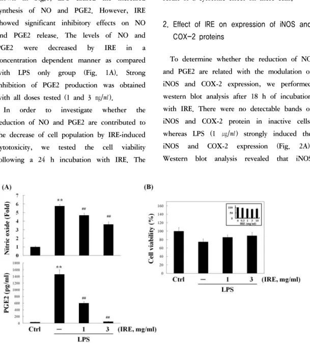
PGE2 in inflammatory conditions, we monitored the production of NO and PGE2 in culture medium exposed to 1 ㎍/㎖ of LPS in the presence or absence of IRE for 24 h. As shown in Fig.1, LPS caused the increased synthesis of NO and PGE2. However, IRE showed significant inhibitory effects on NO and PGE2 release. The levels of NO and PGE2 were decreased by IRE in a concentration dependent manner as compared with LPS only group (Fig. 1A). Strong inhibition of PGE2 production was obtained with all doses tested (1 and 3 ㎎/㎖).
In order to investigate whether the reduction of NO and PGE2 are contributed to the decrease of cell population by IRE-induced cytotoxicity, we tested the cell viability following a 24 h incubation with IRE. The
Fig. 1. Effect of IRE on the production of NO and PGE2 in LPS-activated Raw264.7 cells.
Cells were treated with 1 and 3 ㎎/㎖ of IRE for 24 h, and then the medium were collected for NO and PGE2 measurement (A). Cell viability (B) was assessed by MTT assay after incubation with IRE plus LPS for 24 h.
Inset shows the effect of IRE alone on cell viability. Data represent the mean ± S.D. with three separate experiments (significant compared to control, **P<0.01; significant compared to LPS alone, ##P<0.01).
results suggested that IRE did not affect the cell viability at the concentrations used (Fig.
1B). Therefore, inhibition of LPS-stimulated NO and PGE2 production by IRE was not the result of a cytotoxic effect on these cells.
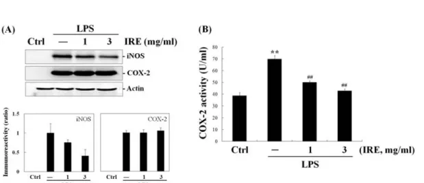
2. Effect of IRE on expression of iNOS and COX-2 proteins
To determine whether the reduction of NO and PGE2 are related with the modulation of iNOS and COX-2 expression, we performed western blot analysis after 18 h of incubation with IRE. There were no detectable bands of iNOS and COX-2 protein in inactive cells, whereas LPS (1 ㎍/㎖) strongly induced the iNOS and COX-2 expression (Fig. 2A).
Western blot analysis revealed that iNOS
expression by IRE was in parallel with the comparable inhibition of NO release. IRE exhibited down-regulatory effect on iNOS protein expression. However, IRE failed to show any significant reduction in the expression of COX-2 protein. These data indicated that reduction of PGE2 production by IRE occurs through other mechanisms except inhibition of COX-2 expression.
Because IRE had no effect on expression of COX-2 protein in spite of its inhibitory activity on PGE2 production, we further monitored COX-2 activity in cell-free extracts.
As shown in Fig. 2B, LPS (1 ㎍/㎖, 18 h) markedly elevated COX-2 activity as compared to control. IRE displayed a dose-related inhibition of COX-2 activity. Moreover, a significant inhibition down to almost basal levels was achieved with 3 ㎎/㎖ of IRE. From inhibit PGE2 synthesis and release by interfering enzymatic action of COX-2 rather
Fig. 2. Reduction of iNOS expression and COX-2 activity by IRE.
The expressions of iNOS and COX-2 protein (A) were monitored 18 h after IRE treatment. COX-2 enzyme activity (B) was measured using ELISA kit as described in materials and methods section. Data represent the mean ± S.D. with three separate experiments (significant compared to control, **P<0.01; significant compared to LPS alone, ##P<0.01).
these results, it was suggested that IRE can than by involving with COX-2 expression.
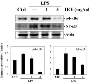
3. Inhibition of NF-κB and p-I-κ Bα expression by IRE
Next we investigated whether IRE affects NF-κB expression, because NF-κB plays a crucial role in regulating inflammation by involving production of NO and PGE2 in LPS-stimulated macrophage cells. Strong induction of NF-κB was observed by LPS (1 ㎍/㎖, 1 h). Although the treatment of the cells with 1.0
㎎/㎖ of IRE slightly reduced the nuclear NF- κB expression, significant decreasing amount of NF-κB protein was found in 3 ㎎/㎖ of IRE treated group (Fig. 3).
As stated earlier, activation of NF-κB is
mediated by phosphorylation of its inhibitor I-
κB. Thus, we further monitored
phosphorylation of I-κBα, the most abundant
isotype of I-κB
8). LPS (1 ㎍/㎖, 15 min) increased expression of p-I-κBα, but IRE showed inhibitory effect by reducing LPS- inducible p-I-κBα expression level (Fig. 3). The band of p-I-κBα was markedly disappeared after treatment of 3 ㎎/㎖ of IRE. These results suggest that IRE may have down- regulatory effect on NO and PGE2 production by interfering with the dissociation of I-κBα from NF-κB/I-κBα complex and resulting in the reduction of nuclear translocation of NF-κB.
Fig. 3. Effect of IRE on the expressions of p-I-κB and NF-κB.
Cells were stimulated for 15 min or 1 h with IRE plus LPS. The bands of p-I-κB (15 min) and NF-κB (1 h) were monitored from total and nuclear cell extracts, respectively. Bar graphs show the densitometry of blot from all experiments in each group.
4. Inhibitory effects of IRE on inflammatory cytokines
TNF-α and IL-1β are frequently encountered inflammatory cytokines. These cytokines are involved in a variety of immunological functions and interact with a variety of target
cells leading to the progression of many inflammatory diseases
19). We determined whether IRE displays anti-inflammatory activity by eliminating these cytokines. The levels of cytokines were measured in the culture media after the incubation with LPS (1 ㎍/㎖) and IRE for 12 h. IRE showed a concentration dependent inhibition of TNF-α and IL-1β production (Fig. 4).
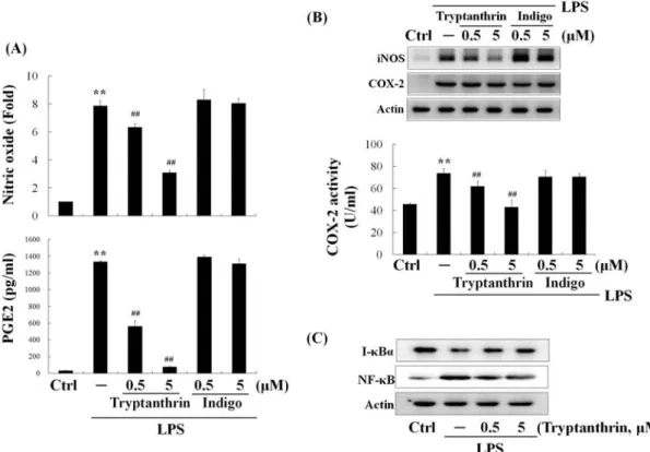
5. Inhibitory effects on NO and PGE2 production by tryptanthrin, but not by indigo
Tryptanthrin and indigo are the major
alkaloids of Isatidix Radix with various
pharmaceutical activities. To assess the effects
of thryptanthrin and indigo on the production
of inflammatory mediators, each alkaloid with
concentration of 0.5 and 5 μM was treated to
the cells for 24 h and NO and PGE2 levels in
culture media were measured. Tryptanthrin led
to the reduction of NO and PGE2 in a
dose-dependent manner while indigo did not
display any inhibitory effects (Fig. 5A). These
results were derived from the suppression of
iNOS expression and COX-2 activity by
tryptanthrin (Fig. 5B). On the other hand,
tryptanthrin had no effect on COX-2
expression as if IRE did. Since the effects of
IRE on NO and PGE2 were related to the
NF-κB pathway, we next examined the effect
of tryptanthrin on the expression of nuclear
NF-κB and total I-κBα protein. Nuclear
translocation of NF-κB is associated with the
proteolytic degradation of I-κBα. As shown
in Fig. 5C, western blot analysis showed that
Fig. 4. Inhibitory effects of IRE on production of cytokines in LPS-stimulated Raw264.7 cells.
Cells were incubated with IRE plus LPS for 12 h. TNF-α (A) and IL-1β (B) in the medium were determined by ELISA kit. Data represent the mean ± S.D. with three separate experiments (significant compared to control, **P
<0.01; significant compared to LPS alone, ##P<0.01).
Fig. 5. Inhibition of NO and PGE2 production by tryptanthrin through the suppression of NF-κB nuclear translocation.
Cells were treated with 0.5 and 5 μM of tryptanthrin or indigo for 24 h, and the levels of NO and PGE2 (A) were determined from the cultured medium. COX-2 activity and expressions of iNOS and COX-2 (B) were measured. Raw264.7 cells were treated with tryptathrin for 15 min or 1 h, and the bands of I-κB (15 min) and NF-κB (1 h) were monitored by Western blot (C). Data represent the mean ± S.D. with three separate experiments (significant compared to control, **P<0.01; significant compared to LPS alone, ##P<0.01).