ISSN 1225-6552, eISSN 2287-7630 http://dx.doi.org/10.7853/kjvs.2013.36.1.23
< Original Article >
Veterinary Service
Available online at http://kjves.org
*Corresponding author: Shien-Young Kang, Tel. +82-43-261-2598, Fax. +82-43-267-2595, E-mail. [email protected]
Molecular characterization of avian rotavirus isolated in Korea
Jun-Hui Wang, Bon-Sang Koo, In-Pil Mo, Shien-Young Kang*
College of Veterinary Medicine, Chungbuk National University, Cheongju 361-763, Korea (Received 11 January 2013; revised 6 March 2013; accepted 25 March 2013)
Abstract
An avian rotavirus (AvRV-2) was isolated from feces of broilers suffering from acute gastroenteritis in 2011. It was the first avian rotavirus isolated in Korea. To investigate the molecular characteristics of AvRV-2, the VP4, VP6, VP7 and NSP4 gene nucleotide sequences were determined and compared with those of rotavirus strains available in the GenBank database. The phylogenetic tree of VP7 gene showed that AvRV-2 had a high degree of nucleotide sequence homology (93.4% to 94.7%) with those of rotaviruses belonging to genotype G19 cluster. The phylogenetic tree of the VP4 gene revealed a high degree of nucleotide sequence homology (95.8% to 95.9%) with genotype P[30] rotaviruses iso- lated from chickens. The VP6 and NSP4 gene nucleotide sequences showed the highest identities with those of avian strains with 95.3% to 96.4% and 90.3% to 92.2%, respectively. Genetic characterization of the VP4, VP6, VP7 and NSP4 showed that AvRV-2 strain was most closely related to chicken rota- virus strains from Germany and Japan. Comparative nucleotide sequences and phylogenetic analysis in- dicated that avian rotavirus isolated from broilers belonged to genotype G19P[30] and it was the first report on avian rotavirus infection in Korea.
Key words : Avian rotaviruses, VP4, VP6, VP7, NSP4 gene
INTRODUCTION
Rotaviruses are classified in the genus Rotavirus within the family Reoviridae. They have 11 segments of double-stranded RNA genome and consist of six struc- tural proteins (VP1-4, VP6 and VP7) and six non- structural proteins (NSP1-6). A mature infectious virion has a diameter of approximately 100 nm and includes three layers with no lipid-containing envelope (Raming et al, 2005). The inner core is mainly formed by VP2 which surrounds genomic RNA. VP1 and VP3, minor components of the core, are presumed to have a link with RNA polymerase and the guanylyl transferase, re- spectively (Ito et al, 2001). The intermediate layer is formed by VP6, which was the first rotavirus protein used for classification. The outer layer consists of VP4 and VP7. They also independently elicit neutralizing an-
tibodies which contain the serotype-specific epitopes that were defined P (protease sensitive) serotype and G (gly- coprotein) serotype, respectively (Gorziglia et al, 1990).
However, antigenic characterization by virus neutraliza- tion test is time-consuming and requires virus collec- tions and proper immunological reagents that are not available in all the laboratories. And due to the increas- ing cases of nucleotide sequences, the antigenic sero- types were replaced by VP4 and VP7 genotypes. So far, at least 27G (G1-G27) and 35P (P[1]-P[35]) genotypes of rotaviruses have been identified globally. Recently, new classification strategy was proposed by determining the percentage identity cut-off values of each nucleotide sequence of eleven rotavirus segmented genomes (Ma- tthijnssens et al, 2011). Nonstructural proteins may play a role in viral replication and morphogenesis, but the ef- fects remain to be studied in more detail. The nonstruc- tural proteins interact with nucleic acid except for NSP4, which is the only nonstructural protein that does
not bind to RNA. It has been reported that NSP4 plays an important role in viral morphogenesis, pathogenesis and enterotoxin activity (Ball et al, 1996; Estes and Kapikian, 2007).
The rotaviruses include at least seven group (A-G).
Group A rotaviruses are important enteric pathogens in human, mammalian and avian species. Group A, B and C rotaviruses are currently found in both human and an- imal species, whereas group D, E, F and G have been isolated only in animal to date (Estes and Kapikian, 2007). Group A, D, F and G have been detected in sev- eral avian species including chicken, turkey and pigeon (Hines et al, 1995). Rotavirus infection in avian species was first reported in 1977 (Bergeland et al, 1977).
Avian rotaviruses have been detected in Argentina, Belgium, Brazil, Germany, Japan, the United Kingdom, and the United States (Bellinzoni et al, 1987;
Meulemans et al, 1985; Gusmal et al, 1994; Elschner et al, 2005; Takase et al, 1986; McNulty et al, 1978;
Yason and Schat, 1985). Avian rotaviruses cause enter- itis and diarrheal disease in avian species. Clinical signs are associated with depression, dehydration, impaired appetite, poor weight gains and increased mortality.
However, clinical symptoms of avian rotavirus infection are not obvious and subclinical infection is also common. So, diagnosis of avian rotavirus infection is necessary to the laboratory confirmation.
In this study, avian rotavirus was first isolated from feces of broilers suffering from acute gastroenteritis. The nucleotide sequences of major structural proteins (VP4, VP7 and VP6) and nonstructural protein (NSP4) were determined and compared with those of reference rotavi- rus strains from GenBank database to determine the ge- netic relatedness of the virus.
MATERIALS AND METHODS
Virus isolation
The fecal samples were collected from 8-day-old broiler chickens with severe diarrhea, which were sub- mitted to the Avian Disease Laboratory, College of Veterinary Medicine, Chungbuk National University, for
diagnosis in 2011. The fecal samples were diluted at 1:10 in phosphate buffered saline (PBS, pH 7.2) and centrifuged at 3,000 rpm for 20 min. The supernatant was filtrated with 0.2 μm syringe filter. Avian rotavirus was isolated from filtered supernatants using MA104 cells as described previously (Kang et al, 1986a). The AEQ strain, which was isolated from turkey in USA and provided by Dr. LJ saif (Ohio State University), was used as a reference avian rotavirus.
Virus identification
Isolated virus was identified as avian rotavirus by electron microscope (EM), electropherotyping and in- direct immunofluorescence antibody (IFA) test.
EM
Tissue culture-infected viral fluid was centrifuged at 2,500 rpm for 20 min after freeze-thawed three times.
The supernatant was centrifuged at 25,000 rpm for 2 h in a SW 41 Ti Rotor (Beckman, USA). The pellet was diluted with 0.5 ml of distilled water and negatively sta- ined with phosphotungstate (pH 7.2). The viral particles were observed using an electron microscopy (Libra/20, Carl Zeiss, Germany).
Electropherotyping
The viral RNA was extracted with QIAamp Viral RNA Mini Kit (Qiagen, Germany) and electrophero- typing was performed on SDS-polyacrylamide gel elec- trophoresis (SDS-PAGE) as described previously (Otto et al, 1999). After finishing SDS-PAGE, the gel was stained with 1% ethidium bromide and observed under the UV illuminator.
IFA test
IFA test was performed as described previously (Kang et al, 1989). In brief, MA104 cells were inoculated with isolated virus. When CPE appeared after 2∼3 days, in- fected MA104 cells were fixed with 80% acetone for 10 min and washed three times with PBS. Rotavirus VP6-specific
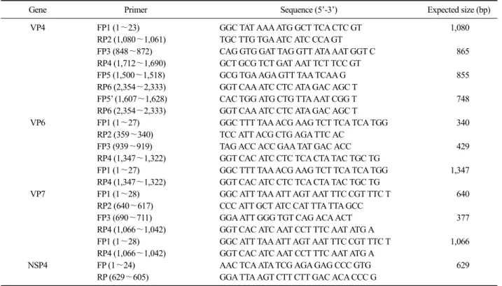
Table 1. Oligonucleotide primers used in this study
Gene Primer Sequence (5’-3’) Expected size (bp)
VP4 FP1 (1∼23) GGC TAT AAA ATG GCT TCA CTC GT 1,080
RP2 (1,080∼1,061) TGC TTG TGA ATC ATC CCA GT
FP3 (848∼872) CAG GTG GAT TAG GTT ATA AAT GGT C 865
RP4 (1,712∼1,690) GCT GCG TCT GAT AAT TCT TCC GT
FP5 (1,500∼1,518) GCG TGA AGA GTT TAA TCAA G 855
RP6 (2,354∼2,333) GGT CAA ATC CTC ATA GAC AGC T
FP5’ (1,607∼1,628) CAC TGG ATG CTG TTA AAT CGG T 748
RP6 (2,354∼2,333) GGT CAA ATC CTC ATA GAC AGC T
VP6 FP1 (1∼27) GGC TTT TAA ACG AAG TCT TCA TCA TGG 340
RP2 (359∼340) TCC ATT ACG CTG AGA TTC AC
FP3 (939∼919) TAG ACC ACC GAA TAT GAC ACC 429
RP4 (1,347∼1,322) GGT CAC ATC CTC TCA CTA TAC TGC TG
FP1 (1∼27) GGC TTT TAA ACG AAG TCT TCA TCA TGG 1,347
RP4 (1,347∼1,322) GGT CAC ATC CTC TCA CTA TAC TGC TG
VP7 FP1 (1∼28) GGC ATT TAA ATT AGT AAT TTC CGT TTC T 640
RP2 (640∼617) CCC ATT GCT ATC CAT TTA TTA GCC
FP3 (690∼711) GGA ATT GGG TGT CAG ACA ACT 377
RP4 (1,066∼1,042) GGT CAC ATC AAT CCT TTC AAT ATG A
FP1 (1∼28) GGC ATT TAA ATT AGT AAT TTC CGT TTC T 1,066
RP4 (1,066∼1,042) GGT CAC ATC AAT CCT TTC AAT ATG A
NSP4 FP (1∼24) AAC TCA ATA TCG AGA GAG CCC GTG 629
RP (629∼605) GGA TTA AGT CTT CTT GAC ACA CCC G
monoclonal antibody (25A11) was applied and incubated at 37oC for 1 h. After washing three times with PBS, the FITC-conjugated affinipure goat anti-mouse IgG+
IgM (Jackson, USA) was applied and incubated at 37oC for 1 h. Finally, after washing three times with PBS, 80% glycerin was applied and the fluorescence was ob- served under a fluorescence microscope (Olympus, Japan).
Reverse transcription polymerase chain reaction (RT-PCR)
Viral RNA was extracted from avian rotavirus in- fected cell culture supernatant using the QIAamp viral RNA mini Kit (QIAGEN, USA). To determine the com- plete nucleotide sequence of VP4, VP7, VP6 and NSP4 gene, primers used for RT-PCR of each gene were syn- thesized based on previously reported data of NCBI GenBank by commercial company (Cosmo Genetech Company, Korea) (Table 1). cDNA was synthesized us- ing the Prime Script 1st strand cDNA Kit (TakaRa, Japan). PCR was performed using PCR Thermal Cycler Dice (TakaRa, Japan). The 50 μl reaction mixtures in-
cluded 10 μl of cDNA template, 6 μl of dNTP (2.5 mM each), 5 μl of 10× ExTaq buffer, 2 μl of forward and reverse primer (10 pmole/μl), 0.5 μl of Taq poly- merase (5 unit/μl) and 24.5 μl of RNase free distilled water. The PCR condition for amplification of VP4, VP7, VP6 and NSP4 gene was 95oC for 5min, 35 cy- cles of 95oC for 35s (denaturation), 52∼56oC for 35s (annealing) and 75oC for 1 min (extension), and a final incubation at 75oC for 10 min.
Nucleotide sequences and phylogenetic analysis
The PCR products were acquired with electrophoresis on 1.5% agarose gel and purified using Expin Gel SV Kit (GeneAll Biotechnology, Korea). The nucleotide se- quences of VP4, VP7, VP6 and NSP4 gene were com- pared with those of reference rotavirus strains available in the NCBI GenBank database using MegAlign pro- gram (DNASTAR, USA) and the phylogenetic trees were constructed based on Clustal W method (Thom- pson et al, 1994).
Fig. 1. Electron microscopic detection of avian rotavirus AvRV-2.
Fig. 2. Electropherotypic RNA migration patterns of genomic RNA. Lane A: Porcine rotavirus OSU strain, Lane B: Avian rotavirus AvRV-2 strain, Lane C: Avian rotavirus AEQ strain.
Nucleotide sequence accession numbers The nucleotide sequences of VP4, VP6, VP7 and NSP4 gene of avian rotavirus (AvRV-2) were submitted to GenBank under the accession numbers: JQ085405, JQ085406, JQ085407, and JQ085408, respectively.
RESULTS
Virus isolation and identification
The MA-104 cells inoculated with the supernatant of processed fecal samples showed CPE from the second passage. Purified virus was confirmed by EM, SDS-PAGE electropherotyping and IFA. Characteristic rotavirus morphology was shown by EM (Fig. 1). SDS-PAGE electropherotyping result exhibited a RNA migration pattern of 5:1:3:2, which was typical for avian group A rotaviruses (Fig. 2). To add weight to isolated virus that belonged to avian group A rotaviruses, IFA was per- formed using a group A rotavirus VP6-specific mono- clonal antibody. The IFA results confirmed that isolated rotavirus belonged to group A rotaviruses (Fig. 3).
Analysis of VP4 gene nucleotide sequences AvRV-2 VP4 gene was 2,359 bp nucleotides in length and coded for 771 amino acids. Amino acids of AvRV-2 strain were shorter (2 to 6 amino acids) than those of human and mammalian rotaviruses. Comparing the nucleotide sequence of VP4 gene with those of ref- erence rotavirus strains available in GeneBank database, nucleotide sequence homology of the AvRV-2 was 50.2% to 95.9%. The VP4 gene nucleotide sequences of AvRV-2 strain showed a low homology (50.2% to 63.9%) with those of human and mammalian rotaviru- ses, while it had nucleotide sequence identities of 76.5%
to 96.9% with those of avian rotavirus strains. Especial- ly, AvRV-2 strain had a high degree of nucleotide se- quence identities (95.8% to 95.9%) with avian rotavi- ruses isolated from chickens, which belonged to geno- type P[30] (Fig. 4).
Analysis of VP7 gene nucleotide sequences AvRV-2 VP7 gene was 1,067 bp nucleotides in length and coded for 330 amino acids. Amino acids of AvRV-2
Fig. 3. Cytopathic effects (A) and Immunofluorescences (B) of avi- an rotavirus AvRV-2 in MA104 cells (× 400).
Fig. 4. Phylogenetic analysis of VP4 gene nucleotide sequence of AvRV-2. Bo: bovine, Ch: chicken, Eq: equine, Fe: feline, Gu: guanaco, Hu: human, La: lapine, Mu: murine, Ov:
ovine, Pi: pigeon, Po: porcine, Rh-rhesus, Si: simian, Tu:
turkey. The number in parentheses indicates the P genotype and rotavirus strains in box were isolated from avian species.
The AvRV-2 strain was marked in the black arrow.
Fig. 5.Phylogenetic analysis of VP7 gene nucleotide sequence of AvRV-2. Bo: bovine, Ch: chicken, Eq: equine, Fe: feline, Gu: guanaco, Hu: human, La: lapine, Mu: murine, Pi: pi- geon, Po: porcine, Rh-rhesus, Sg: sugar glider, Si: simian, Tu: turkey. The number in parentheses indicates the G gen- otype and rotavirus strains in box were isolated from avian species. The AvRV-2 strain was marked in the black arrow.
strain were longer (3 amino acids) than those of human and mammalian rotaviruses. Phylogenetic tree showed that VP7 genotypes of avian rotaviruses included G7, G17, G18, G19, G22, and G23 (Fig. 5). Comparing the nucleotide sequence of VP7 gene with those of refer- ence avian rotavirus strains available in GeneBank data- base, nucleotide sequence homology of the AvRV-2 was 69.5% to 94.7%. Especially, AvRV-2 strain has high nucleotide sequence identities of 93.4% to 94.7% with
genotype G19 strains that had been isolated from chick- ens in Germany and Japan.
Analysis of VP6 gene nucleotide sequences AvRV-2 VP6 gene was 1,352 bp nucleotides in length and coded for 398 amino acids. The numbers of amino acids of AvRV-2 was the same as other avian, human and mammalian rotaviruses. Phylogenetic tree re- vealed that avian rotaviruses were divided into two I genotypes, I4 and I11 and AvRV-2 was closely related (95.3% to 96.4%) to I11genotype (Fig. 6). Avian rotavi- ruses isolated from turkey and chicken belonged to gen- otype I4 and I11 respectively, except Ch-2 strain. Ch-2 strain was isolated from chickens and was closely re- lated (92.6∼96.6%) to genotype I4, while it had low nucleotide sequence identities (78.0∼79.3%) with geno- type I11. The rotaviruses isolated from pigeon also be- longed to genotype I4.
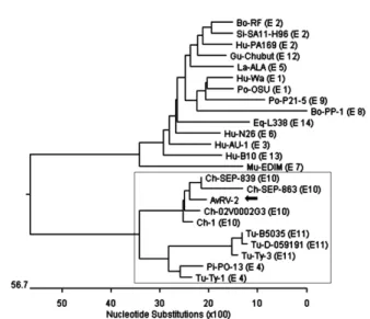
Fig. 7.Phylogenetic analysis of NSP4 gene nucleotide sequence of AvRV-2. Bo: bovine, Ch: chicken, Eq: equine, Gu: guanaco, Hu: human, La: lapine, Mu: murine, Pi: pigeon, Po: por- cine, Si: simian, Tu: turkey. The number in parentheses in- dicates the E genotype and rotavirus strains in box were iso- lated from avian species. The AvRV-2 strain was marked in the black arrow.
Fig. 6. Phylogenetic analysis of VP6 gene nucleotide sequence of AvRV-2. Bat: bats, Bo: bovine, Ch: chicken, Eq: equine, Fe:
feline, Gu: guanaco, Hu: human, Pi: pigeon, Po: porcine, Si:
simian, Tu: turkey. The number in parentheses indicates the I genotype and rotavirus strains in box were isolated from avi- an species. The AvRV-2 strain was marked in the black arrow.
Analysis of NSP4 gene nucleotide sequences AvRV-2 NSP4 gene was 662 bp nucleotides in length and coded for 169 amino acids. The length of amino acids of AvRV-2 was shorter than those of turkey (1 amino acid), human (7 amino acids) and mammalian (7 amino acids) rotavirus strains. Phylogenetic tree ex- hibited that avian rotaviruses were divided into three E genotypes, E4, E10 and E11 and NSP4 gene nucleotide sequence of AvRV-2 strain had high degree of nucleo- tide identities (90.3% to 92.2%) with those of avian ro- taviruses from chickens, E10 cluster (Fig. 7). AvRV-2 strain was 76.6∼78.0% identical to those of rotaviruses isolated from turkey which belonged to genotype E11.
DISCUSSION
Since rotavirus infection in avian species was first re- ported in 1977, it became prevalent among several spe- cies of domesticated birds (Hines et al, 1995; McNulty and Reynolds, 2008; Minamoto et al, 1988). Rotaviruses isolation from bovine, porcine and human had been re- ported in Korea (Park et al, 2006; Jeong et al, 2009; Oh et al, 1983). However, rotavirus infection from avian species was not yet reported. Runting-stunting syndrome in broiler chickens was continuous problems for chicken
producers throughout the world. The specific enteric agents for this syndrome were not known. But several viruses such as avian rotaviruses, astroviruses and re- oviruses were detected in chickens showing enteric symptoms. In this study, virus isolated from the feces of 8-days old broiler chickens suffering from acute gastro- enteritis was confirmed as avian rotavirus by EM, elec- tropherotyping and IFA. Isolated rotavirus AvRV-2, was seen wheel-like appearances and about 70∼75 nm in diameter under the electron microscopy. Wheel-like ap- pearances of virus particles in EM was the character- istics of rotaviruses. Based on the migration of 11 RNA segments, genomic RNA migration patterns of AvRV-2 were 5:1:3:2 and were similar with those of AEQ strain used as a reference avian group A rotavirus. From the electropherotyping results, we could assumed that AvRV-2 was a avian group A rotavirus. The genome segments of avian group A rotaviruses had a migration pattern of 5:1:3:2, while avian group D rotaviruses had a migra- tion pattern of 5:2:2:2 (Kang et al, 1986b). On the other hand, mammalian group A rotaviruses had a migration pattern of 4:2:3:2 (McNulty et al, 1981). However, the migration patterns of the genomic segments might be in- fluenced by antigenic shift, drift and rearrangements (Estes and Kapikian, 2007). The AvRV-2 was further
confirmed as a group A rotavirus using a group A rota- virus specific monoclonal antibody by IFA. Monoclonal antibody, 25A11 used in this study was known to be group A rotavirus VP6-specific (Kang et al, 1989).
After the avian rotavirus was confirmed, molecular characterization of VP4, VP7, VP6 and NSP4 genes were analyzed by comparing the nucleotide sequences with those of other rotavirus strains. The avian rotavirus genotypes of VP4 included P[17], P[30], P[31] and P[35]. AvRV-2 strain located in the same cluster (P[30]) with those of chicken strains and had no direct relation with those of mammalian and human rotavirus.
In this study, avian rotaviruses belonged to one of the 6 G genotypes (G7, G17, G18, G19, G22, G23). Intere- stingly, all avian rotaviruses including AvRV-2 isolated from chicken belonged to the same G19 genotype ex- cept Ch-2, which belonged to the G7 genotype. Other avian rotaviruses isolated from different avian species belonged to the different G genotypes except for turkey.
Rotaviruses isolated from turkey belonged to the G7, G17 and G22 genotypes. It suggested that cross-infec- tion with avian rotaviruses between chicken and turkey had been reported and the reassortment between these avian rotaviruses might be occurred (Yason and Schat, 1986; Schumann et al, 2009). At the same time, AvRV-2 and other chicken rotavirus strains belonged to the geno- type I11 except for Ch-2 strain, which was closely re- lated with turkey strains belonging to the genotype I4.
On the other hand, chicken avian rotavirus strains in- cluding AvRV-2 located in the same E cluster (E10), while turkey strains located in the cluster E11. Based on above results, the genotypes of AvRV-2 strain were re- lated with those of chicken strains and it suggested that the reassortment between AvRV-2 strain and other spe- cies did not yet occurred. Reassortment between mam- malian and avian rotaviruses were reported (Wani et al, 2003). Reassortment among bovine, porcine and human rotavirus strains was also reported in Korea (Park et al, 2011). However, in Korea, it was not known whether the potential reassortments between avian and mamma- lian rotavirus strains occurred or not. Continued surveil- lance studies for avian rotaviruses will be necessary to verify it.
The phylogenetic trees based on VP4, VP7, VP6 and
NSP4 showed that AvRV-2 was clustered into chicken rotaviruses isolated previously in Germany and Japan, which belonged to the genotype G19P[30]. Further stud- ies were needed to confirm whether AvRV-2 came from foreign strains or not. Prevalences of avian rotavirus in- fection in chicken farms in Korea should be elucidated.
Worldwide collaboration including sharing informations of avian rotaviruses is crucial for effective monitoring of any significant genetic changes that could lead to production of high pathogenic avian rotaviruses.
ACKNOWLEDGMENTS
This work was supported by the research grant of the Chungbuk National University in 2011.
REFERENCES
Ball JM, Tian P, Zeng CQ, Morris AP, Estes MK. 1996. Age-de- pendent diarrhea induced by a rotaviral nonstructural glycoprotein. Sci 272: 101-104.
Bellinzoni R, Mattion N, Vallejos L, La Torre JL, Scodeller EA.
1987. Atypical rotavirus in chickens in Argentina. Res Vet Sci 43: 130-131.
Bergeland ME, McAdaragh JP, Stotz I. 1977. Rotaviral enteritis in turkey poults. Proc 26th West Poult Dis Conf: 129- 130.
Elschner M, Hotzel H, Reetz J, Diller R, Otto P. 2005. Isolation, identification and characterization of group A rotavirus from a chicken: the inner capsid protein sequence shows only a distant phylogenetic relationship to most other avian group A rotaviruses. J Vet Med Infect Dis Vet Publish Health 52: 211-213.
Estes MK, Kapikian AZ. 2007. Rotaviruses. pp. 1917-1957. In:
Knipe DM, Howley PM, Griffin DE, Lamb RA, Martin MA , Roizman B, Straus SE(ed.). Fields Virology. 5th ed. Lippincott Williams and Wilkins, Pennsylvania.
Gorziglia M, Larralde G, Kapikian AZ, Chanock RM. 1990.
Antigenic relationships among human rotaviruses as de- termined by outer capsid protein VP4. Proc Natl Acad Sci 87: 7155-7159.
Gusmal RH, Mascarenhas JD, Gabbay YB, Linhares AC. 1994.
Nosocomial transmission of an avian-like rotavirus strain among children in Belém, Brazil. J Diarrhoeal Dis Res 12: 129-132.
Hines ME, Styer EL, Baldwin CA, Cole JR Jr. 1995. Combined adeovirus and rotavirus enteritis with Escherichia coli septicemia in an emu chick (Dromaius novaehollandiae).
Avian Dis 39: 646-651.
Ito H, Sugiyama M, Masubuchi K, Mori Y, Minamoto N. 2001.
Complete nucleotide sequence of a group A avian rotavi- rus genome and a comparison with its counterparts of mammalian rotaviruses. Virus Res 75: 123-138.
Jeong YJ, Park SI, Hosmillo M, Shin DJ, Chun YH, Kim HJ, Kwon HJ, Kang SY, Woo SK, Park SJ, Kim GY, Kang MI, Cho KO. 2009. Detection and molecular character- ization of porcine group C rotaviruses in South Korea.
Vet Microbiol 138: 217-224.
Kang SY, Nagaraja KV, Newman JA. 1986a. Primary isolation and identification of avian rotaviruses from turkeys ex- hibiting signs of clinical enteritis in a continuous MA104 cell line. Avian Dis 30: 494-499.
Kang SY, Nagaraja KV, Newman JA. 1986b. Electropherotypic analysis of rotaviruses isolated from turkeys. Avian Dis 30: 794-801.
Kang SY, Saif LJ, Miller KL. 1989. Reactivity of VP4-specific monoclonal antibodies to a serotype 4 porcine rotavirus with distinct serotypes of human(symptomatic and asym- ptomatic) and animal rotaviruses. J Clin Microbiol 27:
2744-2750.
Matthijnssens J, Ciarlet M, McDonald SM, Attoui H, Banyai K, Brister JR, Buesa J, Esona MD, Estes MK, Gentsch JR, Iturriza-Gomara M, Johne R, Kirkwood CD, Martella V, Mertens PP, Nakagomi O, Parreno V, Rahman M, Ru- ggeri FM, Saif LJ, Santos N, Steyer A, Taniguchi K, Patton JT, Desselberger U, Van Tanst M. 2011. Uni- formity of rotavirus strain nomenclature proposed by the Rotavirus Classification Working Group (RCWG). Arch Virol 156: 1397-1413.
McNulty MS, Allan GM, Stuart JC. 1978. Rotavirus infection in avian species. Vet Rec 103: 319-320.
McNulty MS, Allan GM, Todd D, McFerran JB, McCracken RM.
1981. Isolation from chickens of a rotavirus lacking the rotavirus group antigen. J Gen Virol 55: 405-413.
McNulty MS, Reynolds DL. 2008. Rotavirus Infections. pp.
338-350. In: Saif YM, Fadly AM, Glisson JR, Mc- Dougald LR, Nolan LK, Swayne DE(ed.). Diseases of Poultry. 12th ed. Blackwell publishing, Oxford.
Meulemans G, Peeters JE, Halen P. 1985. Experimental infection of broiler chickens with rotavirus. Br Vet J 141: 69-73.
Minamoto N, Oki K, Kinjo T, Suzuki Y. 1988. Isolation and characterization of rotavirus from feral pigeon in mam- malian cell cultures. Epidemiol Infect 100: 481-492.
Oh BH, Moon SJ, Lee KS. 1983. Clinical, laboratory and epi- demiological features of human rotavirus (HRV) gastro- enteritis. Kor J Pediatr Soc 26: 959-966.
Otto P, Schulze P, Herbst W. 1999. Demonstration of group C rotaviruses in fecal samples of diarrheic dogs in Ger- many. Arch Virol 144: 2467-2473.
Park SH, Saif LJ, Jeong C, Lim GK, Park SI, Kim HH, Park SJ, Kim YJ, Jeong JH, Kang MI, Cho KO. 2006. Molecular characterization of novel G5 bovine rotavirus strains. J Clin Microbiol 44: 4101-4112.
Park SI, Matthijnssens J, Saif LJ, Kim HJ, Park JG, Alfajaro MM, Kim DS, Son KY, Yang DK, Hyun BH, Kang MI, Cho KO. 2011. Reassortment among bovine, porcine and human rotavirus strains results in G8P[7] and G6P[7]
strains isolated from cattle in Korea. Vet Microbiol 152:
55-66.
Raming RF, Ciarlet M, Mertens PPC, Dermody TS. 2005. Rota- virus. pp. 484-496. In: Fauquet CM, Mayo MA, Mani- loff J, Desselberger U, Ball LA(ed.). Virus Taxonomy:
Eighth Report of the International Committee on Taxo- nomy of Viruses. Elsevier, California.
Schumann T, Hotzel H, Otto P, Johne R. 2009. Evidence of inter- species transmission and reassortment among avian group A rotaviruses. Virology 386: 334-343.
Takase K, Nonaka F, Sakaguchi M, Yamada S. 1986. Cytopathic avian rotavirus isolated from duck faeces in chicken kid- ney cell cultures. Avian Pathol 15: 719-730.
Thompson JD, Higgins DG, Gibson TJ. 1994. CLUSTAL W:
Improving the sensitivity of progressive multiple se- quence alignment through sequence weighing, position specific gap penalties and weight matrix choice. Nucleic Acids Res 22: 4673-4680.
Wani SA, Bhat MA, Ishag SM, Ashrafi MA, Buchh AS, Haq M.
2003. Detection of a mammalian-like group A rotavirus in diarrhoeic chicken. Vet Microbiol 94: 13-18.
Yason CV, Schat KA. 1985. Isolation and characterization of avi- an rotaviruses. Avian Dis 29: 499-508.
Yason CV, Schat KA. 1986. Pathogenesis of rotavirus infection in turkey poults. Avian Pathol 15: 421-435.