아시아에서 분리된 viral haemorrhagic septicaemia virus (VHSV) isolates의 계통분석학적 비교
안상중†⋅조미영⋅지보영⋅박명애3) 국립수산과학원 수산생물방역과
Phylogenetic Analysis of Viral Haemorrhagic Septicaemia Virus (VHSV) Isolates from Asia
Sang Jung Ahn†, Mi Young Cho, Bo-Young Jee and Myoung Ae Park
Aquatic Life Disease Control Division, National Fisheries Research & Development Institute (NFRDI), Busan 619-705, Korea
Viral haemorrhagic septicaemia virus (VHSV), the causative agent of viral haemorrhagic septicaemia (VHS), is an epidemic virus of cultured olive flounder Paralichthys olivaceus in Korea. In the present study, the entire glycoprotein (G) gene including several hypervariable regions from 36 isolates of diverse geographic and host origin and 8 Korean VHSV isolates from cultured olive flounder were analyzed. Phylogenetic analysis indicated that most of Asian VHSV belong to the genotype IVa group, suggesting that they originated from a common ancestral virus. Comparative sequence analysis of the complete G protein from Korean VHSV isolates revealed 3 Korean strain-specific nucleotide residues (nucleotide number of G-region: A755, T834 and T1221). These results suggest that Korean VHSV originated from a common ancestor, but these regional specific nucleotide sequences suggest that genetic differences of VHSV are more related to geographic areas than to host fish species.
Key words :Viral haemorrhagic septicaemia virus (VHSV), Glycoprotein, Olive flounder, Paralichthys olivaceus
무지개송어를 비롯한 담수 연어과 어류에 감염되 어 심각한 바이러스 질병을 야기하는 병원체로 알려 진 viral haemorrhagic septicaemia virus (VHSV)는 다 른 어류바이러스인 infectious hematopoietic necrosis virus (IHNV)와 hirame rhabdovirus (HIRRV)와 함께 family Rhabdoviridae, genus Novirhabdovirus에 속하 는 약 11,000 bp의 negative-strand RNA virus로 6개의 유전자들, nucleocapsid (N), phosphoprotein (P), matrix protein (M), glycoprotein (G), non-virion protein (NV)
†Corresponding author: Sang Jung Ahn
Tel: +82-51-720-3047, Fax: +82-51-720-3039 E-mail: [email protected]
와 polymerase (L) 유전자들이 3’-N-P-M-G-NV-L-5’
의 순으로 구성되어 있다 (Schütze et al., 1999; van Regenmortel et al., 2000; Trdo et al., 2005).
VHSV는 1963년 덴마크에서 무지개송어로부터 처음 분리된 이후 (Jensen, 1965; Wolf, 1988; Smail, 1999), 1979년 유럽의 Atlantic Ocean 및 North Sea에서 Atlantic cod Gadus morhua로부터 분리되고 (Jensen
et al., 1979), turbot Scophthalmus maximus (Schlotfeldt
et al., 1991), Atlantic herring Clupea harengus (Dixon
et al., 1997), rockling Rhinonemus cimbrius 및 whiting
Merlangius melangus (Mortensen et al., 1999) 등에서
분리되어 담수 및 해수어류 모두에 질병을 유발하는병원체라는 것이 밝혀졌다. VHSV는 유럽지역 뿐만 아니라 1980년대 후반에는 북미지역의 서부해안에 서 해수 어류와 소하성 연어에서 분리되어 해양환경 에 바이러스가 넓게 분포되어 있음을 확인하였다 (Brunson et al., 1989; Hopper, 1989; Winton et al., 1989; Meyers et al., 1992; Meyers et al., 1994; Meyers and Winton, 1995; Meyers et al., 1999).
동아시아지역에서는 일본 와카사만의 야생 넙치 에서 처음으로 검출된 후 (Takano et al., 2000), 양식넙 치에서 VHSV에 의한 피해가 보고되었다 (Isshiki et
al., 2001). 국내에서는 유사한 시기에 양식 넙치에서
rhabdovirus성 질병이 유행하여, 이 원인체를 조사한 결과 북미 지역 및 일본에서 분리한 VHSV와 동일한 유전형인 것으로 확인되었다 (Kim et al., 2003). 이후,다양한 해산 어류 및 담수어류에서 분리되었으며,
양식넙치에서 해마다 지속인인 폐사를 일으키는 병 원체로 보고되고 있다 (Kim et al., 2004; Kim et al., 2009; Cho et al., 2012).
현재, VHSV는 세계동물보건기구 (World Organization for Animal Health, OIE)의 수생동물위생규약 (Aquatic Animal Health Code)에 의해 관리대상질병 (notifiable disease)으로 지정되어 있으며 (OIE, 2013), 국내에서 도 법정전염병으로 분류되어, 감염 및 질병발생이 확인되면 이동제한과 소독 조치등의 방역조치가 이 루어지는 질병이다. 최근, 다양한 수산생물질병의 발 생과 소비자들의 좋은 먹거리에 대한 수요의 증가로 인해 전 세계적으로 수산생물의 방역 (Aquatic animal Disease Control), 예찰 (Surveillance) 및 모니터링 (Monitoring)의 중요성이 더욱 커졌으며, 이러한 국가 적인 예찰 및 방역활동 뿐만 아니라 지역적 (regional and zonal)이고 구역화 (compartmentalisation)된 수산 생물의 질병 예찰 및 모니터링 연구결과의 분석은 향후 수산생물질병의 역학조사 및 질병 관리에 필요 한 유용한 자료를 제공할 뿐만 아니라 자국 내 특정 질병에 대한 무병(청정)지역을 증명할 수 있는 매우
중요한 역할을 한다 (FAO, 2004). 따라서 VHSV의 국내 분리주의 서열변이의 확인 및 다른 국가 분리주 와의 서열비교와 지속적인 수산질병 모니터링이 더 욱 중요하게 되었다.
전 세계적으로 동정된 VHSV의 N 과 G 유전자의 염기서열을 비교해본 결과, 계통분석학적으로 4 개 의 genotypes (I-IV)와 함께 genotypes I (Ia-Ie) 과 IV (IVa와 IVb)에 몇 개의 subgroup을 포함하고 있다 (Snow et al., 1999; Einer-Jensen et al., 2004; Lumsden
et al., 2007). Genotype I은 유럽의 다양한 담수 및
해수어류종들로부터 분리된 VHSV를 포함하며, Baltic Sea, Skagerrak, Kattegat 와 English Channel의 해수어류들로부터 분리된 많은 VHSV분리주들도 포 함된다 (Dixon et al., 1997; Thiéry et al., 2002;Einer-Jensen et al., 2004; Snow et al., 2004). Genotype II는 Baltic Sea의 해수어류로부터 분리된 분리주들이 포함되며 (Snow et al., 2004), genotype III는 영국과 아일랜드의 North Sea와 North Atlantic으로부터 분리된 분리주들과 노르웨이 서부의 rainbow trout로부터 분리 된 VHSV가 포함된다 (Einer-Jensen et al., 2004; Snow
et al., 2004; López-Vázquez et al., 2006; Dale et al.,
2009). Genotype IV는 북미의 Pacific coast와 Atlantic coast 뿐만 아니라 북미의 Great Lakes 지역과 아시아 지역으로부터 분리된 분리주들을 포함하고 있다 (Nishizawa et al., 2002; Kim et al., 2003; Elsayed et al., 2006; Gagné et al., 2007; Lumsden et al., 2007). 따라서, VHSV의 유전적 거리 및 계통분석 결과를 통해 숙주 어류종보다 지리적위치에 더 관계된다고 보여진다.본 연구에서는 우리나라 양식 넙치에서 분리한 VHSV 분리주들의 glycoprotein (G)의 염기서열을 분 석하여 Korean VHSV 분리주들과 기보고된 일본 및 중국의 분리주들을 계통발생학적으로 비교하여 국 내 특이적인 염기서열을 확인하여, 구역화 (zoining) 된 수산생물 질병의 예찰, 모니터링 및 질병 관리에 필요한 유용한 자료를 제공하려고 한다.
재료 및 방법
Virus 및 Cell line
본 연구에 사용한 VHSV 분리주들의 분리유래는 선행연구의 결과, 2010년 지리적으로 먼 거리의 완도 와 제주도에서 VHSV의 특징적인 증상인 안구돌출 및 출혈, 체색흑화, 복수에 의한 복부팽만 등의 질병 증상을 보였으며, 제주도에서는 약 80%, 완도에서는
약 15%의 양식 넙치의 치어를 폐사시킨
VHSV-IVa-KOR-YGH-Wando 분리주와 VHSV-IVa- KOR-CJA-Jeju 분리주를 기초로 하였다 (Cho et al., 2012). 또한, VHSV-IVa-KOR-GeoJe-201001, VHSV- IVa-KOR-Jeju-201001, VHSV-IVa-KOR-Jeju-200501 3종의 국내 VHSV 분리주에 대한 염기서열은 국립수
산과학원 병리연구과로부터 제공받았다. 바이러스
의 배양은 병어의 조직 마쇄 여과액을 Epithelial papilloma of carp (EPC) cell (Fijan et al., 1983)에 접종 한 후 17 °C에서 14일간 배양하여 세포변성효과 (cytopathic effect, CPE)를 확인하였다. 세포배양액은 10% Fetal Bovine Serum (FBS), 1% antibiotic- antimycotic (Gibco BRL)을 첨가한 Eagle’s minimum essential medium (EMEM)를 사용하였다.
RNA 분리
각각의 VHSV 분리주들을 Flask (25 cm2)에 배양한 EPC cell에 접종한 3일째, CPE가 나타나기 시작한 EPC cells을 수거하여 TRIzolⓇ Reagent (Ambion, USA)을 사용하여 cell에서 total RNA를 분리하였다.
건조한 RNA를 30 μl의 RNase free water (DEPC treated H2O)에 녹인 다음 cDNA 합성 후 RT-PCR에 사용하였다.
cDNA 합성 및 RT-PCR
Viral RNA의 역전사 및 RT-PCR을 이용한 바이러 스의 동정법은 세계동물보건기구 (Office of
International Epizootics, OIE)에 의해 추천되는 Manual of Diagnostic Tests for Aquatic Animals (OIE, 2013)의 VHSV의 분자생물학기법을 이용한 진단법을 기초로 하였 다. VHSV의 검출을 위한 conventional reverse-transcription polymerase chain reaction (RT-PCR)법은 cDNA 합성 후 RT-PCR을 수행하는 two step법으로 진행되었다.
cDNA의 합성 (total volume 20 μl)은 iScript cDNA Synthesis Kit (Bio-Rad, USA)를 이용하여 5 μl의 viral RNA, 4 μl의 5× iScript Reaction Mix, 1 μl의 iScript Reverse Transcriptase, 및 10 μl Nuclease-free water로 25°C에서 5분, 42°C에서 30분, 85°C에서 5분 반응하였다.
합성된 cDNA는 VHSV N-gene의 1-505 base를 포
함하는 부위를 증폭하는 VN Forward
(5’-ATGGAAGGAGGAATTCGTGAAGCG-3’)와 VN reverse (5’-GCGGTGAAGTGCTGCAGTTCCC-3’) primer set를 이용하여 RT-PCR을 수행하였다 (Snow
et al., 2004). 5 μl의 cDNA, 11.125 μl의 UltraPure
TM Distilled Water (Invitrogen, USA), 5 μl 5× Go TaqⓇ Flexi buffer, 2.5 μl 25 mM MgCl2, 0.5 μl VN Forward (10 μM), 0.5 μl VN Reverse (10 μM), 0.25 μl 25 mM (of each) dNTP, 0.125 μl Go TaqⓇ Flexi DNA polymerase (Promega, USA)의 PCR반응 mixture를 94°C에서 5분 동안 initial denaturation을 거쳐 94°C에 서 30초의 denaturation, 55°C에서 30초의 annealing, 68°C에서 60초간 extention 과정을 36회 반복하고 마 지막으로 68°C에서 7분간 final extension 과정을 DNA Engine TetradⓇ 2 Peltier Thermal cycler (Bio-Rad, USA)를 이용해 수행하였다. PCR 산물은 1.5%agarose gel electrophoresis와 QIAxcel Advanced capillary gel electrophoresis system (QIAGEN, Germany)을 이용해 분석하였고, QIAquick gel extraction kit (Qiagen, USA)를 이용해 정제하여 ABI 3500xL Genetic Analyzer (Applied Biosystems, USA) 를 이용해 염기서열을 분석하였다.
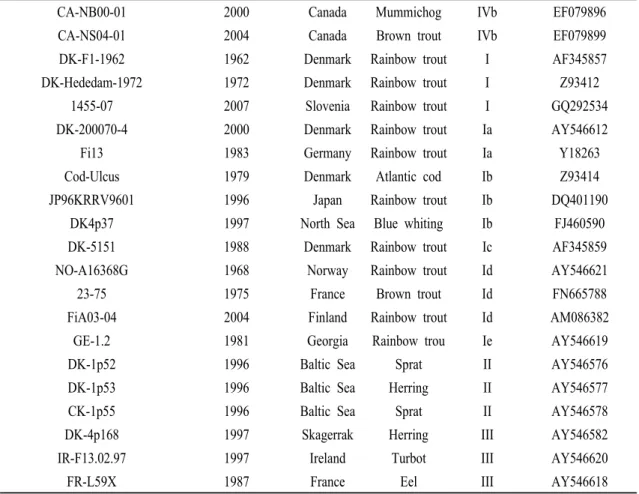
Virus isolate Year of isolation Origin Host species Genotype Accession number
VHSV-IVa-KOR-YGH-Wando 2010 Korea Olive flounder IVa JQ651388VHSV-IVa-KOR-CJA-Jeju 2010 Korea Olive flounder IVa JQ651393 VHSV-IVa-KOR-FYeosu05 2005 Korea Olive flounder IVa KF477302 VHSV-IVa-KOR-KJ2008 2008 Korea Olive flounder IVa JF792424 VHSV-IVa-KOR-GeoJe-201001 2010 Korea Olive flounder IVa unpublished
VHSV-IVa-KOR-Jeju-201001 2010 Korea Olive flounder IVa unpublished VHSV-IVa-KOR-Jeju-200501 2005 Korea Olive flounder IVa unpublished VHSV_IVa_KOR-KR2002 2002 Korea Olive flounder IVa AY167587 VHSV-IVa-CHN-PORV 2005 China Olive flounder IVa KC685626 VHSV-IVa-JPN-JF00Ehi1 2000 Japan Olive flounder IVa AB490792 VHSV-IVa-JPN-JP99Obama25 1999 Japan Olive flounder IVa DQ401191 VHSV-strain KRRV9822 1998 Japan Olive flounder IVa AB179621
Makah 1988 USA Coho salmon IVa U28747
BC93-372 1993 Canada Pacific herring IVa DQ401186
U13653 2005 Canada Drum IVb HQ453209
Table 1. GenBank accession numbers for the G sequences of VHSV isolates used to generate Fig. 1 Glycoprotein (G)-유전자의 PCR
VHSV 분리주들의 지역특이적인 서열의 확인을 위해 서열변이가 심한 G-유전자 (Benmansour et al., 1997)의 full length open reading frames (ORFs)의 서열을 VHSV- G-ORF-F1 (5‘-ATGGAATGGAATACTTTTTTCTTGGTG-3’)와 VHSV-G-ORF-R1 (5’-TCAGACCGTCTGACTTCTGGAGAACTGC-3‘) primer set를 디자인하여 PCR을 수행하였으며, 1524bp 의 PCR 산물을 얻을 수 있었다. 증폭된 PCR산물은 pGEMⓇ-T Easy vector (Promega, USA)를 이용해 클로 닝하여, ExprepTM Plasmid SV DNA prep kit (GeneAll Biotechnology Co., Ltd., Korea)를 이용해 정제된 plasmid DNA의 염기서열을 확인하였다.
염기서열 비교 및 계통분석
Nucleotide 서열과 추정 아미노산 서열은 BioEdit Sequence Alignment Editor (Hall, 1999)의 CLUSTAL W alignment model (Thompson et al., 1994)통해 양방
향 염기서열 데이터를 교차분석 후 단임 염기서열 (contig)을 확보하여, National Center for Biotechnology Information (http://www.ncbi.nlm.nih.gov/)의 BLAST (Basic Local Alignment Search Tool) 검색을 통해 유사 도 분석을 수행하였다. VHSV 분리주들의 G-유전자 염기서열을 이용한 분자계통분석은 Clustal X software (Thompson et al., 1997)를 이용하여 정렬하였고 분석 방법으로 근린결합분석 (NJ, neighbor-joining analysis, 1000 rounds of bootstrap)을 수행하였고 MEGA4 program (Tamura et al., 2007)을 이용해 염기서열간의 유전적 거리와 계통도 (phylogenetic tree)를 얻었다.
본 연구에서 비교한 8개의 Korean VHSV 분리주와 다른 나라의 28개의 VHSV 분리주들에 대한 정보는 Table 1에 나타내었다. G-region의 특이서열의 sequence logo는 Weblogo 3를 이용해 분석하였다 (Crooks et
al., 2004).
CA-NB00-01 2000 Canada Mummichog IVb EF079896
CA-NS04-01 2004 Canada Brown trout IVb EF079899
DK-F1-1962 1962 Denmark Rainbow trout I AF345857
DK-Hededam-1972 1972 Denmark Rainbow trout I Z93412
1455-07 2007 Slovenia Rainbow trout I GQ292534
DK-200070-4 2000 Denmark Rainbow trout Ia AY546612
Fi13 1983 Germany Rainbow trout Ia Y18263
Cod-Ulcus 1979 Denmark Atlantic cod Ib Z93414
JP96KRRV9601 1996 Japan Rainbow trout Ib DQ401190
DK4p37 1997 North Sea Blue whiting Ib FJ460590
DK-5151 1988 Denmark Rainbow trout Ic AF345859
NO-A16368G 1968 Norway Rainbow trout Id AY546621
23-75 1975 France Brown trout Id FN665788
FiA03-04 2004 Finland Rainbow trout Id AM086382
GE-1.2 1981 Georgia Rainbow trou Ie AY546619
DK-1p52 1996 Baltic Sea Sprat II AY546576
DK-1p53 1996 Baltic Sea Herring II AY546577
CK-1p55 1996 Baltic Sea Sprat II AY546578
DK-4p168 1997 Skagerrak Herring III AY546582
IR-F13.02.97 1997 Ireland Turbot III AY546620
FR-L59X 1987 France Eel III AY546618
결과 및 고찰
본 연구에서는 총 8개의 국내 VHSV의 분리주들인 VHSV-IVa-KOR-YGH-Wando, VHSV-IVa-KOR-CJA-Jeju (Cho et al., 2012), VHSV-IVa-KOR-FYeosu05 (Kim
et al., 2013), VHSV-IVa-KOR-GeoJe-201001,
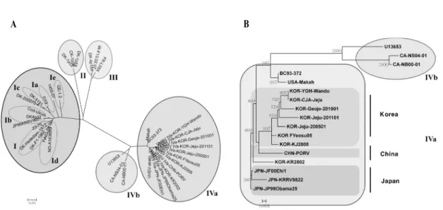
VHSV-IVa-KOR-Jeju-201001, VHSV-IVa-KOR-Jeju- 200501과 HSV-IVa-KOR-KR2002 (Kim et al., 2011) 분리주들과 GenBank로부터 얻은 28개의 외국 VHSV 분리주들 (Table 1)의 G 유전자의 서열을 비교분석하 였다 (Fig. 1). 이전 보고서들과 동일하게 계통도상으 로 4개의 VHSV의 그룹이 4 genotypes (I∼IV)에 속하 는 것으로 나타났다 (Snow et al., 1999; Einer-Jensenet al., 2004; Lumsden et al., 2007). 모든 국내 VHSV
분리주들은 genotype IVa에 포함되었으며, VHSV genotype IV는 북미의 Pacific coast, Great Lakes와 아 시아지역의 분리주가 포함된 IVa와 북미의 Atlantic coast의 분리주들이 포함된 IVb로 소그룹화되었다 (Elsayed et al., 2006; Gagné et al., 2007) (Fig. 1A).
Fig. 1B의 계통도에 따르면, 2000년대 초반에 국내에 서 동정된 VHSV 분리주 (VHSV_IVa_ KOR-KR2002) 는 일본의 VHSV 분리주들과 더 가까운 것을 확인할 수 있다. 일본 분리주들의 경우 이전 연구를 통해 두가지 다른 기원의 VHSV를 확인한 바 있다 (Nishizawa et al., 2002). 하나는 일본연안의 토착형 바이러스 분리주들과 전통적인 유럽 genotype Ib 그룹 에 속하는 분리주 (JP96KRRV9601)가 함께 동정되었 다. 하지만 국내분리주들의 경우에는 현재까지 다른
A
B
Fig. 1. Phylogenetic analysis using the VHSV isolates. (A) VHSV have identified 4 main genotypes (I to IV), with several subgroups within Genotypes I (minimum Ia to Ie) and IV (IVa and IVb). (B) Phylogenetic position of the Korean VHSV isolates.
genotype이 동정되지 않고 genotype IVa만 한국에 유 입되었음을 나타내고 있다. 그리고 일본 및 중국에서 분리된 대부분 분리주들 역시 genotype IVa 계열에 포함되어, 아시아 지역의 VHSV의 경우 genotype IVa 계열의 공통적인 ancestor virus로부터 파생되었음을 예상할 수 있다. 또한, VHSV의 숙주 종 간의 서열변 이의 다양성보다는 지리적 위치와 관련된 유전적 다 양성을 보이고 있다. 이는 각 지역별 VHSV 분리주들 의 특이적 염기서열의 존재 가능성을 나타내어 주고 있다.
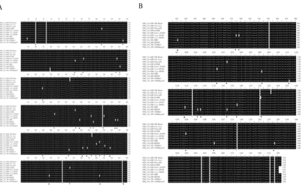
아시아에서 분리된 VHSV 분리주들의 nucleotide 서열들을 multiple sequence alignment를 통해 비교해 본 결과, 국내 VHSV 분리주들은 일본과 중국 분리주 들 (VHSV-IVa-JPN-JF00Ehi1, VHSV-IVa-JPN- JP99Obama25과 VHSV-IVa-CHN-PORV)과 98.0% 이 상의 nucleotide 서열의 상동성을 나타내었다. 아미노 산 레벨에서는 국내 분리주들과 일본과 중국의 VHSV 분리주들과는 4∼5개의 변이가 발견되었으 며, 이는 다른 보고서에서 발표된 것처럼 G-단백질의 single amino acid substitution에 의한 VHSV 분리주의 고병원성과 저병원성 차이의 가능성을 가지고 있다
(Betts and Stone, 2000; Campbell et al., 2009). VHSV의 G 단백질은 숙주의 interferon (IFN) response의 유도 에 관여한다고 알려져 있으며 (Boudinot et al., 2004;
Tafalla et al., 2007), rhabdovirus의 경우에는 G protein 이 감염초기에 attachment와 fusion (Matlin et al., 1982)
에 관여한다 보고되었고, 무지개송어의 저병원성
VHSV의 경우 숙주의 IFN response의 억제보단 초기 감염 및 복제와 더 관련이 있는 것으로 알려져 있어 (Campbell et al., 2011), 각국의 분리주들이 각기 다른 병원성 및 항원특이성을 가질 가능성이 있다. VHSV 는 바이러스 분류 국제위원회 (International Committee on Taxonomy of Viruses, ICTV)의 분류법 에 따라 에볼라 바이러스 (Ebola virus)와 인플루엔자 바이러스 (influenza virus)와 같은 group V로 분류된 다. 또한, single-stranded RNA genome으로 다른 RNA virus들과 마찬가지로 빠른 진화를 야기하는 error-prone replication mechanism을 가지기 때문에 서 열변이가 심해 유전적 다양성과 감염기작에 대한 연 구에 용이한 반면, 새로운 항원성을 가진 바이러스들 이 자주 출현한다 (Holmes, 2009; Snow, 2011). 따라 서, 지속적인 서열변이의 모니터링 및 국제적인 조사
A
B
Fig. 2. Multiple sequence alignment analysis of Asian VHSV isolates. Korean strain-specific nucleotide residues (*) of G region, Japanese strain-specific residue (○), Chinese strain-specific residues (▲), and regional specific nucleotide sequences in Asian VHSV isolates (●) were indicated.
와 정보의 공유가 필요하다.
지역별 VHSV 분리주들의 특이적 염기서열의 확 인을 위해 multiple sequence alignment를 통해 아시아 의 VHSV 분리주들의 nucleotide 서열들을 비교해본 결과, G-단백질의 8 부위 (G34, C528, A755, T834, T951, T1147, T1221, T1336)에서 국내 VHSV 분리주 들의 특이적인 서열을 확인하였다 (Fig. 2). 또한, 중국 유래 VHSV 분리주의 특이적 염기서열 11 부위 (C498, C545, C548, C642, G648, T678, A738, C804, C834, G851, C1139)와 일본 유래 VHSV 분리주의 특이적 염기서열 1 부위 (C179)를 확인할 수 있었다.
VHSV의 국내분리주간의 염기서열 유사도는 99.1~100%였으며, 국내 분리주와 중국 분리주는 98.1~98.7%, 일본 분리주와는 98.5~99.2%의 상동성 을 보였다. 이러한 바이러스의 서열변이의 다양성을 진화의 관점으로 보면, 일단 종이 형성되면 오랜 기간
동안 변하지 않다가 갑자기 크게 변한다는 단속평형 설 (또는 도약평형설, punctuated equilibrium theory)과 종의 형성은 일정하게 일어나며 작은 변화들이 축적 을 통해 오랜 세월을 거쳐 변하게 된다는 점진적진화 론 (progressive evolution, gradualism)으로 많은 연구 를 통해 설명하고 있다. 본 연구결과는 이전의 VHSV 의 연구에서 North American과 European VHSV 분리 주들이 동일한 지리적 위치와 각기 다른 VHSV가 분리된 시기를 통해 nucleotide mutation을 진화적 안 정기 (phase of genetic stasis; slow evolution)와 분화기 (phase of stepwise evolution; fast evolution)로 설명한 진화의 단속평형설로도 설명되지만 (Benmansour et
al., 1997), 다른 한편으로는 각각의 국내 분리주의
nucleotide 서열의 변이와 함께 지역적 변이 역시 확인 할 수 있어 점진적 진화의 관점에서도 설명이 가능하 다. 본 연구에서 확인된 아시아에서 분리된 VHSV분리주들의 특이적 염기서열의 변이양상은 향후 VHSV의 서열변이양상에 대한 연구와 모니터링 및 검역에 중요한 유전적 마커로 활용될 수 있을 것이다.
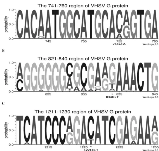
Genotype IVa의 Asian VHSV 분리주들을 포함한 전 세계적으로 동정된 VHSV의 genotype들 (I-IV)의 G-region의 국내 VHSV 분리주들의 특이적인 서열을 확인하기 위하여 총 40종의 분리주들의 염기서열을 분석하여 Asian VHSV의 분리주들과의 비교결과 국 내 VHSV 분리주의 특이적 부위였던 8 부위 (G34,
C528, A755, T834, T951, T1147, T1221, T1336) 중 3 부위의 국내VHSV 분리주 특이적 염기서열들 (A755, T834, T1221)을 확인하였고, 4종의 일본분리주 들만의 특이적 염기서열은 존재하지 않았다. 중국분리주 의 경우는 비교가능한 분리주의 서열이 1종 뿐이라 추가 적인 연구가 필요할 것이다. WebLogo 3 (http://weblogo.
threeplusone.com/) (Crooks et al., 2004)를 이용해 G-region 서열 logo를 이용한 positional probability consensus와 치 환된 염기서열을 각각 확인하였다 (Fig. 3).
A
B
C
Fig. 3. Korean strain-specific G-region sequence logos. Each nucleotide positional probability consensus was generated using WebLogo 3 (http://weblogo. threeplusone.com/). Korean strain-specific nucleotide sequences (*) of G region in 40 VHSV isolates were indicated with nucleotide substitutions.
본 연구에서 수행된 VHSV 분리주들의 계통분석 및 염기서열의 분석 결과 2005년 이후에 조사된 아시 아 국가의 VHSV의 분리주들은 각 지역의 특징적인 염기서열 부위를 포함하고 있으며, 이러한 VHSV의 유전적 특성분석에 대한 연구와 질병의 발생 및 감염 기작에 대한 이해와 연구결과는 국가적으로는 물론 이고 국제적인 연구 및 모니터링과 같은 수산생물질 병 관리에 유용한 자료한 자료가 될 것이다.
요 약
바이러스성 출혈성 패혈증을 일으키는 VHSV는 국내의 넙치양식에 심각한 피해를 주고 있다. 본 연구 에서는 우리나라에서 분리한 8종의 VHSV 분리주들 의 glycoprotein (G)의 염기서열을 분석하여 Korean VHSV 분리주들과 기보고된 일본 및 중국의 분리주 들을 계통발생학적으로 비교하여 G-protein의 8 부위 (G34, C528, A755, T834, T951, T1147, T1221, T1336) 에서 국내 VHSV 분리주들의 특이적인 서열을 확인 하였고, 전 세계적으로 분리된 VHSV의 분리주들과 의 비교결과 8 부위 중 3 부위 (A755, T834, T1221)의 국내VHSV 분리주 특이적 염기서열들을 확인하였 다. 또한 중국 유래 VHSV 분리주의 특이적 염기서열 11 부위 (C498, C545, C548, C642, G648, T678, A738, C804, C834, G851, C1139)와 일본 유래 VHSV 분리주 의 특이적 염기서열 1부위 (C179)를 확인하였다. 이 상의 연구결과는 국내 특이적인 염기서열을 확보하 여, 구역화된 수산생물 질병의 예찰, 모니터링 및 질 병 관리에 유용하게 사용될 것으로 사료된다.
감사의 글
본 연구는 국립수산과학원 (수산생물방역 체계구 축, RP-2013-AQ-209)의 지원에 의해 운영되었습니다.
참고문헌
Benmansour, A., Basurco, B., Monnier, A.F., Vende, P., Winton, J.R. and de Kinkelin, P.: Sequence variation of the glycoprotein gene identifies three distinct lineages within field isolates of viral haemorrhagic septicaemia virus, a fish rhabdovirus. J. Gen. Virol., 78: 2837-2846, 1997.
Betts, A.M., Stone, D.: Nucleotide sequence analysis of the entire coding regions of virulent and avirulent strains of viral haemorrhagic septicaemia virus. Virus Genes 20: 259-262, 2000.
Boudinot, P., Bernard, D., Boubekeur, S., Thoulouze, M., Bremont, M., Benmansour, A.: The glycoprotein of a fish rhabdovirus profiles the virus-specific T-cell repertoire in rainbow trout.
J. Gen. Virol., 85: 3099-3108, 2004.
Brunson, R., True, K. and Yancey, J.: VHS virus isolated at Makah National Fish Hatchery. Am. Fish Soc. Newsl., 17: 3-4, 1989.
Campbell, S., Collet, B., Einer-Jensen, K., Secombes, C.J., Snow, M.: Identifying potential virulence determinants in viral haemorrhagic septicaemia virus (VHSV) for rainbow trout. Dis. Aquat.
Org., 86: 205-212, 2009.
Campbell, S., McBeath, A., Secombes, C., Snow, M., Collect, B.: Interferon response following infection with genetically similar isolates of viral haemorrhagic septicaemia virus (VHSV) exhibiting contrasting virulence in rainbow trout. Fish Shellfish Immunol., 30: 287-294, 2011.
Cho, M.Y., Lee, U.H., Moon, C.H., Bang, J.D., Jee,
B.Y., Cha, S.J., Kim, J.W., Park, M.A., Do, J.W., Park, J.W.: Genetically similar VHSV isolates are differentially virulent in olive flounder Paralichthys olivaceus. Dis. Aquat.
Org., 101: 105-114, 2012.
Crooks, G.E., Hon, G., Chandonia, J.M., Brenner, S.E.:
WebLogo: A sequence logo generator. Genome Research, 14: 1188-1190, 2004.
Dale, O.B., Ørpetveit, I., Lyngstad, T.M., Kahns, S., Skall, H.F., Olesen, N.J., Dannevig, B.H.:
Outbreak of viral haemorrhagic septicaemia (VHS) in seawater-farmed rainbow trout in Norway caused by VHS virus Genotype III.
Dis. Aquat. Org., 85: 93-103, 2009.
Dixon, P.F., Feist, S., Kehoe, E., Parry, L., Stone, D.M., Way, K.: Isolation of viral haemorrhagic septicaemia virus from Atlantic herring Clupea harengus from the English Channel. Dis. Aquat.
Org., 30: 81-89, 1997.
Einer-Jensen, K., Ahrens, P., Forsberg, R. and Lorenzen, N.: Evolution of the fish rhabdovirus viral haemorrhagic septicaemia virus. J. Gen. Virol., 85: 1167-1179, 2004.
Elsayed, E., Faisal, M., Thomas, M., Whelan, G., Batts, W. and Winton, J.: Isolation of viral haemorrhagic septicaemia virus from muskellunge, Esox
masquinongy (Mitchill), in Lake St Clair,
Michigan, USA reveals a new sublineage of the North American genotype. J. Fish Dis., 29:611-619, 2006.
FAO (Food and Agriculture Organization): Surveillance and zoning for aquatic animal diseases. FAO Fisheries Technical Paper 451, Rome, 2004.
Fijan, N., Sulimanovic, D., Bearzotti, M., Muzinic, D., Zwillenberg, L.O., Chilmonczyk, S., Vautherot, J.F., de Kinkelin, P.: Some properties of the Epithelioma papulosum cyprini (EPC) cell line from carp Cyprinus carpio. Ann. Inst. Pasteur Virol., 134: 207-220, 1983.
Gagné, N., MacKinnon, A.M., Boston, L., Souter, B., Cook-Versloot, M., Griffiths, S. and Olivier, G.: Isolation of viral haemorrhagic septicaemia virus from mummichog, stickleback, striped bass and brown trout in eastern Canada. J. Fish Dis., 30: 213-223, 2007.
Hall, T.A.: BioEdit: a user-friendly biological sequence alignment editor and analysis program for Windows 95/98/NT. Nucleic Acids Symp., 41:
95-98, 1999.
Holmes, E.C.: The evolutionary genetics of emerging viruses. Annu. Rev. Ecol. Evol. Syst., 40:
353-372, 2009.
Hopper, K.: The isolation of VHSV from Chinook salmon at Glenwood Springs, Orcas Island, Washington.
Am. Fish Soc. Newsletter, 17: 1, 1989.
Isshiki, T., Nishizawa, T., Kobayashi, T., Nagano, T.
and Miyazaki, T.: An outbreak of VHSV (viral hemorrhagic septicemia virus) infection in farmed Japanese flounder Paralichthys olivaceus in Japan. Dis. Aquat. Org., 47: 87-99, 2001.
Jensen, N.J., Bloch, B., Larsen, J.L.: The ulcus-syndrome in cod (Gadus morhua) III. A preliminary virological report. Nord. Vet-Med., 31: 436-442, 1979.
Jensen, M.H.: Research on the virus of Egtved disease.
Ann. NY Acad. Sci., 126: 422-426, 1965.
Kim, S.M., Lee, J.I., Hong, M.J., Park, H.S., Park, S.I.:
Genetic relationship of the VHSV (viral hemorrhagic septicemia virus) isolated from cultured olive flounder, Paralichthys olivaceus in Korea. J. Fish Pathol., 16: 1-12, 2003.
Kim, S.M., Park, S.I.: Detection of viral hemorrhagic septicemia virus (VHSV) in wild marine fishes in the coastal region of Korea. J. Fish Pathol., 17: 1-10, 2004.
Kim, W.S., Kim, S.R., Kim, D., Kim, J.O., Park, M.A., Kitamura S., Kim, H., Kim, D., Han, H., Jung, S., Oh, M.J.: An outbreak of VHSV (viral hemorrhagic septicemia virus) infection in farmed olive flounder Paralichthys olivaceus in Korea. Aquaculture 296: 165-168, 2009.
Kim, W.S., Jung, S.J., Kim, J.O., Kim, D.W., Kim, J.H.
and Oh, M.J.: Genetic positioning of Korean viral hemorrhagic septicemia virus (VHSV) from cultured and wild marine fishes. J. Fish Pathol., 24: 1-9, 2011.
Kim, J.O., Kim, W.S., Nishizawa, T., Oh, M.J.: Complete genome sequence of viral hemorrhagic septicemia virus isolated from an olive flounder in South Korea. Genome Announc., 1:
e00681-13, 2013.
López-Vázquez, C., Raynard, R.S., Bain, N., Snow, M., Bandin, I., Dopazo, C.P.: Genotyping of marine viral haemorrhagic septicaemia virus isolated from the Flemish Cap by nucleotide sequence analysis and restriction fragment length polymorphism patterns. Dis. Aquat. Org. 73:
23-31, 2006.
Lumsden, J.S., Morrison, B., Yason, C., Russell, S.,
Young, K., Yazdanpanah, A., Huber, P., Al-Hussinee, L., Stone, D., Way, K.: Mortality event in freshwater drum Aplodinotus grunniens from Lake Ontario, Canada, associated with viral haemorrhagic septicemia virus, Type IV.
Dis. Aquat. Org., 76: 99-111, 2007.
Matlin, K.S., Reggio, S.H., Helenius, A., Simons, K.:
Pathway of vesicular stomatitis virus entry leading to infection. J. Mol. Biol., 156: 609-631, 1982.
Meyers, T.R. and Winton, J.R.: Viral hemorrhagic septicemia virus in North America. Annu. Rev.
Fish Dis., 5: 3-24, 1995.
Meyers, T.R., Sullivan, J., Emmenegger, E., Follet, J., Short, S., Batts, W.N., Winton, J.R.: Identification of viral hemorrhagic septicemia virus isolated from Pacific cod Gadus macrocephalus in Prince William Sound, Alaska, USA. Dis. Aquat. Org., 12: 167-175, 1992.
Meyers, T.R., Short, S., Lipson, K., Batts, W.N,. Winton, J.R., Wilcock, J., Brown, E.: Association of viral hemorrhagic septicemia virus with epizootic haemorrhagaes of the skin in Pacific herring Clupea harengus pallasi from Prince William Sound and Kodiak Island, Alaska, USA.
Dis. Aquat. Org., 19: 27-37, 1994.
Meyers, T.H., Short, S., Lipson, K.: Isolation of the North American strain of viral hemorrhagic septicemia virus (VHSV) associated with epizootic mortality in two new host species of Alaskan marine fish. Dis. Aquat. Org., 38:
81-86, 1999.
Mortensen, H.F., Heuer, O.E., Lorenzen, N., Otte, L.
and Olesen, N.J.: Isolation of viral haemorrhagic septicaemia virus (VHSV) from wild marine fishspecies in the Baltic Sea, Kattegat, Skagerrak and the North Sea. Virus Res., 63: 95-106, 1999.
Nishizawa, T., Iida, H., Takano, R., Isshiki, T., Nakajima, K. and Muroga, K.: Genetic relatedness among Japanese, American and European isolates of viral hemorrhagic septicemia virus (VHSV) based on partial G and P genes. Dis. Aquat.
Org., 48: 143-148, 2002.
OIE: Viral haemorrhagic septicaemia. In: Manual of diagnostic tests for aquatic animals. World Organisation for Animal Health, Paris. 2013.
Schlotfeldt, H.J., Ahne, W., Jørgensen, P.E.V. and Glende, W.: Occurrence of viral haemorrhagic septicaemia in turbot (Scophthalmus
maximus)-a natural outbreak. Bull. Eur. Assoc.
Fish Pathol., 11: 105-107, 1991.
Schütze, H., Mundt, E., Mettenleiter, T.C.: Complete genomic sequence of viral haemorrhagic septicemia virus, a fish rhabdovirus. Virus Genes 19: 59-65, 1999.
Smail, S.A.: Viral haemorrhagic septicaemia. In Woo, P.T.K. and et Bruno, D.W., editors. Fish diseases and disorders, Vol. 3: Viral, bacterial and fungal infections. CAB International, New York, USA.
pp.123-146, 1999.
Snow, M,, Cunningham, C.O., Melvin, W.T., Kurath, G.: Analysis of the nucleoprotein gene identifies distinct lineages of viral haemorrhagic septicaemia virus within the European marine environment. Virus Res., 63: 35-44, 1999.
Snow, M., Bain, N., Black, J., Taupin, V., Cunningham,
C.O,. King, J.A., Skall, H.F., Raynard, R.S.:
Genetic population structure of marine viral haemorrhagic septicaemia virus (VHSV). Dis.
Aquat. Org., 61: 11-21, 2004.
Snow, M.: The contribution of molecular epidemiology to the understanding and control of viral diseases of salmonid aquaculture. Vet. Res., 42: 56, 2011.
Tafalla, C., Chico, V., Pérez, L., Coll, J., Estepa, A.:
In vitro and in vivo differential expression of
rainbow trout (Oncorhynchus mykiss) Mx isoforms in response to viral hemorrhagic septicemia virus (VHSV) G gene, poly I: C and VHSV. Fish Shellfish Immunol., 23:210-221, 2007.
Takano, R., Nishizawa, T., Arimoto, M. and Muroga, K.: Isolation of viral haemorrhagic septicaemia virus (VHSV) from wild Japanese flounder,
Paralichthys olivaceus. Bull. Eur. Assoc. Fish
Pathol., 20: 186-192, 2000.Tamura, K., Dudley, J., Nei, M., Kumar, S.: MEGA4:
Molecular Evolutionary Genetics Analysis (MEGA) software version 4.0. Mol. Biol. Evol., 24: 1596-1599, 2007.
Thiéry, R., de Boisséson, C., Jeffroy, J., Castric, J., de Kinkelin, P., Benmansour, A.: Phylogenetic analysis of viral haemorrhagic septicaemia virus (VHSV) isolates from France (1971-1999). Dis.
Aquat. Org., 52: 29-37, 2002.
Thompson, J.D., Higgins, D.G., Gibson, T.J.: CLUSTAL W: improving the sensitivity of progressive multiple sequence alignment through sequence weighting, position-specific gap penalties and weight matrix choice. Nucleic Acids Res., 22:
4673-4680, 1994.
Thompson, J.D., Gibson, T.J., Plewniak, F., Jeanmougin, F., Higgins, D.G.: The ClustalX windows interface: flexible strategies for multiple sequence alignment aided by quality analysis tools. Nucleic Acids Res., 25: 4876-4882, 1997.
Trdo, N., Benmansour, A., Calisher, C., Dietzgen, R.G., Fang, R.X., Jackson, A.O., Kurath, G., Nadin-Davis, S., Tesh, R.B. and Walker, P.J.:
Family Rhabdoviridae. In Fauquet, C.M., Mayo, M.A., Maniloff, J., Desselberger, U. and Ball, L.A., editors. Virus taxonomy: Eighth report of the international committee on taxonomy of viruses. Elsevier/Academic Press, London, United Kingdom. pp. 623-644, 2005.
van Regenmortel, M.H.V., Fauquet, C.M., Bishop,
D.H.L., Carstens, E.B.O.: Virus taxonomy: the classification and nomenclature of viruses. The Seventh International Report of the International Committee on Taxonomy of Viruses. Academic Press, San Diego, CA, 2000.
Winton, J., Batts, W.N., Nishizawa, T.: Characterization of the first North American isolates of viral hemorrhagic septicaemia virus. Am. Fish Soc.
Fish Health Section Newsl., 17: 2-3, 1989.
Wolf, K.: Viral hemorrhagic septicemia. In Wolf, K., editor. Fish viruses and fish viral diseases.
Cornell University Press, Ithaca, New York, USA. pp. 217-249, 1988.
Manuscript Received : October 21, 2013 Revised : November 21, 2013 Accepted : December 11, 2013