NAD(P)H Quinone Oxidoreductase 1 (NQO1) as a Cancer Therapeutic Target
Eun Jung Park and Taeg Kyu Kwon*
Department of Immunology, School of Medicine, Keimyung University, Daegu 704-701, Korea
Received December 19, 2013 /Revised January 3, 2014 /Accepted January 8, 2014NAD(P)H quinone oxidoreductase 1 (NQO1) is a flavoprotein that catalyzes the two electron reduc- tion of diverse substrates, including quinones. It uses NADH or NADPH as a cofactor for enzymatic machinery. In the metabolism of quinones, NQO1 has two conflicting functions because of the differ- ent stability of converted hydroquinones. The stable form of hydroquinone is excreted from cells by conjugation with glutathione or glucuronic acid. The unstable form of hydroquinone induces cell death by induction of oxidative stress and DNA damage. Certain quinones known as bio-reductive agents have a cytotoxic function following reduction by NQO1. Bio-reductive agents, such as β-lapa- chone or mitomycin C, induce the depletion of NAD(P)H and the generation of oxidative stress in an NQO1-dependent manner. NQO1 is highly expressed in several cancer tissues. Therefore, NQO1 is a good therapeutic target for cancer treatment with bio-reductive agents.
Key words : Cancer, cell death, NAD(P)H Quinone Oxidoreductase 1 (NQO1), oxidative stress, β- lapachone
*Corresponding author
*Tel : +82-53-580-3882, Fax : +82-53-580-3795
*E-mail : [email protected]
This is an Open-Access article distributed under the terms of the Creative Commons Attribution Non-Commercial License (http://creativecommons.org/licenses/by-nc/3.0) which permits unrestricted non-commercial use, distribution, and reproduction in any medium, provided the original work is properly cited.
Journal of Life Science 2014 Vol. 24. No. 1. 98~103 DOI : http://dx.doi.org/10.5352/JLS.2014.24.1.98
서 론
NAD(P)H quinone oxidoreductase 1 (NQO1)은 2개의 전 자를 사용하여 다양한 기질들을 촉매하는 효소이다[36].
NQO1의 발현을 정상 조직과 그에 상응하는 조직이 암화된 경우로 나누어 비교했을 때 암 조직에서 NQO1의 발현 및 활 성이 높게 나타난다[3, 9]. NQO1이 암 세포 및 조직에서 높은 발현을 나타낸다는 특징 때문에 암 치료를 위한 유효한 표적 으로 여겨지며, β-lapachone, mitomycin C, EO9 등의 약물이 NQO1을 통해 작용하는 생체환원제 물질로 강력한 항암 효과 를 가진다고 알려져 있다[2, 11, 32, 45]. 본 보고에서는 NQO1 과 생체환원제 물질의 NQO1을 통한 항암 효과 및 그 작용 기전에 대하여 정리하였다.
본 론
NAD(P)H quinone oxidoreductase 1 (NQO1) NQO1의 발현 및 세포 내의 분포
Rat에서는 플라보 단백질에 속해 있는 NQO는 서로 다른 3가지의 형태로 존재하며, mouse에서도 2 종류의 NQO가 유 사한 기능을 가지고 있으며, 인간에서도 2 종류의 NQO가 존
재한다[19, 51, 53, 57, 64]. Rat의 간, 인간의 지방 조직 등에서 세포질에 존재하는 NQO1의 특징과 유전적 상동성을 조사한 보고에 의하면 NQO1은 공통적으로 274개 잔기의 단백질로 존재하며 84% 가량 유전적 유사성을 가진다[24, 25, 60, 66].
인간에서 NQO1과 NQO2를 인코딩하는 유전자와 그에 따른 단백질은 각각 54%, 49%의 유사성을 가진다고 알려져 있는데, 이는 두 단백질을 인코딩하는 유전자의 개시코돈(ATG)은 동 일하지만 NQO2의 경우 카르복시 말단 43개 아미노산이 적은 것으로 확인되었다[24]. NQO1은 세포 내에서 대부분이 세포 질에 존재하며 약 5-10% 정도가 미토콘드리아, 골지체 등의 막에 부착된 형태로 존재한다[15, 67]. 32 kDa의 분자가 이량체 를 구성하며 효소 활성을 나타내기 위해서는 보조인자로 NADH나 NADPH를 사용한다[1, 3].
NQO1의 기능
Ernster에 의해 NQO1이 밝혀졌을 때, NQO1은 미토콘드리
아의 전자 전달에 중요한 역할을 하는 것으로 여겨졌다[16,
55]. 그렇지만 NQO1이 미토콘드리아 호흡 체인의 구성요소
가 아님이 밝혀지면서 그 기능 또한 새롭게 모색되었고,
Martius에 의해 NQO1이 비타민 K1의 대사에 관여할 것이라
는 주장이 제기되었지만 이후 비타민 K1은 NQO1의 기질이
아닌 것으로 확인되었다[55]. 1980년대 초반, 각기 다른 2개의
그룹에 의해 나프토퀴논(naphthoquinone)인 menadione을 통
해 연구한 보고들에서 처음으로 quinone 계통 물질의 해독작
용에 NQO1이 관여한다는 것이 밝혀졌다[37, 62]. NQO1
knockout mouse에서 menadione의 독성이 증가됨을 통해
NQO1의 quinone 해독 작용이 증명되었으며[52], benzopyr-
ene과 7,12-dimethylbenzanthracene에 의한 피부 암화가 NQO1
결핍 mouse에서 보고 되었다[39, 40]. 또한 NQO1의 mena-
- Review -
Fig. 1. Double, protection and detoxification roles of NQO1.
Reduction of quinone by NQO1 forms two types of hydroquinone. Unstable hydroquinone induces alkyla- tion of intracellular molecules and generates reactive oxygen species. Stable hydroquinone is excreted by con- jugated with glutathione or glucuronic acid.
Fig. 2. Structure of β-lapachone. Chemical structure of β- lapachone.
dione으로 인한 세포 독성의 보호 기능은 햄스터 난소 세포주 인 CHO 및 인간 배아 신장 세포주 HEK293에서도 확인되었 다[12, 44]. 이러한 NQO1의 세포 보호 작용과는 반대로 qui- none의 대사 과정에서 불안정한 형태의 hydroquinone의 발 생을 통해 강한 세포 독성을 유발한다는 것이 밝혀진 바 있다 [11, 43, 49]. 이에 대해서는 NQO1을 통한 생체환원제의 항암 효과 단락에서 더 논의하고자 한다.
NQO1의 정상 조직과 암에서의 발현
NQO1는 모든 진핵 생물에 존재하는 단백질이며 포유류에 서 다양한 장기와 조직에서 발현된다[3, 16]. 인간의 경우 간에 서는 그 발현이 나타나지 않는 반면 위에서는 높은 발현을 나타낸다[9, 41, 59]. 서론에서 언급한 바와 같이 NQO1의 발현 은 암화된 조직에서 높게 나타나는데 이전의 보고에 따르면 인간의 정상 간 조직과 간암 조직 및 간암 주변부의 조직에서 의 NQO1의 발현을 비교한 결과, 암 조직과 그 주변부에서 NQO1의 발현이 20-50배 높게 나타났다[9]. 이러한 발현 양상 의 특징으로 인해 NQO1은 암 극복의 유효한 표적으로 여겨지 고 연구가 되고 있다.
NQO1을 통한 생체환원제의 항 암 작용 NQO1을 통한 생체환원제의 작용 기전
세포 내에서 NQO1과 경쟁적으로 작용하는 사이토크롬 p450 환원효소 혹은 사이토크롬 b5 환원효소는 NQO1과는 달리 quinone을 반응성이 높은 semiquinone으로 전환시킨다 [17, 26]. Semiquinone은 강한 활성 산소종(reactive oxygen species, ROS)의 생성을 야기함으로 세포에 산화적 손상을 야 기한다[21, 29]. 이와는 다르게 NQO1은 quinone의 물질대사 과정에서 반응성이 높은 semiquinone으로의 전환을 야기하 지 않고 hydroquinone으로 전환시키기 때문에 세포 보호능력 을 지니는 것으로 여겨졌다[28, 54]. 이는 NQO1에 의해 대사 된 안정된 형태의 친수성 hydroquinone이 세포 내에 존재하 는 글루타치온이나 글루쿠론산과 결합하여 세포 밖으로 배출 되기 때문에 세포 독성을 야기하지 않는 것으로 알려져 있다.
그러나 NQO1의 이러한 작용과는 반대로 자연적으로 유래된 혹은 합성된 quinone 물질들의 대사 과정에서 불안정한 형태 의 hydroquinone의 발생도 확인되었으며, β-lapachone, mito- mycin C, EO9 등의 quinone 구조를 가지는 물질들은 NQO1 에 의한 대사를 통해 강한 세포 독성을 유발한다는 것이 밝혀 져 있다[8, 43, 49]. 이러한 물질은 생체환원 물질로 명명되며 이들의 대사과정에서 발생되는 불안정한 hydroquinone은 세 포 생장에 필수적 요소인 DNA와 같은 물질의 알킬화를 야기 하거나 반응성이 높은 활성산소종의 발생을 야기하여 세포 독성을 초래함으로 암 세포에서는 강력한 항 암 능력을 나타 내는 것이 밝혀졌다[4, 58]. 이를 통해 NQO1의 기능은 해독작 용에만 국한된 것이 아니라 각각의 다른 기질에 대하여 해독 또는 독성을 야기하는 양면적인 성격을 가지고 있음이 확인되
었으며 이는 암 치료를 위한 표적으로 제시되었다(Fig. 1).
β -Lapachone
생체환원제 물질 중 하나인 β-lapachone (Fig. 2)은 남아메
리카에서 서식하는 lapacho 나무(학명: Tabeuia avellanedae)의
껍질에서 추출한 천연 물질인 lapachol의 유도체로 고대로부
터 다양한 질병을 치료하기 위한 민간요법으로 사용되어져
왔다[46, 56]. 여러 보고에 따르면 β-lapachone은 항진균, 항바
이러스, 항박테리아, 항염증, 항관절염 및 항암 효과가 입증된
바 있다[6, 23, 42, 46, 63]. 그중 β-lapachone은 암세포에서
apoptosis, necrosis 또는 autophagy 매개 세포 사멸을 야기함
으로 항 암 효과를 나타낸다는 것이 많은 다양한 암세포주,
전립선암[13, 30, 34], 유방암[5, 49, 50], 골수암[18, 35], 뇌암[47,
65], 대장암[7, 22], 폐암[4, 68], 간암[31, 69] 등의 다양한 암에
서 나타난다고 알려져 있다. β-Lapachone은 topoisomerase
I의 억제를 통해 DNA 손상을 복구하는 것을 막음으로 apop-
tosis를 야기한다고 알려졌으며[14, 69], H
2O
2생성으로 산화적
손상을 야기하여 세포 사멸을 야기한다는 것이 보고된 바 있
다[5]. 또한 β-lapachone에 의해 p53과 무관한 경로를 통해
세포 주기 정지를 야기함으로 apoptosis 또는 necrosis가 유도
Fig. 3. Cell death mechanism of β-lapachone. NQO-1-mediated reduction of β-lapachone generates hydroquinone form of β-lapachone. Hydroquinone spontaneously reduced to semiquinone and original quinone (futile cycle).
Severe depletion of NADH/NADPH and production of reactive oxygen species (oxidative stress) by semi- quinone as a result of futile cycle induces cell death. (Q:
quinone, HQ: hydroquinone, SQ: semiquinone)
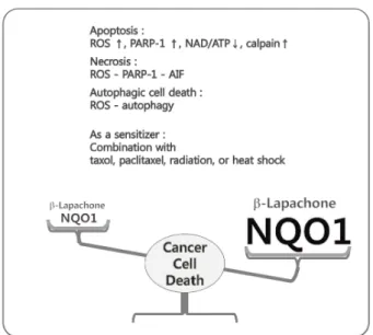
Fig. 4. β-Lapachone-induced cell death modes. Summary of β- lapachone-induced cell death modes.
됨이 보고된 바 있으며[22, 38], 본 연구팀에 의해서 뇌암 세포 주인 U87 MG 세포에서 β-lapachone에 의한 활성산소의 생성 이 autophagy를 통한 세포 사멸을 야기한다는 것이 보고된바 있다[47]. 또한 β-lapachone은 sensitizer로써 방사선 조사, taxol이나 pacritaxel과 같은 항암제 및 heat shock 등과 병행하 여 처리할 시 세포 사멸을 증대시키는 효과가 입증된 바 있다 [10, 33, 48, 61]. 2000년에는 Boothman과 그의 팀에 의해 β -lapachone에 의한 암 세포 사멸이 NQO1 의존적으로 일어난 다는 것이 알려졌다[49]. 이들은 β-lapachone이 quinone 구조 의 물질로 NQO1의 기질로써 작용할 것이라 여겼고, 유방암 세포주인 MCF-7, WS8, MDA-MB-468 세포에 dicoumarol을 통해 NQO1을 저해한 경우 β-lapachone에 의한 세포 사멸이 억제됨을 확인하였다. 또한 여러 유방암 세포주에서 NQO1의 발현량을 비교하였고 NQO1의 발현이 높은 세포주일수록 β -lapachone에 의한 세포 사멸이 증대됨을 확인하였다. 이 같은 현상은 β-lapachone이 NQO1, 사이토크롬 p450 환원 효소, 사이토크롬 b5 환원 효소에 의한 'futile cycle'이라고 불리는 과정을 통해 반복적으로 hydroquinone, semiquinone, qui- none으로의 전환이 이루어져 보조인자인 NAD(P)H의 결핍으 로 인한 세포 내 에너지 고갈과 불안정한 hydroquinone 및 semiquinone에 의한 산화적 스트레스를 야기함으로 암세포 에서 세포사멸을 야기하기 때문이다(Fig. 3). 최근의 보고에 따르면 유방암 세포주에서 β-lapachone에 의한 산소의 고갈, 활성산소의 생성이 야기되며 이는 catalase와 NQO1 저해제인 dicoumarol에 의해 조절되며 NQO1의 발현이 없는 세포주에 서는 미미하게 나타났다[5]. β-Lapachone에 의한 NQO1 의존 적인 산화적 스트레스는 DNA 손상 복구 효소인 PARP-1 (poly [ADP-ribose] polymerase 1)의 급격하고 강한 활성을 야기하며 그 하부 조절인자인 AIF (Apoptosis-inducing fac- tor)의 핵으로의 이동을 야기함으로 programmed necrosis를
야기하여 항암 작용을 가지는 것으로 확인되었다(Fig. 4). 뿐만 아니라 β-lapachone의 항암 효과는 전 임상 연구를 통해 항암 제로서의 가능성에 대한 연구가 이루어지고 있다[20, 27].
결 론
NQO1은 암에서 그 발현이 두드러지게 나타난다는 특징으 로 인해 암 치료의 표적으로 각광받게 되었다. 세포 보호의 기능과 세포 독성 야기의 기능을 모두 가지는 NQO1은 특정 quinone 물질의 대사 과정에서 생체환원효소로 작용하여 산 화적 스트레스 및 DNA 손상을 통해 세포 독성을 야기한다.
Quinone 계통의 물질 중 β-lapachone은 NQO1을 통해 작용 하는 생체환원제 물질로 정상 세포 및 조직 대비 암에서 상대 적으로 높은 발현을 나타내는 NQO1을 표적으로 여러 다양한 암세포, 동물 모델 및 전임상 연구에서 뛰어난 항암 효과를 나타내었으며, 기존의 항암 요법과의 병행 사용 또한 가능한 물질이다. NQO1과 β-lapachone을 비롯한 생체환원제 물질에 대한 기존의 연구는 암 세포 사멸 기전에 대한 연구가 대부분 을 차지한다. 따라서 기존의 연구를 통해 밝혀진 분자생물학 적 기전을 바탕으로 암의 형성, 전이, 혈관 신생 및 암 주변 미세환경 등에 대한 연구가 추가적으로 이루어진다면 NQO1 을 통한 현실적인 암 제어 및 극복이 이루어질 것으로 기대된 다.
감사의 글
본 연구는 한국연구재단의 중견연구자 지원사업의 지원을
받아 수행되었습니다(No. 2011-0016239).
References
1. Amzel, L. M., Bryant, S. H., Prochaska, H. J. and Talalay, P. 1986. Preliminary crystallographic X-ray data for an NAD(P)H:quinone reductase from mouse liver.
J Biol Chem
261, 1379.2. Begleiter, A., El-Gabalawy, N., Lange, L., Leith, M. K., Guziec, L. J. and Guziec, F. S., Jr. 2009. A model for NAD (P)H: quinoneoxidoreductase 1 (NQO1) targeted indivi- dualized cancer chemotherapy.
Drug Target Insights
4, 1-8.3. Belinsky, M. and Jaiswal, A. K. 1993. NAD(P)H: quinone oxidoreductase1 (DT-diaphorase) expression in normal and tumor tissues.
Cancer Metastasis Rev
12, 103-117.4. Bey, E. A., Bentle, M. S., Reinicke, K. E., Dong, Y., Yang, C. R., Girard, L., Minna, J. D., Bornmann, W. G., Gao, J.
and Boothman, D. A. 2007. An NQO1- and PARP-1-medi- ated cell death pathway induced in non-small-cell lung can- cer cells by beta-lapachone.
Proc Natl Acad Sci USA
104, 11832-11837.5. Bey, E. A., Reinicke, K. E., Srougi, M. C., Varnes, M., Anderson, V. E., Pink, J. J., Li, L. S., Patel, M., Cao, L., Moore, Z., Rommel, A., Boatman, M., Lewis, C., Euhus, D.
M., Bornmann, W. G., Buchsbaum, D. J., Spitz, D. R., Gao, J. and Boothman, D. A. 2013. Catalase abrogates beta-la- pachone-induced PARP1 hyperactivation-directed pro- grammed necrosis in NQO1-positive breast cancers.
Mol Cancer Ther
12, 2110-2120.6. Boveris, A., Docampo, R., Turrens, J. F. and Stoppani, A.
O. 1978. Effect of beta-lapachone on superoxide anion and hydrogen peroxide production in Trypanosoma cruzi.
Biochem J
175, 431-439.7. Choi, B. T., Cheong, J. and Choi, Y. H. 2003. beta- Lapachone-induced apoptosis is associated with activation of caspase-3 and inactivation of NF-kappaB in human colon cancer HCT-116 cells.
Anticancer Drugs
14, 845-850.8. Choudry, G. A., Stewart, P. A., Double, J. A., Krul, M. R., Naylor, B., Flannigan, G. M., Shah, T. K., Brown, J. E. and Phillips, R. M. 2001. A novel strategy for NQO1 (NAD (P)H:
quinone oxidoreductase, EC 1.6.99.2) mediated therapy of bladder cancer based on the pharmacological properties of EO9.
Br J Cancer
85, 1137-1146.9. Cresteil, T. and Jaiswal, A. K. 1991. High levels of expression of the NAD(P)H: quinone oxidoreductase (NQO1) gene in tumor cells compared to normal cells of the same origin.
Biochem Pharmacol.
42, 1021-1027.10. D'Anneo, A., Augello, G., Santulli, A., Giuliano, M., di Fiore, R., Messina, C., Tesoriere, G. and Vento, R. 2010. Paclitaxel and beta-lapachone synergistically induce apoptosis in hu- man retinoblastoma Y79 cells by downregulating the levels of phospho-Akt.
J Cell Physiol
222, 433-443.11. Danson, S., Ward, T. H., Butler, J. and Ranson, M. 2004.
DT-diaphorase: a target for new anticancer drugs.
Cancer Treat Rev
30, 437-449.12. De Haan, L. H., Boerboom, A. M., Rietjens, I. M., van Capelle, D., De Ruijter, A. J., Jaiswal, A. K. and Aarts, J.
M. 2002. A physiological threshold for protection against
menadione toxicity by human NAD(P)H:quinone oxidor- eductase (NQO1) in Chinese hamster ovary (CHO) cells.
Biochem Pharmacol
64, 1597-1603.13. Don, M. J., Chang, Y. H., Chen, K. K., Ho, L. K. and Chau, Y. P. 2001. Induction of CDK inhibitors (p21(WAF1) and p27(Kip1)) and Bak in the beta-lapachone-induced apoptosis of human prostate cancer cells.
Mol Pharmacol
59, 784-794.14. Dubin, M., Fernandez Villamil, S. H. and Stoppani, A. O.
2001. Cytotoxicity of beta-lapachone, an naphthoquinone with possible therapeutic use.
Medicina (B Aires)
61, 343-350.15. Ernster, L., Danielson, L. and Ljunggren, M. 1962. DT diaphorase. I. Purification from the soluble fraction of rat-liver cytoplasm, and properties.
Biochim Biophys Acta
58, 171-188.16. Ernster, L. and Navazio, F. 1958. Soluble diaphorase in ani- mal tissue.
Acta Chem Scand
12, 595-602.17. Gonzalez, F. J. 1988. The molecular biology of cytochrome P450s.
Pharmacol Rev
40, 243-288.18. Gupta, D., Podar, K., Tai, Y. T., Lin, B., Hideshima, T., Akiyama, M., LeBlanc, R., Catley, L., Mitsiades, N., Mitsiades, C., Chauhan, D., Munshi, N. C. and Anderson, K. C. 2002. Beta-lapachone, a novel plant product, over- comes drug resistance in human multiple myeloma cells.
Exp Hematol
30, 711-720.19. Höjeberg, B., Blomberg, K., Stenberg, S. and Lind, C. 1981.
Biospecific adsorption of hepatic DT-diaphorase on immobi- lized dicoumarol: I. Purification of cytosolic DT-diaphorase from control and 3-methylcholanthrene-treated rats.
Archives of biochemistry and biophysics
207, 205-216.20. Hartner, L., Rosen, L., Hensley, M., Mendelson, D., Staddon, A., Chow, W., Kovalyov, O., Ruka, W., Skladowski, K. and Jagiello-Gruszfeld, A. 2007. Phase 2 dose multi-center, open-label study of ARQ 501, a checkpoint activator, in adult patients with persistent, recurrent or metastatic leio- myosarcoma (LMS).
J Clin Oncol
25, 20521.21. Hodnick, W. F., Kalyanaraman, B., Pritsos, C. A. and Pardini, R. S. 1988. The production of hydroxyl and semi- quinone free radicals during the autoxidation of redox ac- tive flavonoids.
Basic Life Sci
49, 149-152.22. Huang, L. and Pardee, A. B. 1999. beta-lapachone induces cell cycle arrest and apoptosis in human colon cancer cells.
Mol Med
5, 711-720.23. Jackson, J., Higo, T., Hunter, W. and Burt, H. 2008. Topoiso- merase inhibitors as anti-arthritic agents.
Inflammation Res
57, 126-134.24. Jaiswal, A. K., Burnett, P., Adesnik, M. and McBride, O. W.
1990. Nucleotide and deduced amino acid sequence of a hu- man cDNA (NQO2) corresponding to a second member of the NAD(P)H: quinone oxidoreductase gene family.
Extensive polymorphism at the NQO2 gene locus on chro- mosome 6.
Biochemistry
29, 1899-1906.25. Jaiswal, A. K., McBride, O. W., Adesnik, M. and Nebert, D. W. 1988. Human dioxin-inducible cytosolic NAD(P) H:
menadione oxidoreductase. cDNA sequence and local- ization of gene to chromosome 16.
J Biol Chem
263, 13572- 13578.26. Joseph, P. and Jaiswal, A. K. 1994. NAD(P)H: quinone oxi- doreductase1 (DT diaphorase) specifically prevents the for- mation of benzo[a]pyrene quinone-DNA adducts generated by cytochrome P4501A1 and P450 reductase.
Proc Natl Acad Sci USA
91, 8413-8417.27. Kawecki, A., Adkins, D., Cunningham, C., Vokes, E., Yagovane, D., Dombal, G., Koralewski, P., Hotko, Y. and Vladimirov, V. 2007. A phase II study of ARQ 501 in pa- tients with advanced squamous cell carcinoma of the head and neck.
J Clin Oncol
25, 16509.28. Keyes, S. R., Fracasso, P. M., Heimbrook, D. C., Rockwell, S., Sligar, S. G. and Sartorelli, A. C. 1984. Role of NADPH:
cytochrome c reductase and DT-diaphorase in the bio- transformation of mitomycin C1.
Cancer Res
44, 5638-5643.29. Kikuchi, T., Sugiura, Y. and Komiyama, T. 1981. Generation of semiquinone radical from carbazilquinone by NADPH- cytochrome P-450 reductase.
Biochem Pharmacol
30, 1717- 1718.30. Kumi-Diaka, J. 2002. Chemosensitivity of human prostate cancer cells PC3 and LNCaP to genistein isoflavone and be- ta-lapachone.
Biol Cell
94, 37-44.31. Lai, C. C., Liu, T. J., Ho, L. K., Don, M. J. and Chau, Y.
P. 1998. beta-Lapachone induced cell death in human hep- atoma (HepA2) cells.
Histol Histopathol
13, 89-97.32. Lewis, A. M., Ough, M., Hinkhouse, M. M., Tsao, M. S., Oberley, L. W. and Cullen, J. J. 2005. Targeting NAD(P)H:
quinone oxidoreductase (NQO1) in pancreatic cancer.
Mol Carcinog
43, 215-224.33. Li, C. J., Li, Y. Z., Pinto, A. V. and Pardee, A. B. 1999. Potent inhibition of tumor survival in vivo by beta-lapachone plus taxol: combining drugs imposes different artificial check- points.
Proc Natl Acad Sci USA
96, 13369-13374.34. Li, C. J., Wang, C. and Pardee, A. B. 1995. Induction of apop- tosis by beta-lapachone in human prostate cancer cells.
Cancer Res
55, 3712-3715.35. Li, Y., Li, C. J., Yu, D. and Pardee, A. B. 2000. Potent in- duction of apoptosis by beta-lapachone in human multiple myeloma cell lines and patient cells.
Mol Med
6, 1008-1015.36. Lind, C., Cadenas, E., Hochstein, P. and Ernster, L. 1990.
DT-diaphorase: purification, properties, and function.
Methods Enzymol
186, 287-301.37. Lind, C., Hochstein, P. and Ernster, L. 1982. DT-diaphorase as a quinone reductase: a cellular control device against semiquinone and superoxide radical formation.
Arch Bio- chem Biophys
216, 178-185.38. Liu, T. J., Lin, S. Y. and Chau, Y. P. 2002. Inhibition of poly(ADP-ribose) polymerase activation attenuates beta-la- pachone-induced necrotic cell death in human osteosarcoma cells.
Toxicol Appl Pharmacol
182, 116-125.39. Long, D. J., 2nd, Waikel, R. L., Wang, X. J., Perlaky, L., Roop, D. R. and Jaiswal, A. K. 2000. NAD(P)H: quinone oxidor- eductase 1 deficiency increases susceptibility to benzo(a) pyrene-induced mouse skin carcinogenesis.
Cancer Res
60, 5913-5915.40. Long, D. J., 2nd, Waikel, R. L., Wang, X. J., Roop, D. R.
and Jaiswal, A. K. 2001. NAD(P)H: quinone oxidoreductase
1 deficiency and increased susceptibility to 7,12-dimethyl- benz[a]-anthracene-induced carcinogenesis in mouse skin.
J Natl Cancer Inst
93, 1166-1170.41. Martin, L. F., Patrick, S. D. and Wallin, R. 1987. DT-diaphor- ase in morbidly obese patients.
Cancer Lett
36, 341-347.42. Menacho-Marquez, M. and Murguia, J. R. 2006. Beta-la- pachone activates a Mre11p-Tel1p G1/S checkpoint in bud- ding yeast.
Cell Cycle
5, 2509-2516.43. Mikami, K., Naito, M., Tomida, A., Yamada, M., Sirakusa, T. and Tsuruo, T. 1996. DT-diaphorase as a critical determi- nant of sensitivity to mitomycin C in human colon and gas- tric carcinoma cell lines.
Cancer Res
56, 2823-2826.44. Nishiyama, T., Izawa, T., Usami, M., Ohnuma, T., Ogura, K. and Hiratsuka, A. 2010. Cooperation of NAD(P)H: qui- none oxidoreductase 1 and UDP-glucuronosyltransferases reduces menadione cytotoxicity in HEK293 cells.
Biochem Biophys Res Commun
394, 459-463.45. Ough, M., Lewis, A., Bey, E. A., Gao, J., Ritchie, J. M., Bornmann, W., Boothman, D. A., Oberley, L. W. and Cullen, J. J. 2005. Efficacy of beta-lapachone in pancreatic cancer treatment: exploiting the novel, therapeutic target NQO1.
Cancer Biol Ther
4, 95-102.46. Pardee, A. B., Li, Y. Z. and Li, C. J. 2002. Cancer therapy with beta-lapachone.
Curr Cancer Drug Targets
2, 227-242.47. Park, E. J., Choi, K. S. and Kwon, T. K. 2011. beta- Lapachone-induced reactive oxygen species (ROS) gen- eration mediates autophagic cell death in glioma U87 MG cells.
Chem Biol Interact
189, 37-44.48. Park, H. J., Choi, E. K., Choi, J., Ahn, K. J., Kim, E. J., Ji, I. M., Kook, Y. H., Ahn, S. D., Williams, B., Griffin, R., Boothman, D. A., Lee, C. K. and Song, C. W. 2005. Heat-in- duced up-regulation of NAD(P)H: quinone oxidoreductase potentiates anticancer effects of beta-lapachone.
Clin Cancer Res
11, 8866-8871.49. Pink, J. J., Planchon, S. M., Tagliarino, C., Varnes, M. E., Siegel, D. and Boothman, D. A. 2000. NAD(P)H: quinone oxidoreductase activity is the principal determinant of be- ta-lapachone cytotoxicity.
J Biol Chem
275, 5416-5424.50. Pink, J. J., Wuerzberger-Davis, S., Tagliarino, C., Planchon, S. M., Yang, X., Froelich, C. J. and Boothman, D. A. 2000.
Activation of a cysteine protease in MCF-7 and T47D breast cancer cells during beta-lapachone-mediated apoptosis.
Exp Cell Res
255, 144-155.51. Prochaska, H. J. and Talalay, P. 1986. Purification and char- acterization of two isofunctional forms of NAD(P)H: qui- none reductase from mouse liver.
J Biol Chem
261, 1372-1378.52. Radjendirane, V., Joseph, P., Lee, Y. H., Kimura, S., Klein-Szanto, A. J., Gonzalez, F. J. and Jaiswal, A. K. 1998.
Disruption of the DT diaphorase (NQO1) gene in mice leads to increased menadione toxicity.
J Biol Chem
273, 7382-7389.53. Raftell, M. and Blomberg, K. 1980. Immunochemical studies on two DT diaphorase active antigens isolated from rat liver cytosol by affinity chromatography.
Arch Biochem Biophys
199, 165-171.54. Ross, D., Kepa, J. K., Winski, S. L., Beall, H. D., Anwar, A. and Siegel, D. 2000. NAD(P)H: quinone oxidoreductase
초록:암 치료 표적으로의 NAD(P)H Quinone Oxidoreductase 1 (NQO1) 박은정 · 권택규*
(계명대학교 의과대학 면역학교실)
NQO1은 플라보 단백질 계통의 2 전자 환원 효소이며 NADH 또는 NADPH를 보조인자로 quinone 계통의 화 합물을 hydroquinone으로 환원 한다. 암에서 NQO1은 그에 상응하는 정상 조직과 비교하였을 때 비교적 높은 발현을 나타낸다. NQO1의 다양한 기능 중 quinone 물질 대사는 두 가지 형태의 상반되는 기능을 가진다. 이것은 quinone으로부터 전환된 hydroquinone의 상태적 안정성과 불안정성에 기인하며, 불안정한 hydroquinone의 생성 은 산화적 손상 야기 및 DNA 손상은 세포의 운명을 바꾸어 놓게 된다. 따라서 암에서 그 발현이 높은 NQO1을 표적으로 작용하는 생체환원 물질은 암 세포 사멸을 강하게 유도하게 되어 암 치료의 가능성을 보여주고 있다.
항암 표적 분자로서 NQO1 특징과 NQO1을 통해 작용하는 생체환원물질 β-lapachone의 항암 효과와 기전에 대 하여 살펴보았다.
1 (NQO1): chemoprotection, bioactivation, gene regulation and genetic polymorphisms.
Chem Biol Interact
129, 77-97.55. Ross, D. and Siegel, D. 2004. NAD(P)H: quinone oxidor- eductase 1 (NQO1, DT-diaphorase), functions and pharma- cogenetics.
Methods Enzymol
382, 115-144.56. Schaffner-Sabba, K., Schmidt-Ruppin, K. H., Wehrli, W., Schuerch, A. R. and Wasley, J. W. 1984. beta-Lapachone:
synthesis of derivatives and activities in tumor models.
J Med Chem
27, 990-994.57. Segura-Aguilar, J., Kaiser, R. and Lind, C. 1992. Separation and characterization of isoforms of DT-diaphorase from rat liver cytosol.
Biochim Biophys Acta
1120, 33-42.58. Seow, H. A., Penketh, P. G., Belcourt, M. F., Tomasz, M., Rockwell, S. and Sartorelli, A. C. 2004. Nuclear over- expression of NAD(P)H: quinone oxidoreductase 1 in Chinese hamster ovary cells increases the cytotoxicity of mi- tomycin C under aerobic and hypoxic conditions.
J Biol Chem
279, 31606-31612.59. Siegel, D. and Ross, D. 2000. Immunodetection of NAD(P) H:quinone oxidoreductase 1 (NQO1) in human tissues.
Free Radic Biol Med
29, 246-253.60. Smith, D., Martin, L. and Wallin, R. 1988. Human DT-dia- phorase, a potential cancer protecting enzyme. Its purifica- tion from abdominal adipose tissue.
Cancer Lett
42, 103-112.61. Song, C. W., Chae, J. J., Choi, E. K., Hwang, T. S., Kim, C., Lim, B. U. and Park, H. J. 2008. Anti-cancer effect of bio-reductive drug beta-lapachon is enhanced by activating NQO1 with heat shock.
Int J Hyperthermia
24, 161-169.62. Thor, H., Smith, M. T., Hartzell, P., Bellomo, G., Jewell, S.
A. and Orrenius, S. 1982. The metabolism of menadione (2-methyl-1,4-naphthoquinone) by isolated hepatocytes. A study of the implications of oxidative stress in intact cells.
J Biol Chem
257, 12419-12425.63. Tzeng, H. P., Ho, F. M., Chao, K. F., Kuo, M. L., Lin-Shiau, S. Y. and Liu, S. H. 2003. beta-Lapachone reduces endotox- in-induced macrophage activation and lung edema and mortality.
Am J Respir Crit Care Med
168, 85-91.64. Vasiliou, V., Ross, D. and Nebert, D. W. 2006. Update of the NAD(P)H:quinone oxidoreductase (NQO) gene family.
Hum Genomics
2, 329-335.65. Weller, M., Winter, S., Schmidt, C., Esser, P., Fontana, A., Dichgans, J. and Groscurth, P. 1997. Topoisomerase-I in- hibitors for human malignant glioma: differential modu- lation of p53, p21, bax and bcl-2 expression and of CD95- mediated apoptosis by camptothecin and beta-lapachone.
Int J Cancer
73, 707-714.66. Williams, J. B., Lu, A. Y., Cameron, R. G. and Pickett, C.
B. 1986. Rat liver NAD(P)H: quinone reductase.
Construction of a quinone reductase cDNA clone and regu- lation of quinone reductase mRNA by 3-methylcholan- threne and in persistent hepatocyte nodules induced by chemical carcinogens.
J Biol Chem
261, 5524-5528.67. Winski, S. L., Koutalos, Y., Bentley, D. L. and Ross, D. 2002.
Subcellular localization of NAD(P)H: quinone oxidor- eductase 1 in human cancer cells.
Cancer Res
62, 1420-1424.68. Woo, H. J. and Choi, Y. H. 2005. Growth inhibition of A549 human lung carcinoma cells by beta-lapachone through in- duction of apoptosis and inhibition of telomerase activity.
Int J Oncol
26, 1017-1023.69. Woo, H. J., Park, K. Y., Rhu, C. H., Lee, W. H., Choi, B.
T., Kim, G. Y., Park, Y. M. and Choi, Y. H. 2006. Beta-la- pachone, a quinone isolated from Tabebuia avellanedae, in- duces apoptosis in HepG2 hepatoma cell line through in- duction of Bax and activation of caspase.