□ 원 저 □
NCI-H157 폐암 세포주에서 Caspase Cascade 활성을 통한 Arsenic Trioxide와 Sulindac 병합요법의 세포고사효과
‡원광대학교 의과대학 내과학교실 원광의과학연구소
김학렬, 양세훈, 정은택
=Abstract=
Inducing Apoptosis of NCI-H157 Human Lung Carcinoma Cells via Activation of Caspase Cascade by Combination Treatment
with Arsenic Trioxide and Sulindac
Hak Ryul Kim, M.D., Sei Hoon Yang, M.D., Eun Taik Jeong, M.D.
Department of Internal Medicine, College of Medicine Wonkwang University, Iksan, Korea
Arsenic trioxide(As2O3) was introduced into the treatment of refractory or relapsed acute promyelocytic leukemia. Some investigators have reported that arsenic trioxide had induced apoptosis in a variety of solid human tumor cell lines, including non-small cell lung cancer. Non-steroidal anti-inflammatory drugs(NSAIDs) are powerful chemopreventive agents for gastrointestinal cancers and the growth of established tumors are reduced by inducing apoptosis. It's also reported that NSAIDs enhanced tumor response to chemotherapeutic drugs or radiation. In this study, we aimed to determine whether combination of arsenic trioxide with sulindac augmented its apoptotic potential in NCI-H157 human lung cancer cells.
The human lung cancer cell line NCI-H157 was treated with arsenic trioxide and sulindac. Cell viability was measured by the MTT assay. Apoptosis was measured by nuclear staining and flow cytometric analysis. The catalytic activity of the caspase families were measured by the fluorogenic cleavage of biosubstrates. The western blotting were also performed to define the mechanical basis of apoptosis.
Combination treatment of arsenic trioxide and sulindac decreased the viability of NCI-H157 human lung cancer cells in a dose-dependent manner. The catalytic activity of caspase-3, 8 and 9 proteases
‡본 연구는 2003년도 원광대학교 교내 연구비 지원에 의해 이루어짐 Address for correspondence :
Sei Hoon Yang, M.D.
Department of Internal Medicine, Wonkwang University Hospital, 344-2 Shinyong-dong, Iksan, Jeonbuk, 570-711, Korea.
Phone : 063-850-1078 Fax : 063-855-2025 E-mail : [email protected]
서 론
Arsenic(비소)은 토양, 물, 공기 및 생명체에 자연 적으로 존재하는 화학물로 환경적 또는 직업적으 로 노출 시에 피부병변, 말초혈관장애, 말초신경장 애, 간 손상, 폐나 기타부위에 악성종양을 유발한 다1,2. 고대 그리스 및 로마시대부터 질병의 치료 효과와 중독이 알려져 왔으며, 또한 중국 전통의 비방으로 수세기 동안 치료제로 사용되어 왔다.
1992년 중국에서 arsenic trioxide(As2O3, 비소삼산 화물)가 재발성 또는 불응성 급성전골수성백혈병 (acute promyelocytic leukemia, APL)의 완전관해 를 유도하는데 매우 효과적이었다는 고무적인 연 구 결과를 바탕으로3, 2001년 미국의 다기관 임상 연구에 기존에 치료를 받고 재발한 40명 환자 중 34명(85%)에서 35일 만에 완전관해에 도달하였음 을 보고하여 이 질환에 매우 효과적인 치료제로 주목받았다4. In vitro 연구에서 백혈병이외에도 비 소세포폐암을 포함한 식도암, 신경아세포종, 두경 부암, 자궁경부암, 방광암, 위암, 다발성골수종 세 포주에서 비소가 세포고사를 유도한다고 보고하고 있으나, 많은 연구에도 불구하고 고형암세포에 대 한 세포고사 기전은 아직 분명하게 밝혀지지 않은 실정이다5-10.
비스테로이드성 항염증제(nonsteroidal anti-infl ammatory drugs, NSAIDs)는 대장암을 비롯한 유 방암, 폐암 등에서 암화학예방 효과가 있음이 역학
적 연구에서 보고 되고 있다11-13. 특히 NSAIDs를 지속적으로 경구 복용할 경우 대장암에 걸릴 위험 도가 40-50% 감소됨이 2001년 미국 임상 암학회 에서 보고된 바 있다. 이러한 예방효과는 주로 cyclooxygenase(COX) 억제가 대표적으로 관계하 는 것으로 알려져 있으나14,15, 그 외에도 암세포 성 장의 억제와 세포고사 유도로 인해 암 예방효과가 있는 것으로 보고 되고 있다16-18. 최근 화학요법이 나 방사선치료에 NSAIDs를 병합치료시 종양의 반 응성이 증가함이 알려졌으나 정확한 기전은 역시 밝혀지지 않은 실정이다19-21.
이에 저자들은 폐암세포주에서 arsenic trioxide 와 sulindac의 병합요법이 세포고사능을 배가시키 는지 여부를 알아보고자 하였다.
재료 및 방법
1. 재 료
1) 세포주
사람의 편평상피암 세포에서 유래한 NCI-H157 세 포는 한국 세포주 은행(서울대학교)로부터 분양 받 아 계대배양하면서 실험을 실시하였다.
2) 시 약
실험에 필요한 RPMI 1640, 항생제, trypsin 및 우 태아 혈청(fetal bovine serum: FBS)은 GIBCO were increased after combination treatment. Consistently PARP was cleaved from 116kDa to 85kDa fragments, and the expression of ICAD was decreased by time-dependent manner. Also combination treatment increased the expression of Fas and Fas/L.
Combination therapy of arsenic trioxide with sulindac augments cell death and induces apoptosis via the activation of caspase cascade in NCI-H157 human lung carcinoma cells.
(Tuberculosis and Respiratory Diseases 2004, 56:381-392)
Key words : Arsenic trioxide, Sulindac, Apoptosis, Lung Cancer Cell line.
BRL사(Grand Island, NY, USA)에서 구입하였고, Sulindac, arsenic trioxide(As2O3), Methylthiazol- 2-yl-2,5-diphenyl, tetrazolium bromide(MTT), pro pidium iodide(PI), bicinchoninic acid(BCA), dime thyl sulfoxide(DMSA)는 Sigma사(St. Louis, USA) 제품을 사용하였다. Caspase-3 protease의 기질인 Ac-DEVD-7-amino-4-methylcoumarin(AMC), caspase-8 protease의 기질인 Ac-IETD-7-amino- 4-tri-fluoromethylcoumarin(AFC), caspase-9 pro tease의 기질인 Ac -LEHD-AFC은 Calbiochem사 (CA, USA)로부터 구입하였고, poly(ADP-ribose) polymerase(PARP), inhibitor of caspase-3-acti vated DNase(ICAD), Fas, Fas/L에 대한 항체는 Santa Cruz사(CA, USA)에서 구입하여 사용하였 다. 또한 anti-rabbit IgG conjugated horse-radish peroxidase와 enhanced chemiluminescence kit(ECL kit)는 Amersham사(Buckinghamshine, England)에 서 구입하였다.
2. 방 법
1) NCI-H157 세포주배양 및 시약처리
NCI-H157세포주를 37℃, 5% CO2 배양기에서 10
% 우태아 혈청이 포함된 RPMI 1640 배양액으로 배양하였으며, 24시간 간격으로 배양액을 교체하여 log phase에 있는 세포에 As2O3와 sulindac을 농도 별로 단독 또는 병합처리하고 48시간 후에 세포성 장 억제, 세포고사 현상 및 이에 연관된 생화학 및 분자생물학적 실험을 수행하였다.
2) 세포 생존율 측정
세포(1×10⁵ cells/mL)는 세포 배양판(24-well plate) 에 1mL씩 분주하여 12시간이상 CO2 배양기 안에 서 안정시킨 후, 실험에 필요한 시약을 처리한 다 음, MTT 용액(5mg/ml in PBS)을 배양액 최종부 피의 1/10이 되게 첨가하였다. 4시간 후 MTT가 들어있는 배양액을 모두 버리고 DMSO 500ml을 세포배양판에 넣고 충분히 녹인 후 96 well plate
에 100ml을 넣고 분광광도계(ELISA reader, Mole cular Devices Co., Sunnyvale, CA, USA)을 이용 하여 살아있는 세포에 의해 생성된 보라색 for mazan을 595nm파장에서 흡광도로서 측정하였다.
3) Hoechst 33342 염색
세포핵의 형태학적 변화를 조사하기 위해 As2O3와 sulindac을 처리한 세포를 4% 포름알데히드(for maldehyde) 용액에서 10분간 고정시킨 후 pho sphate buffered saline(PBS, pH 7.4)로 2회 세척하 고 Hoechst 33342 염색약을 생리식염수에 10 μM 이 되게 희석하여 1시간 염색한 후 다시 생리식염 수에 희석하여 형광현미경(Leica MPS 60, Ger many)으로 관찰하였다.
4) 유식세포 분석
세포고사 현상의 확인을 위해 propidium iodide(PI) 로 DNA를 염색한 후 형광의 세기를 측정하였다.
NCI-H157 폐암세포에 As2O3와 sulindac을 처리한 후 일정시간 후에 포집하여 PBS로 두 번 세척하 였다. 세척된 세포에 PBS 300 μl를 넣고 세포의 DNA는 PI solution(0.1% Triton X-100, 20 μg/ml PI, 200 μg/ml RNase) 600 μl을 넣어 20분간 반응 시킨 후 FACS Vantage flow cytometer를 이용하 여 sub-G0/G1으로 나타나는 세포고사를 분석하였 으며, 그 정보의 분석은 Cell Quest softwave (Becton Dickinson)을 이용하였다.
5) Caspase계 cystein protease 활성도 측정 As2O3와 sulindac을 처리한 후 포집된 NCI-H157 세포(1×10⁶)를 4℃에서 15분 lysis buffer(1% Tri tonX-100, 0.32M sucrose, 10 mM Tris/HCl, pH 8.0, 5 mM EDTA, 2 mM dithiothreitol [DTT], 1 mM PMSF, 1 μg/ml aprotinin, 1μg/ml leupeptin) 로 용해하고 13,000 rpm으로 15분간 원심 분리하였 다. 원심 분리하여 얻은 세포 파쇄액은 bicincho ninic acid(BCA, Sigma Co. MO, USA)법으로 단 백질을 정량하고, 세포 파쇄액을 분석 완충용액 (100 mM HEPES, pH 7.5, 10% sucrose, 0.1%
Chaps, 2 mM DTT, 1mM PMSF, 1 μg/ml apro tinin, 1μg/ml leupeptin)에 희석된 형광기질과 37℃
에서 30분간 반응시킨 후 fluorometer(Molecular Devices Co, USA)로 측정하였다. 먼저 caspase-3 protease의 효소 활성의 측정은 형광기질인 Ac- DEVD-7-amino-4-methylco-umarin(AMC) (Calbiochem Co. CA, USA) 50 μM을 이용하였고 형광기질의 proteolytic cleavage를 측정하여 caspase활성을 결정하였다. 이때의 파장은 excita tion wavelength(380nm)와 emission wavelength (460nm)를 사용하였다. 또한 Caspase-8 protease 의 효소활성은 Ac-IETD-7-amino-4-trifluorome thylcoumarin(AFC)(Calbiochem Co. Germany) 50 μM을 기질로 사용하였고, Caspase-9 protease 효 소적 활성은 Ac-LEHD-AFC(Calbiochem) 50 μM 을 기질로 이용하였으며 이들의 proteolytic cle avage를 400nm(excitation wavelength)와 505nm (emission wavelength)의 파장 하에서 측정하였다.
6) Western Blot
배양된 세포에 As2O3와 sulindac을 처리한 후 포 집하여 차가운 Hank's balanced salt solution (HBSS, pH 7.4)으로 2회 세척하였다. 얻어진 세포 는 파쇄용액(50mM HEPES pH 7.4, 150mM Nacl, 1% deoxycholate, 1mM EDTA, 1mM PMSF, 1μ g/ml aprotinin)과 4℃에서 30분간 반응하였다. 세 포 파쇄액은 13,000 rpm에서 20분간 원심분리한 후 상층액을 BCA 용액을 이용하여 단백질을 정량 하였다. 동량의 세포 파쇄액(단백질: 200 μg)은 2 배의 sample buffer와 혼합하여 100℃에서 5분간 가열한 후 12.5% SDS-PAGE를 시행하였다. 전기 영동이 끝난 gel의 단백질은 semi-dry 방법으로 실온에서 단위 면적당 0.8mA 전하를 2시간동안 걸어주어 nitrocellulose membrane상에 이동시켰 다. Nitrocellulose membrane은 blocking buffer (5% skim milk)와 상온에서 1시간 반응하여 비특 이적 항체결합을 예방하였다. PARP, ICAD, Fas, Fas/L에 대한 항체는 0.01%(v/v)의 Tween-20이
포함된 3% skim milk/TBS에 1:1000으로 희석하여 상온에서 3시간 반응 후 이차항체인 anti-rabbit IgG conjugated horse-radish peroxidase와 1시간 반응하였다. Nitrocellulose membrane은 TBS로 3 번 세척한 후 ECL kit를 사용하여 ECL 필름에 노 출하였다.
9) 결과 산출 방법
표시된 결과는 3번 이상의 독립적인 실험결과이며 이들의 평균(mean)과 표준편차(standard deviation, S.D.)를 산출하여 표시하였다. 실험결과의 통계처 리는 Student's t-test에 준하여 처리하였으며
p-0.05인 경우를 유의한 것으로 판정하였다.
결 과
1. As2O3와 sulindac의 병합요법이 폐암세포주 의 생존율에 미치는 영향
As2O3와 sulindac의 병합요법이 NCI-H157 폐암세 포주의 생존율에 미치는 변화를 알아보았다. As2O3
은 2.5 μM의 농도로 단독처리하고, sulindac 2.5, 5, 10, 15, 20 μM의 농도로 단독처리군과 As2O3 2.5 μ M과의 병합처리군으로 나누어 48시간 후 세포활 성도를 MTT 방법으로 측정하였다. 생존율의 결과 는 As2O3 단독처리시 90.9%였고, sulindac 단독처 리시에도 농도 의존적으로 99%에서 79%로 생존 율 감소를 보인데 비해, 병합요법시에는 농도 의존 적으로 59%에서 24%로 단독요법에 비해 유의한 생존율의 감소를 보였다(Fig. 1).
2. As2O3와 sulindac의 병합요법에 의한 세포고사
병합요법에 의한 세포사가 세포고사 기전에 의해 매개되었는지 여부를 보기위해, 세포고사 현상의 형태학적 특징을 관찰하였다. 폐암세포주의 DNA 분절을 Hoechst 33342 염색으로 확인하였고, PI 염 색법으로 폐암세포핵의 세포주기를 분석하였다.
As2O3와 sulindac을 처리한 폐암세포주의 핵을 Hoechst 33342 염색 시 대조군의 세포핵의 모양이 타원형의 균일한 형광염색을 나타내었고, 단독요법 시에는 미미한 변화만을 보였으나 병합요법시에는 세포수가 적어지고 핵이 여러 조각으로 분절된 형 광절편이 관찰되었다(Fig. 2A). 또한 유식세포기를 이용하여 세포주기를 분석한 결과 세포고사가 일 어난 세포들의 낮은 분자량을 가진 DNA 단편들이 세포막의 변화로 이동하여 G1 주기 전에 나타나는 sub-G0/G1 DNA 분획은 대조군에서 5.81%였으나, 병합처리군에서 21.08%로 현저히 증가하였다(Fig.
2B). 이상의 결과로써 As2O3와 sulindac의 병합요 법에 의한 세포죽음이 세포고사에 의하여 매개됨 을 알 수 있었다.
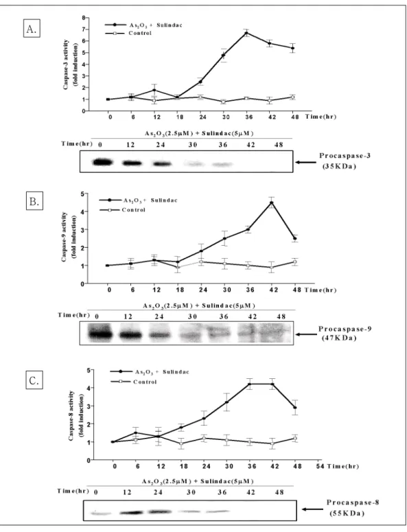
3. As2O3와 sulindac의 병합요법이 caspase 활 성화에 미치는 영향
As2O3와 sulindac의 병합요법에 의한 폐암세포의 세포고사가 caspase의 활성화에 의한 것인지를 확
인하기 위하여 caspase-3, 8, 9 protease의 효소적 활성을 이들 효소의 형광기질(fluorogenic sub strate)을 이용하여 조사하였고, caspase protease 의 활성에 의한 세포내 단백질의 변화를 western blotting으로 확인하였다. Caspase-3 protease의 활 성은 24시간 후부터 증가하기 시작하여 36시간째 에 6.8배까지 증가하였고, caspase-8 protease의 활 성은 18시간 후부터 증가하여 36시간째 4.3배까지 증가된 효소활성을 보였으며, caspase-9 protease 의 활성은 24시간 후부터 증가하기 시작하여 42시 간째 4.6배까지 증가된 효소활성을 보였다. 또한 Western blotting상에서 procaspase-3, 8, 9 pro tease의 발현이 이들 단백질의 분절로 인하여 시간 의존적으로 각각 발현이 감소함을 관찰하였다(Fig.
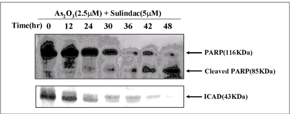
3). 또한 caspase family 단백질 중 하위단계를 보기 위해 세포내 caspase-3 protease의 기질 단백질인 PARP와 ICAD cleavage 정도를 western blotting 을 통해 확인하였다. PARP는 핵 안에 존재하면서 손상된 DNA의 복구에 관련된 단백질로서 caspase- 3 protease에 의해 116kDa 크기의 단백질이 절단되 Fig. 1. Combination treatment of arsenic trioxide and sulindac decreased the viability of NCI-H157
human lung cancer cells in a dose-dependent manner. The cell viability was determined by a MTT assay at 48hours after culture. The data represent the mean±S.D. of three independent experiments.
어 85kDa의 단편으로 절단됨을 확인하였다. 또한 ICAD도 핵 안에 존재하면서 caspase-3에 의해 ICAD가 잘리면 CAD가 활성화되어 손상된 DNA 를 잘라 절편을 만들고 세포고사가 일어나는데, caspase-3 protease에 의해 43kDa크기의 단백질이 절단되어 시간 의존적으로 발현이 감소됨을 확인하 였다(Fig. 4). 이상의 결과로 폐암세포주에서 As2O3
와 sulindac의 병합요법은 caspase-3, 8, 9의 효소활 성을 유도하였고, 이러한 효소의 활성화는 세포내
PARP와 ICAD 단백질의 분절을 초래하여 폐암세포 주의 세포고사를 유도함을 알 수 있었다.
4. As2O3와 sulindac의 병합요법이 Fas와 Fas/
L 단백질 발현에 미치는 영향
Caspase-8 protease의 활성 증가가 세포고사 신호 전달체계의 상위 death receptor인 Fas와 Fas/L의 영향을 받는지 알아보고자 As2O3와 sulindac의 병 Fig. 2. (A) Nuclear morphological investigation of combination treated NCI-H157 cells by Hoechst
staining. Cells were treated with As2O3 and/or sulindac for 48hrs and nuclear morphology was investigated by Hoechst 33342 dye staining under fluorescence microscopy. (B) Combination treatment of As2O3 and sulindac increased sub-G0/G1 fraction in NCI-H157 cells. Cells were treated with As2O3 and sulindac for 48hrs. Then, cellular DNA were stained with PI staining solution and analyzed by flow cytometry. The data represent the one of three independent experiments.
Control As2O3 Sulindac As2O3 + Sulindac
Fig. 3. Combination treatment of As2O3 and sulindac increased the catalytic activity of caspase-3,8,9 protease and cleaved procaspase-3, 8, 9 protease of NCI-H157 lung cells in a time-dependent manner. The data represent the mean±S.D. of quadraplicates.
A.
B.
C.
합요법으로 처리한 폐암세포주에서 이들 단백질의 발현을 Western blotting으로 조사하였다. 그 결과 12시간처리 군부터 Fas 단백질의 발현이 증가하였 고, Fas/L 단백질의 발현은 24시간처리 군부터 시간 의존적으로 발현이 증가함을 확인하였다(Fig. 5).
고 찰
세포고사의 이상은 암 발생, 항암치료에 대한 내성 발현, 자가면역질환의 발생 및 퇴행성 질환의 병리 현상에 중요한 역할을 한다고 보고되고 있다22,23. Fig. 4. Combination treatment of As2O3 and sulindac induced the clevage of PARP and ICAD in
NCI-H157 cells. The cells were treated with 2.5μM As2O3 and 5μM sulindac for various periods. The equal amounts of protein from cell lysate were subjected on 12.5% SDS-PAGE, transferred onto nitrocellulose membrane and immunoblotted with anti-PARP and anti-ICAD antibodies. The data represent the one of three independent experiments.
Fig. 5. Combination treatment of As2O3 and sulindac induced the expression of Fas and Fas/L proteins in NCI-H157 cells. The cells were treated with 2.5μM As2O3 and 5μM sulindac for the indicated periods. The same amounts of protein from lysates were subjected on 15%
SDS-PAGE. The membrane was immunoblotted with Fas and Fas/L antibodies. The immunoreactive bands were visualized by ECL kit. The data represent the one of three independent experiments.
항암화학요법은 정상세포 뿐만 아니라 암세포에 작용하는 특성을 이용하고 있으며, 이러한 암세포 사멸의 일부는 세포고사에 의해 매개됨이 밝혀지 면서 이에 대한 생화학적 그리고 분자생물학적인 접근이 활발히 이루어지고 있다.
As2O3는 농도에 따라 APL세포에 두 가지 방식 으로 작용하는데24, 먼저 0.1-0.5 μM의 저농도에서 는 분화의 유도로 인해 As2O3가 직접 PML 또는 PML/RARα를 분해하는 작용을 한다25. 또한 1-2 μ M의 농도에서는 세포고사 유도로 고사유발신호에 의해 미토콘드리아 막전위의 변화가 일어나고 이 에 따라 막의 투과성이 변화되어 cytochrome C와 다른 고사유도인자가 유리되어 caspase의 활성화 가 일어난다7,26,27. 이밖에도 활성산소의 활성화, JNK의 활성화, tubulin의 불활성화, p53의 유도 등
28,29
이 고사유도의 기전으로 제시되고 있으나 미토 콘드리아의 기능변화나 Fas와 Fas/L의 발현 등에 대한 구체적인 고사 기전은 밝혀져 있지 않다. 임 상적으로 As2O3의 독성으로는 오심과 설사, 기침, 피로감, 빈맥, 피부발진, 체중증가와 체액저류, 백 혈구증가증, APL 분화증후군, QTc 연장, 말초혈관 병증, 고혈당증 등이 있다. As2O3는 체외실험에서 림프종이나 다른 고형종양세포에서도 용량 의존적 으로 고사를 유도함이 밝혀져 치료제로서 주목을 받았으나 APL에서 사용되는 As2O3의 치료농도가 0.5-2.0 μM인 반면 5-10 μM의 고농도가 필요하기 때문에 부작용으로 인해 실제 임상적으로 적용하 지 못하고 있는 실정이다.
NSAIDs가 최근 생물학적 치료, 방사선치료, 기 존의 항암제들과의 병합요법시 상승효과가 있음이 보고되고 있다. Torrance 등30은 sulindac과 EKI- 569(EGFR tyrosine kinase inhibitor)의 병합치료시 장 종양을 감소시키는데 단독치료에 비해 상승효 과가 있다고 하여 COX-2와 EGFR 신호전달과정 모두를 억제함으로서 암의 예방과 치료에 새로운
분자생물학적인 치료방법으로 관심을 끌고 있다.
Duffy 등31은 폐암과 백혈병 세포주에서 NSAIDs 가 anthracycline과 vinca alkaloid계의 항암제의 세포독성을 증가시켰다고 하였고, Soriano 등32은 sulindac과 paclitaxel이나 cisplatin의 병합요법이 소세포와 비소세포폐암 모두에서 종양의 성장억제 효과를 배가시켰으며, Hida 등33도 비소세포폐암에 서 nimesulide와 irinotecan, taxotere, VP-16, cisplatin이나 SM5887과의 병합치료시 상승작용이 있음을 보고함으로서 암 치료시 NSAIDs의 효과를 증명하였다.
본 연구에서 NCI-H157 폐암세포에 As2O3와 sulindac을 병합치료시 단독치료군에 비해 생존율을 의미 있게 감소시켰고, 이러한 세포죽음은 Hoe chst 33342 염색을 통한 염색사의 응축과 핵 분절 유도확인과 유식세포 분석에 의한 sub-G0/G1
DNA분획의 증가현상으로 세포고사에 의해 매개됨 을 확인할 수 있었다. 또한 세포고사의 유도에는 caspase 3, 8, 9를 통한 활성화와 이에 의한 PARP 와 ICAD의 절단을 보여 caspase cascade가 관여 함을 알 수 있었다. 또한 caspase-8 protease의 활 성화에는 Fas와 Fas/L 단백질의 발현증가가 활성 화를 유도하였음을 알 수 있었다. 결론적으로 Fas/
FasL 신호전달계에 의한 caspase-8 protease의 활 성화를 통해 caspase-3 protease의 활성화가 초래 되어 세포고사가 발생하였음을 시사하였다.
As2O3는 재발성 또는 불응성 급성전골수성백혈병 의 치료제로 쓰이는 항암제로서 다른 다양한 암세 포에서 세포고사를 유도하나 백혈병에 비해 상대적 으로 높은 농도가 필요하여 실제적인 치료에 제한 이 있었다. 그러나 폐암세포주에 대해 sulindac과의 병합치료를 통해 그들의 세포고사를 배가시킴으로 서 백혈병치료시의 치료농도로 낮춤으로서 이에 따른 부작용을 감소시켜 향후 치료제로서의 활용 가능성을 제공할 수 있으리라 판단된다.
요 약
연구배경 :
Arsenic trioxide(As2O3)은 재발성 또는 불응성 급 성전골수성백혈병의 치료제로 쓰이는 항암제로서 비소세포폐암을 포함한 다른 암세포주에도 효과가 있는 것으로 되어있다. NSAIDs는 항암 예방약제 로 사용되고 있고, 세포고사를 통해 다른 항암제나 방사선치료의 반응성을 강화시키는 것으로 알려져 있다. 저자들은 NCI-H157 세포주에서 As2O3와 sulindac의 병합치료가 그것들의 세포고사를 배가 시키는지 여부를 알아보고자 하였다.
방 법 :
세포 독성은 MTT 방법으로 측정하였고, 세포고사 를 알아보기 위해 핵산 염색과 유식세포 분석을 시행하였다. 세포고사의 기전을 보기 위해 caspase family의 활성을 보았고, PARP와 ICAD의 분절을 western blotting으로 확인하였다. 또한 Fas와 Fas-L의 발현유무를 western blotting을 통해 관 찰하였다.
결 과 :
NCI-H157 폐암세포에 As2O3와 sulindac을 병합치 료시 단독치료군에 비해 생존율이 의미 있게 감소 하였고, 이러한 세포사는 핵산염색을 통한 염색사 의 응축과 핵 분절 유도와 유식세포 분석에 의한 sub-G0/G1 DNA분획의 증가현상을 통해 세포고사 에 의해 매개됨을 알 수 있었다. 세포고사의 유도 에는 caspase 3, 8, 9를 통한 활성화와 이에 의한 PARP와 ICAD의 절단을 확인하였다. 또한 caspase-8 protease의 활성화에는 Fas와 Fas/L 단 백질의 발현증가가 유도되었음을 알 수 있었다.
결 론 :
NCI-H157 폐암세포주에 As2O3와 sulindac의 병합 요법은 Fas/FasL 신호전달계의 활성화와 caspase 단백질 활성화 의해 세포고사가 유도되었다.
참 고 문 헌
1. Chen CJ, Wu MM, Lee SS, Wang JD, Cheng SH, Wu HY. Atherogenicity and carcino genecity of high-arsenicartesian well water:
Multiple risk factors and related malignant neoplasm of blackfoot disease. Arterio sclerosis 1988;8:452-60.
2. Chen CJ, Chen CW, Wu MM, Kuo TL.
Cancer potential in liver, lung, bladder and kidney due to ingested inorganic arsenic in drinking water. Br J Cancer 1992;66:888-92.
3. Sun HD, Ma L, Hu XC, Zhang TD.
Treatment of acute promyelocytic leukemia by Ailing-1 therapy with use of syndrome differentiation of traditional Chinese medicine.
Chin J Comb Trad Chin Med West Med 1992;12:170-1.
4. Soignet SL, Frankel SR, Douer D, Tallman MS, Kantarjian H, Calleja E, et al. United States multicenter study of arsenic trioxide in relapsed acute promyelocytic leukemia. J Clin Oncol 2001;19:3852-60.
5. Ling YH, Jiang JD, Holland JF, Perez-Soler R. Arsenic trioxide produces polymerization of microtubules and mitotic arrest before apoptosis in human tumor cell lines. Mol Pharmacol 2002;62:529-38.
6. Shen ZY, Tan LJ, Cai WJ, Shen J, Chen C, Tang XM, et al. Arsenic trioxide induces apoptosis of oesophageal carcinoma in vitro.
Int J Mol Med 1999;4:33-7.
7. Akao Y, Nakagawa Y, Akiyama K. Arsenic trioxide induces apoptosis in neuroblastoma cell lines through the activation of caspase 3
in vitro. FEBS Lett 1992;445:59-62.
8. Seol JG, Park WH, Kim ES, Jung CW, Hyun JM, Kim BK, et al. Effect of arsenic trioxide on cell cycle arrest in head and neck cancer cell line PCI-1. Biochem Biophys Res Commun 1999;265:400-4.
9. Zheng J, Deng YP, Lin C, Fu M, Xiao PG, Wu M. Arsenic trioxide incuces apoptosis of HPV16 DNA-immortalized human cervical epithelial cells and selectively inhibits viral gene expression. Int J Cancer 1999;82:286-92.
10. Zhang TC, Cao EH, Li JF, Ma W, Qin JF.
Induction of apoptosis and inhibition of hu man gastric cancer MGC-803 cell growth by arsenic trioxide. Eur J Cancer 1999;35:1258- 63.
11. Thun MJ, Namboodiri NM, Health CW Jr.
Aspirin use and reduced risk of fatal colon cancer. N Engl J Med 1991;325:1593-6.
12. Harris RE, Nambodiri KK, Farrar WB.
Nonsteroidal antiinflammatory drugs and bre ast cancer. Epidemiology 1996;7:203-5.
13. Scheinemachers DM, Everson RB. Aspirin use and lung, colon, and breast cancer inci dence in a prospective study. Epidemiology 1994;5:138-46.
14. Smalley WE, Dubois RN. Colorectal cancer and non-steroidal anti-inflammatory drugs.
Adv Pharmacol 1997;39:1-20.
15. Dannenberg AJ, Zakim D. Chemoprevention of colon-rectal cancer through inhibition of cyclooxygenase-2. Semin Oncol 1999;26:499- 504.
16. Shiff SJ, Qiao L, Tsai LL, Rigas B. Sulindac sulfide, an aspirin-like compound, inhibits proliferation, causes cell cycle quiescence, and
induces apoptosis in HT-29 colon adeno carcinoma cells. J Clin Invest 1995;96:491- 503.
17. Chan TA, Morin PJ, Vogelstein B, Kinzler KW. Mechanism underlying nonsteroidal antiinflammatory drug-mediated apoptosis.
Proc Natl Acad Sci 1998;95:681-6.
18. Klampfer L, Cammenga J, Wisniewski HG, Nimer SD. Sodium salicylate activates caspases and induces apoptosis of myeloid leukemia cell lines. Blood 1999;93:2386-94.
19. Milas L. Cyclooxygenase-2 (COX-2) enzyme inhibitors as potential enhancers of tumor radioresponse. Semin Radiat Oncol 2001;11:
290-9.
20. Koki AT, Leahy KM, Masferrer JL. Potential utility of COX-2 inhibitors in chemopre vention and chemotherapy. Expert Opin In vest Drugs 1999;8:1623-38.
21. Milas L, Kishi K, Hunter N, Mason K, Masferrer JL, Tofilon PJ. Enhancement of tumor response to gamma-radiation by an inhibitor of cyclooxygenase-2 enzyme. J Natl Cancer Inst 1999;91:1501-4.
22. Williams GT. Apoptosis in the immune system. J Pathol 1994;173:1-4.
23. Cohen JJ. Apoptosis: the physiologic pathway of cell death. Hosp Pract 1993;28:35-43.
24. Chen GQ, Shi XG, Tang W, Xiong SM, Zhu J, Cai X, et al. Use of arsenic trioxide (As2O3) in the treatment of acute promye lcytic leukemia(APL): I. As2O3 exerts dose- dependent dual effects on APL cells. Blood 1997;89:3345-53.
25. Chen GQ, Zhu J, Shi XG, Ni JH, Zhong HJ, Si GY, et al. In vitro studies on cellular and
molecular mechanisms of arsenic trioxide in the treatment of acute promyelocytic leu kemia: arsenic trioxide induces NB4 cell apoptosis with downregualation of Bcl-2 expression and modulation of PML-RARα /PML proteins. Blood 1996;88:1052-61.
26. Uslu R, Sanli UA, Sezgin C, Karabulut B, Terzioglu E, Omay SB, et al. Arsenic trio xide-mediated cytotoxicity and apoptosis in prostate and ovarian carcinoma cell lines.
Clin Cancer Res 2000;6:4957-64.
27. Jung BH, Park C, Kim HR, Park MR. Arse nic trioxide induces apoptosis of HL-60 cells via activation of intrinsic caspase protease with mitochondrial dysfunction. Cancer Res and Treat 2002;34:308-15.
28. Huang S, Huang CF, Lee T. Induction of mitosis-mediated apoptosis by sodium arse nite in Hela S3 cells. Biochem Pharmacol 2000;60:771-80.
29. Park WH, Seol JG, Kim ES, Hyun JM, Jung CW, Lee CC, et al. Arsenic trioxide-mediated growth inhibition in MC/CAR myeloma cells via cell cycle arrest in association with
induction of cyclin-dependent kinase inhibi tor, p21, and apoptosis. Cancer Res 2000;60:
3065-71.
30. Torrance CJ, Jackson PE, Montgomery E, Kinzler KW, Vogelstein B, Wissner A, et al.
Combinatorial chemoprevention of intestinal neoplasia. Nat Med 2000;6:1024-8.
31. Duffy CP, Elliott CJ, O'Connor RA, Heenan MM, Coyle S, Cleary IM, et al. Enhancement of chemotherapeutic drug toxicity to human tumour cells in vitro by a subset of NSAIDs. Eur J Cancer 1998;34:1250-9.
32. Soriano AF, Helfrich B, Chan DC, Heasley LE, Bunn PA Jr, Chou TC et al. Synergistic effects of new chemopreventive agents and conventional cytotoxic agents against human lung cancer cell lines. Cancer Res 1999;59:
6178-84.
33. Hida T, Kozaki K, Muramatsu H, Masuda A, Shimizu S, Mitsudomi T, et al. Cyclooxy genase-2 inhibitor induces apoptosis and enhances cytotoxicity of various anticancer agents in non-small cell lung cancer cell lines. Clin Cancer Res 2000;6:2006-11.