17
The anti-tumor effect of combined treatment with arsenic trioxide and interferone-α on transplanted murine
Lewis lung carcinoma
Deug-Log Seo, Je-Hoon Yang, Chung-Kil Won, Myeong-Ok Kim1, Jong-Hwan Lee2, Soo-Dong Kwark, Phil-Ok Koh*
College of Veterinary Medicine and Institute of Animal Medicine, Gyeongsang National University, Jinju 660-701, Korea
1College of Natural Sciences, Gyeongsang National University, Jinju 660-701, Korea
2College of Veterinary Medicine, Konkuk University, Seoul 143-701, Korea OAccepted: February 15, 2005)
Abstract : In the present study, we expected the anti-tumor effect by combined treatment of arsenic trioxide and interferon (IFN)-α on murine Lewis lung carcinoma (LL2) cells through in vivo study. As a experimental model, LL2 cells (1×10⁶/mouse) were injected subcutaneously into the back region of mice. When the tumor volume reached 100 mm3, mice were treated with 1 mg/kg arsenic trioxide, 50000 IU IFN-α, or arsenic trioxide and IFN-α. The development of tumor cells was significantly inhibited by combined treatment with arsenic trioxide and IFN-α. In arsenic trioxide and IFN-α treated group, apoptotic index was reached a peak valve at 48 hr after the treatment and it was restored to approximately the control level at 8 days. Also, positive signals of Bax and Bad were increased at 48 to 96 hr and decreased at 8 day. Whereas, positive cells of Bcl-2 were steadily decreased at 12 to 48 hr and restored to the background level at 8 days. Our data showed that immunoreactivity of Bcl-2 was decreased at 12 to 48 hr, while positive signals of Bax and Bad were increased in accordance with apoptotic index at these times. In conclusion, our results suggest that the combined treatment with arsenic trioxide and IFN-α significantly inhibited the growth of LL2 tumor cells and induced apoptosis through the up and down-regulation of Bcl-2 gene family.
Key words : arsenic trioxide, IFN-α, apoptosis, Bcl-2, Bax, Bad
Introduction
Arsenic trioxide has been used as a therapeutic agent for the acute promyelocytic leukemia (APL). Especially, it was effective APL patients resistant to all-trans retinoic acid (atRA) or other chemotherapeutic drugs [22].
However, in the majority of patients given long-term, toxic side effects which including skin pigmentation, keratosis, cirrhosis, polyneurites, and gastrointestinal problems were observed. Thus, many researchers have focused on the anti-tumor activity of arsenic trioxide through combination with other types of agents, interferon (IFN)-α, atRA, and chemotherapeutic agents [5, 11, 12, 24, 26, 27]. It was reported that the chronic administration of IFN-α or IFN-β can produce regression
of vascular tumors, including Kaposi’s sarcoma, pulmonary hemangiomatosis and hemangiomas [6, 17, 23].
Furthermore, IFN has functions the anti-proliferative effect as well as an immunomodulatory activity in vitro and in vivo [7]. Recently, a high synergistic effect between arsenic trioxide and IFN was reported in human T-cell lymphotropic virus type 1 (HTLV) infected cells [14]. Previous studies explained that arsenic trioxide and IFN combination therapy induced apoptosis and cell cycle arrest.
To inhibit the tumor cellular growth, the processing of apoptosis is an important mechanism [3, 18]. The induction of apoptosis by arsenic trioxide involves inhibition of glutathione peroxidase (GPx) activity and increasing of cellular H2O2 content. These are followed
*Corresponding author: Phil-Ok Koh
College of Veterinary Medicine and Institute of Animal Medicine, Gyeongsang National University, Jinju 660-701, Korea [Tel: +82-55-751-5809, Fax: +82-55-751-5803, E-mail: [email protected]]
by cytochrome C release, caspases 3 activation, DNA fragmentation, and the classic morphological changes of apoptosis [10, 13]. Also, arsenic trioxide directly induced apoptosis through the down-regulation of Bcl-2 in NB4 cells of APL [4]. Previous studies suggest that the induction of apoptosis is a critical event for the suppression of tumor growth. Especially, a number of cellular proto- oncogenes including Bcl-2 and its family gene are important in the regulation of apoptosis. Also, Bcl-2 gene family regulated the apoptosis of tumor cells in a various cancer [2, 9]. The levels of Bcl-2 are regulated by closely related Bcl-2 gene family members such as Bax, Bad, Bcl-x(L), and Bcl-x(S). In these genes, Bcl- 2 and Bcl-x(L) are known as inhibitor of apoptosis, whereas Bax and Bcl-x(S) are known as inducer of apoptosis [15]. It was reported that change in the ratio of Bcl-2 to Bax expression is the critical determinant of cell fate, cell survival and death. Thus, we suggested the possibility that Bcl-2 gene family, Bcl-2, Bax, and Bad regulated the anti-tumor effect by the combined treatment with arsenic trioxide and IFN-α. However, it has been unknown. The present study was performed to investigate the anti-tumor effect by the combined treatment with arsenic trioxide and IFN-α on murine Lewis lung carcinoma (LL2) cell through in vivo study.
Also, we expected the fact that Bcl-2 gene family regulated the anti-tumor effect by these agents.
Materials and Methods
Tumor cell and animal
The Lewis lung carcinoma cell line which obtained from the American type culture collection (ATCC, Rockville, MD, USA) was cultured in Dulbecco’s modified Eagle medium (DMEM: Gibco, USA) supplemented with 10% heat inactivated fetal bovine serum (Sigma, USA), 100 U/ml penicillin, and 100 U/ml streptomycin (Sigma, USA). The cells were maintained in a humidified atmosphere, at 37oC, 5% CO2 in air.
C57BL/6 male mice (110 heads, 7-8 week old), were bred and maintained in the Asan Institute for Life Sciences specific-pathogenic-free (SPF) mouse colony, were housed in wire-bottomed cages in a room with constant temperature (22oC±1) and humidity (55%) and with a 12 hr light (6 a.m. - 6 p.m.) and dark cycle, with free access to food and tap water. Lewis lung carcinoma cells (1×10⁶/mouse) were injected subcutaneously into the back region of mice, tumor volume was measured
with a digimatic calipers (Mitutoyo, Japan) at three times a week, and calculated by the formula [π/6 (w1×w2×w3)], where w1 represented the largest tumor diameter, w2 represented the smallest tumor diameter and w3 represented the tumor height. When each tumor volume reached 100 mm3, each animal was treated with 1 mg/kg arsenic trioxide and/or 50000 IU IFN- α. The body weight and general physical status of the animals were observed every day.
TUNEL assay
The mice were killed by cervical dislocation at 0, 12, 24, 96 hr and 8 days after drug treatment. Tumor tissues were immediately fixed in 10% neutral buffered formalin solution (pH 7.0), embedded in paraffin and cut into 4µm thick sections. Sections were deparaffinized in xylene, dehydrated through graded alcohol, and washed 0.1 M phosphate-buffered saline (PBS). Sections were incubated 20 mg/ml proteinase K for 40 min. After washes in PBS, sections were incubated with equilibration buffer followed by TdT enzyme (Oncogene, USA) in a humidified chamber at 37oC for 1 hr, and then a stop/
wash buffer was applied for 30 min at 37oC. The sections were incubated with anti-digoxigenin peroxidase (Oncogene, USA) for 30 min at room temperature, counterstained with hematoxylin. With the TUNEL method, five fields of non-necrotic areas were randomly selected in each histological specimen, and the number of apoptotic positive nucleus in each field was calculated as cell numbers per 100 cells.
Immunohistochemical studies of Bcl-2 gene family To determine Bcl-2 gene family, Bcl-2, Bax, and Bad immunoreactivity, immunohistochemistry was performed using an avidin-biotin-peroxidase complex method (Vectastain ABC kit; Vector, USA). Endogenous peroxidase was blocked with a 3% hydrogen peroxide solution for 5 min. The sections were washed in PBS, incubated with normal goat serum to prevent nonspecific binding. Anti- mouse monoclonal Bcl-2, Bax, and Bad antibody (Santa Cruz, USA) were diluted 1:100, 1:200, or 1:100, respectively, were incubated at 4oC overnight. After incubation, the slides were washed in PBS, incubated for 60 min with biotinylated anti-mouse IgG, and then incubated with avidin-biotin-complex according to the manufacturer’s recommendations. After the sections were washed in PBS, the color reaction was performed with 3-amino-9-ethylcarbazole (AEC; Vector, USA). The
sections were counterstained with hematoxylin. Five fields of non-necrotic areas were randomly selected in each histological specimen, and the number of apoptotic positive nucleus in each field was calculated as cell numbers per 100 cells.
Data analysis
Data were evaluated using one-way analysis of variance (ANOVA) and Student’s t-test. Statistical significance was at p<0.05.
Results
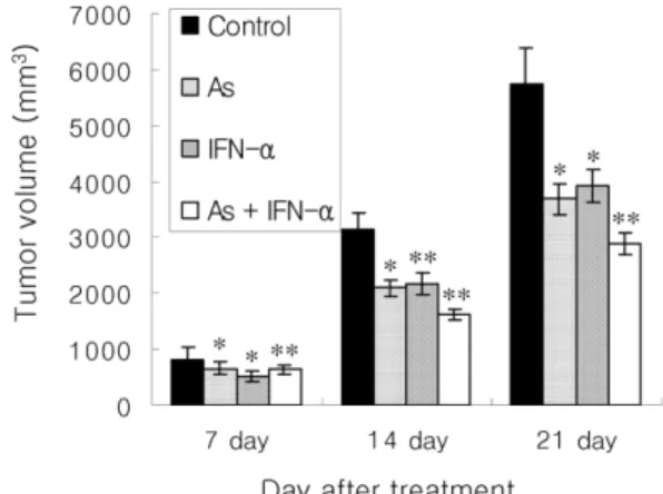
Tumors appeared and developed on the backs of mice about a week after injection of LL2 cell. The development of tumor cells was markedly inhibited by the combination therapy with arsenic trioxide and IFN- α. At 21 days after drug injection, the mean tumor volumes were evaluated at 5747.34±624.62 mm3 in the control group, 3689.48±278.44 mm3 in the arsenic trioxide treated group (64% volume per control, *p<0.05 vs. control), 3923.45±293.76 mm3 in the IFN-α treated group (68% volume per control, *p<0.05 vs. control), and 2895.33±189.68 mm3 in the arsenic trioxide and IFN-α treated group (50% volume per control, **p<0.01 vs. control). Fig. 1 represented that the combination therapy of arsenic trioxide and IFN-α significantly
inhibited the development of tumor.
In combined treatment with arsenic trioxide and IFN-α treated group, the apoptotic cells were observed to be 3.77±0.68% at 0 hr after treatment, 5.03±0.66%
at 12 hr, 6.17±1.08% at 24 hr, 7.03±0.75% at 48 hr, 5.94±0.91% at 96 hr, and 4.11±0.65% at 8 days, respectively. Apoptosis peaked at 48 hr, increased 93%
as compared with the untreated control group. It was restored to approximately the control level at 8 days (Fig. 2).
Positive cells of Bcl-2 were observed to be 10.63
±1.12% at 0 hr, 8.51±1.54% at 12 hr, 7.09±1.23% at 24 hr, 6.65±1.20% at 48 hr, 7.20±1.32% at 96 hr, and 10.80±1.16% at 8 days, respectively (*p<0.05 vs. 0 hr).
The percentage of Bcl-2 positive cells was decreased within 12 hr after treatment with arsenic trioxide and IFN-α. It was continuously decreased at 48 hr and was then restored to the background level by 8 days after treatment (Figs. 2, 3A, and 3B). Whereas, positive cells of Bax were observed to be 1.83±0.13% at 0 hr, 4.40±0.26% at 12 hr, 4.69±0.28% at 24 hr, 4.74±0.26%
at 48 hr, 4.74±0.29% at 96 hr, and 2.80±0.16% at 8 days, respectively (**p<0.01 vs. 0 hr). Positive cells of Bax showed significant change after combined treatment with arsenic trioxide and IFN-α. It was increased and peaked at 48 to 96 hr, was decreased at 8 days after combined treatment (Figs. 2, 3C, and 3D). Furthermore, the percentage of Bad positive signals was observed to be 1.31±0.09% at 0 hr, 2.29
±0.18% at 12 hr, 2.97±0.22% at 24 hr, 3.20±0.26%
Fig. 1. The change of tumor volume by the injection of arsenic trioxide and/ or IFN-α. Tumor volume was measured with a digimatic calipers at 7, 14, 21 days after treatment. The development of tumor cells were significantly inhibited by the combination therapy of arsenic trioxide and IFN-α. Each value is the mean±S.D. for 3 repeated experiments. *p<0.05, **p<0.01 (vs. control).
Fig. 2. The expression of apoptosis and Bcl-2 gene family in LL2 cells treated with arsenic trioxide and IFN-α.
Apoptotic index was examined by TUNEL method and positive cells of Bcl-2 gene family were scored by immunohistochemistry. Each value is the mean±S.D. for 3 repeated experiments. *p<0.05, **p<0.01 (vs. 0 hour).
at 48 hr, 2.62±0.22% at 96 hr, and 1.37±0.12% at 8 days, respectively (**p<0.01 vs. 0 hr). Positive signals of Bad were steadily increased and peaked at 48 hr, was then restored to the background level by 8 days after treatment (Figs. 2, 3E, and 3F).
Discussion
The present study demonstrated that apoptosis was enhanced by combined treatment with arsenic trioxide and IFN-α in transplanted murine Lewis lung carcinoma cell lines, and it was regulated by apoptosis associated genes, Bcl-2, Bad, and Bax. In this study, we were Fig. 3. Expression of Bcl-2 gene family, Bcl-2 (A and B), Bax (C and D), Bad (E and F) in LL2 cells treated with arsenic trioxide and IFN-α by immunohistochemistry. 0 hr (A, C, and E) and 48 hr (B, D, and F) after treated with arsenic trioxide and IFN-α. The bar = 50 µm.
expected two goals. One is the anti-tumor effect by combined treatment with arsenic trioxide and IFN-α on Murine Lewis lung carcinoma (LL2) cell through in vivo study. The other is the change of Bcl-2 gene family by these agents.
Arsenic trioxide was shown to be an effective treatment for the acute promyelocytic leukemia (APL) patients resistant to all-trans retinoic acid (atRA) or other chemotherapeutic drugs [8, 20, 21, 25]. Particularly, low concentration of arsenic trioxide has been reported to induce complete remission in a high proportion of patients with APL without severe toxicity [4, 20, 21].
Jing et al. [10] reported that arsenic trioxide induced the apoptosis in leukemic cells depends on the activity of the enzyme which regulates cellular H2O2 content.
In solid tumor, arsenic trioxide induced the apoptosis and inhibited the tumor growth [25]. It was reported that arsenic trioxide showed an anti-tumor effect and anti-proliferative effect in various cell including human gastric cancer cell lines [11, 19]. Low concentration of arsenic trioxide and IFN-α induced the apoptosis and inhibited tumor cellular growth [3, 18]. The induction of apoptosis by these agents involves inhibition of glutathione peroxidase (GPx) activity, increasing of cellular H2O2 content, releasing of cytochrome C release, and the classic morphological changes of apoptosis [10, 13].
In the previous studies, tumor cell lines were enhanced apoptosis by exposed to arsenic trioxide and/or the other drugs [1, 12, 16, 24]. According to Jing et al.
[11], combined treatment of atRA acid and arsenic trioxide in APL cells was enhanced apoptosis, such that nearly 32% of the cells showed apoptosis. However, single drug treatment showed 10% of the apoptotic cells. In this experiment, the anti-tumor effect of arsenic trioxide was markedly enhanced in the combined treatment with IFN-α as compare to that of single treatment group. Also, our data explained that combined treatment with arsenic trioxide and IFN-α were significantly inhibited the development of tumor cells.
Single treatment of arsenic trioxide or IFN-α was not sufficient to inhibit transplanted LL2. Also, LL2 cells were exposed to 1, 5, 10 mg/kg arsenic trioxide in a dose-dependant manner. However, there was no difference in the growth of tumor cells in each group. As shown in our results, combined treatment with 1 mg/kg arsenic trioxide and 50,000 IU IFN-α decreased the tumor volume up to 50% at 21 days after treatment and enhanced
apoptosis in LL2 tumors. In addition, the positive cell of apoptosis in LL2 tumors increased to 93% as compare with that of untreated group. In this study, anti- tumor effect of arsenic trioxide and IFN-α in tumor tissues coincided with previous studies. Thus, our data demonstrated a synergic effect of the combination of arsenic trioxide and IFN-α on apoptosis in LL2 tumors.
In tumor cells, apoptosis-associated genes were divided to pro-apoptotic regulatory genes and anti-apoptotic regulatory genes. Among these genes, Bcl-2 gene family was known as a critical gene. In Bcl-2 gene family, Bcl-2 inhibits apoptosis while Bax induces apoptosis.
In the previous studies, Jing et al. [11] reported that arsenic trioxide induced the apoptosis through the down regulation of Bcl-2 in tumor cells. But, they did not show the change of Bax and Bad. Our data demonstrated that arsenic trioxide and IFN-α treatment in tumor cells induced the apoptosis through the up regulation of Bax and Bad and the down regulation of Bcl-2. Especially, positive cells of Bcl-2 was significantly decreased at 12 hr after treatment with arsenic trioxide and IFN-α, which steadily decreased with time until at 48 hr. Whereas, the percentage of Bad and Bax positive cells were increased and peaked at 48 to 96 hr after treatment, and were restored to the background level at 8 days. In accordance with the exchange pattern of apoptosis, the expression of Bax and Bad in tumor tissue was highest at the peak time of apoptosis processing. In conclusion, these results suggest that the combination treatment of arsenic trioxide and IFN-α enhanced apoptosis in LL2 cell lines through the up and down-regulation of Bcl-2 gene family.
References
1. Bachleitner-Hofmann T, Gisslinger B, Grumbeck E, Gisslinger H. Arsenic trioxide and ascorbic acid:
synergy with potential implications for the treatment of acute myeloid leukemia. Br J Haematol 2001, 112, 783- 786.
2. Brocheriou I, Carnot F, Briere J. Immunohistochemical detection of Bcl-2 protein in thymoma. Histopathology 1995, 27, 251-255.
3. Bursch W, Lauer B, Timmerman-Trosiener L, Barthel G, Schuppler J, Sculte-Hermann R. Controlled cell death(apoptosis) of normal and putative neoplastic cells in rat liver following withdrawal of tumor promoters.
Carcinogenesis 1984, 5, 53-58.
4. Chen GQ, Shi XG, Tang W, Xiong SM, Cai X, Han ZG, Ni JH, Shi GY, Jia PM. Liu MM, He KL, Niu C, Ma J, Zhang P, Zhang TD, Paul P, Naoe T, Kitamura K, Miller W, Waxman S, Wang ZY, Chen SJ, Chen Z. Use of arsenic trioxide in the treatment of acute promyelocytic leukemia(APL): I. As2O3 experts dose- dependent dual effects on APL cells. Blood 1997, 89, 3345-3353.
5. El-Sabban ME, Nasr R, Dbaibo G, Hermine O, Abboushi N, Quignon F, Ameisen JC, Bex F, Bazarbachi A. Arsenic-interferon-alpha-triggered apoptosis in HTLV-I transformed cells is associated with tax down-regulation and reversal of NF-kappa B activation. Blood 2000, 96, 2849-2855.
6. Ezekowitz RAB, Mulliken JB, Folkman J. Interferon alpha2 a therapy for life threatening hemangiomas of infancy. N Engl J Med 1992, 326, 1456-1463.
7. Gutterman JU. Cytokine therapeutics: Lessons from interferon alpha. Proc Natl Acad Sci USA 1994, 91, 1198-1205.
8. Huang SY, Chang CH, Tang JL, Tien HF, Kuo TL, Huang SF, Yao YT, Chou WC, Chung CY, Wang CH, Shen MG, Chen YC. Acute and chronic arsenic poisoning associated with treatment of acute promyelocytic leukemia. Br J Haematol 1998, 103, 1092-1095.
9. James GC, Elizabeth HR, Laura NH, Andrea JG, Steven PA, David EM, James CC, Tony RF, Russell CC, Thomas LG. Altered Bcl-2 family expression during non-genotoxic hepato-carcinogenesis in mice.
Carcinogenesis 1999, 20, 1590-1599.
10. Jing Y, Dai J, Ruth ME, Chalmers-Redman, Willam G, Willam T, Waxman S. Arsenic trioxide selectively induces acute promyelocytic leukemia cell apoptosis via a Hydrogen peroxide-dependent pathway. Blood 1999, 94, 2102-2111.
11. Jing Y, Wang L, Xia L, Guo-qiang Chen GQ, Chen Z, Wilson H, Wilson M, Waxman S. Combined effect of all-trans retinoic acid and arsenic trioxide in acute promyelocytic leukemia cells in vitro and in vivo.
Blood 2001, 97, 264-269.
12. Kito M, Akao Y, Ohishi N, Yagi K, Nozawa Y.
Arsenic trioxide-induced apoptosis and its enhancement by buthionine sulfoximine in hepatocellular carcinoma cell lines. Biochem Biophys Res Commun 2002, 291, 861-867.
13. Maurizio G, Marcel HM, Koken MK, Chelbi A,
Gerard B, Michel L, Chen Z, de The H. Combined arsenic and retinoic acid treatment enhances dif- ferentiation and apoptosis in arsenic-resistant NB4 cells.
Blood 1998, 91, 4300-4310.
14. Nasr R, Rosenwald A, El-Sabban ME, Arnulf B, Zalloua P, Lepelletier Y, Bex F, Hermine O, Staudt L, Bazarbachi A. Arsenic/interferon specifically reverses 2 distinct gene networks critical for the survival of HTLV-1-infected leukemic cells. Blood 2003, 101, 4576-4582.
15. Oltvai ZN, Milliman CL, Korsmeryer SJ. Bcl-2 heterodimerizes in vivo with a conserved homologue, Bax, that accerates programmed cell death. Cell 1993, 74, 609-619.
16. Park WH, Seol JG, Kim ES, Hyun J M, Jung CW, Lee CC, Kim BK, Lee YY. Arsenic trioxide-mediated growth inhibition in MC/CAR myeloma cells via cell cycle arrest in association with induction of cyclin- dependent kinase inhibitor, p21, and apoptosis. Cancer Res 2000, 60, 3065-3071.
17. Real FX, Oettgen HF, Krown SE. Kaposi’s sarcoma and the equired immunodefeciency syndrome : treatment with high and low doses of recombinant leukocyte a interferon. J Clin Oncol 1986, 4, 544-551.
18. Schulte-Hermann R, Timmerman-Trosnier I, Barthel G. Bursch W. DNA synthesis, apoptosis and phenotypic expression as determinants of growth altered foci in rat liver during phenobarbital promotion. Cancer Res.
1990, 50, 5127-5135.
19. Seol JG, Park WH, Kim ES, Jung CW, Hyun JM, Lee YY, Kim BK. Potential role of caspase-3 and - 9 in arsenic trioxide-mediated apoptosis in PCI-1 head and neck cancer cells. Int J Oncol 2001, 18, 249-255.
20. Shen ZX, Chen GQ, Ni JH, Li XS, Xiong SM, Qiu QA, Zhou L, Fang ZW, Wang YT, Ma J, Zhang P, Zhang TD, Chen SJ, Chen Z, Wang ZY. Use of arsenic trioxide in the treatment of acute promyelocytic leukemia(APL): II. Clinical efficacy and pharmacokinetics in relapsed patients. Blood 1997, 89, 3354-3360.
21. Soignet SL, Maslak P, Wang ZG, Jhanwar S, Calleja E, Dardashti LJ, Corso D, DeBlasio A, Gabrilove J, Scheinberg DA, Pandolfi PP, Warrell RP Jr. Complete remission after treatment of acute promyelocytic leukemia with arsenic trioxide. N Engl J Med 1998, 339, 1341-1348.
22. Tallman MS. Arsenic trioxide: its role in acute promyelocytic leukemia and potential in other hematologic
malignancies. Blood Rev 2001, 15, 133-142.
23. White CW, Sondheimer HM, Crouch EC, Wilson H, Fan LL. Treatment of pulmonary hemangiomatosis with recombinant interferon alpha-2a. N Engl J Med 1989, 320, 1197-1200.
24. Yuksel S, Saydam G, Uslu R, Sanli UA, Terzioglu E, Buyukececi F, Omay SB. Arsenic trioxide and methylprednisolone use different signal transduction pathways in leukemic differentiation. Leuk Res 2002, 26, 391-398.
25. Zhang Y, Nie L. Studies of apoptosis of malignant lymphoma cells induced by arsenic trioxide. Cell Biol
Int 2001, 25, 1003-1006.
26. Zhao W, Wang H, Wu F, Guo W, Qu B, Shen Z, Wang Z. Effects of All-trans-retinoic acid and Arsenic trioxide on the hemostatic disturbance associated with acute promyelocytic leukemia. Thromb Res 2001, 102, 197-204.
27. Zhu Q, Zhang JW, Zhu HQ, Shen YL, Flexor M, Jia PM, Yu Y, Cai X, Waxman S, Lanotte M, Chen DJ, Chen, Zhu, Tong JH. Synergic effects of arsenic trioxide and cAMP during acute promyelocytic leukemia cell maturation subtends a novel signaling cross-talk.
Blood 2002, 99, 1014-1022.