251
1. Introduction
The synthesis of metal nanoparticles has been an active research field during the last few decades [1-3], due to their unique electronic, optical, catalytic, and plasmonic properties, which are related to their size and shape [4-6]. In particular, the biocompatibility and plasmonic properties of Au nanoparticles make them useful for applications in photo-thermal therapy, drug delivery, imaging, and many other fields [7-9]. The interaction between the metal electrons and incident light at the nanoscale level produces the phenomenon known as localized surface plasmon resonance (LSPR). The LSPR and biocompatibility of gold nanoparticles can easily be tuned through surface modification for chemical and biological sensing applications [10].
Therefore, various strategies have been reported for the
synthesis of Au nanoparticles with different shapes and sizes, using top-down and bottom-up methods [11]. The bottom-up methods, in which the atoms are gathered to form the nanostructure, are preferable for the synthesis of nanoparticles. The chemical reduction of metal precursors is a versatile method in which the shape and size of the resulting nanoparticles can be easily tuned by changing the process parameters. Typically, harsh chemicals such as sodium borohydride, hydrazine, and hydroxylamine have been used in these reactions.
The discovery of nanoparticles represented a major milestone in the field of nanotechnology-based materials. More than 1,000 products that contain nanoscale materials, including personal care products, cosmetics, and clothing, are currently on the market (www.nanotechproject.org). The demand for such products is increasing very rapidly; however, meeting this demand for
* To whom correspondence should be addressed.
E-mail: [email protected]; Tel: +82-51-629-6450; Fax: +82-51-629-6429 doi: 10.7464/ksct.2020.26.4.251 pISSN 1598-9712 eISSN 2288-0690
This is an Open-Access article distributed under the terms of the Creative Commons Attribution Non-Commercial License (http://creativecommons.org/licences/
by-nc/3.0) which permits unrestricted non-commercial use, distribution, and reproduction in any medium, provided the original work is properly cited.
청정소재제품기술
Markable Green Synthesis of Gold Nanoparticles Used As Efficacious Catalyst for the Reduction of 4-Nitrophenol
Ashish A. Rokade
1, Seong Il Yoo
2, Youngeup Jin
1, and Seong Soo Park
1,*1Department of Industrial Chemistry, Pukyong National University 45 Yongso-ro, Nam-gu, Busan 48513, Korea
2Department of Polymer Engineering, Pukyong National University 45 Yongso-ro, Nam-gu, Busan 48513, Korea
(Received for review October 22, 2020; Revision received November 19, 2020; Accepted December 3, 2020)
Abstract
The biocompatibility and plasmonic properties of Au nanoparticles make them useful for photothermal therapy, drug delivery, imaging, and many other fields. This study demonstrated a novel, facile, economic, and green synthetic method to produce gold nanoparticles. Gold nanoparticles (AuNPs) with spherical and triangular shapes were effectively synthesized using only Schisandra chenesis fruit extract as the capping and reducing agent. The shape of the AuNPs could be engineered simply by adjusting the molar concentration of HAuCl4 in the reaction mixture. The as-synthesized AuNPs were characterized using UV-VIS spectroscopy, transmission electron microscopy (TEM), X-ray diffraction (XRD), dynamic light scattering (DLS), and energy dispersive X-ray analysis (EDXA). This study revealed that by using the HAuCl4 concentration in the AuNP synthesis, the shape and size of the AuNPs could be controlled by the concentration of HAuCl4 and Schisandra chinensis fruit extract as a surfactant. The as-synthesized AuNPs samples had sufficient colloidal stability without noticeable aggregation and showed the predominant growth of the (111) plane of face-centered cubic gold during the crystal growth. The catalytic efficiency of the AuNPs synthesized using Schisandra chenesis fruit extract was examined by monitoring the catalytic reduction of 4-nitrophenol to 4-aminophenol using Ultraviolet-visible spectroscopy (UV-Vis spectroscopy). The synthesized AuNPs showed good catalytic activity to reduce 4-nitrophenol to 4-aminophenol, revealing their practical usefulness.
Keywords : Au nanoparticles, Green synthesis, Catalysis, 4-Nitrophenol
nanoparticles using conventional method would cause a serious threat to the environment and human health. Green synthesis processes have evolved as facile alternatives to chemical reduction methods [11]. The synthesis of metal nanoparticles through green approaches involves the use of natural resources such as plant extracts and natural polymers. Numerous studies of the direct synthesis of Au and Ag nanoparticles using green reagents have been carried out. For example, Rubus crataegifolius Bge. fruit extract was used to obtain Ag nanoparticles of sizes of 10 ~ 100 nm [12].
Similarly, Rhus chinensis galls were employed in the synthesis of Au and Ag nanoparticles [13,14]. Other green reagents such as tea, lemongrass plants, Aloe vera, the marine algae Ecklonia cava, and starch were also used to produce Ag, Ag2O, and Au nanoparticles [15-17]. In these approach, metal salts (the precursor of nanoparticles) are typically reduced by natural extracts to nucleate nanoparticles. Then small nanoparticles are merged together to form larger ones, the morphology of which could be engineered by the types of natural extracts. More detailed discussion on the green synthesis of metal nanoparticles could be found in the recent review paper [18].
Schisandra chenesis is considered a medicinal plant in modern Chinese medicine. It belongs to the genus Schisandra (family Mangnoliaceae). It is mainly harvested in China, Japan, Korea, and Russia. In Chinese medicine, the healing properties of Schisandra chenesis were associated with its five tastes, i.e., its sweet, sour, bitter, astringent, and salty flavors. It has been reported to be effective in the treatment of chronic cough, dyspnea, nocturnal emission, spermatorrhea, enuresis, frequent urination, diabetes, and insomnia. Recent studies have shown that a polysaccharide from Schisandra chenesis has effects including antioxidant and immunomodulatory functions [19].
This encouraged us to utilize Schisandra chenesis as a reducing and capping agent for the synthesis of Au nanoparticles.
Organic nitro compounds are hazardous to living organisms.
4-Nitrophenol has been classified as a carcinogenic and most toxic pollutant by environmental agencies. 4-Nitrophenol is a by-product of several industries, including the dye and pharmaceutical industries, and methods for its removal from aqueous solution are necessary for environmental safety. The catalytic reduction of 4-nitrophenol to 4-aminophenol is an important method as 4-aminophenol is an intermediate in the production of pharmaceuticals, agrochemicals, dyes, and herbicides [20]. The catalytic reduction of 4-nitrophenol is also perhaps the most frequently used reaction to test the catalytic activity of metal nanoparticles, and can easily be monitored by UV-Vis spectroscopy.
Esumi et al. [21] and Pradhan et al. [22] first identified the suitability of this reaction for evaluating the activity of nanoparticles. There are many reports demonstrating that Au nanoparticles can catalyze a variety of reactions, including redox reactions and organic reactions.
To best of our knowledge, the use of Schisandra chenesis as a reducing and capping agent for the synthesis of Au nanoparticles has not been reported yet. The synthetic method reported in this work is simple, cost effective, eco-friendly, and can be conducted at low temperatures. The synthesis of Au nanoparticles was carried out with varying concentrations of the gold precursor, which was found to affect the shape of Au nanoparticles. The catalytic reduction of 4-nitrophenol was selected as a model reaction to analyze the catalytic activity of the Au nanoparticles, and they were found to show notable catalytic activity for the catalytic reduction of 4-nitrophenol. The synthesis of the Au nanoparticles, and their application to the conversion of hazardous 4-nitrophenol to useful 4-aminophenol, both highlight environmental safety and remediation.
2. Material and Methods 2.1. Materials
Choroauric acid (HAuCl4・3H2O), sodium borohydride, and 4-nitrophenol were purchased from Sigma-Aldrich Korea.
Schisandra chinensis was obtained from a food market in South Korea. Ultrapure triple distilled water was used throughout the study.
2.2. Preparation of Schisandra chinensis fruit extract
Schisandra chinensis fruit extract was prepared by drying 10 g of fruit in the sun for 5 d. After complete drying, the fruits were kept in an oven at 60 ℃ for 5 h. After heating, 10 g of the dried fruits was powdered in an electric grinder, and 1 g of the prepared powder was dissolved in 50 mL of water and boiled at 80 ℃ for 3 h. After cooling, the mixture was centrifuged at 5,000 rpm for 15 min, and the upper liquid was separated. The liquid was further filtered with a 5 µm syringe filter. The prepared extract was used for the reduction of the gold precursor.2.3. Synthesis of Au nanoparticles
In a typical synthesis, 15 mL of 0.5, 1, and 1.5 mM HAuCl4
was mixed with 500 µL of Schisandra chinensis fruit extract in a 20 mL glass vial. The vial was transferred to an oil bath, heated at 55 ℃ for 45 min, and then cooled to room temperature. Finally, after cooling, the reaction solution was centrifuged at 12,500 rpm for 20 min and then redispersed in DI water. Centrifugation was repeated three times to separate the Gold nanoparticles (AuNPs) from the excess Schisandra chenesis fruit extract. The separated AuNPs were characterized and used in the catalytic reduction of 4-nitrophenol.
2.4. Catalytic activity of Au nanoparticles
In a typical reaction, 200 µL of aqueous 4-nitrophenol (2 mM)
and 2 mL of DI water were mixed thoroughly, and 200 µL of freshly prepared NaBH4 solution (1.5 mM) was added to the 4-nitrophenol solution in ambient conditions. After the addition of NaBH4, the color of the solution instantly changed from light yellow to dark yellow due to the formation of 4-nitrophenolate ions, and a sharp peak at 400 nm appeared in its UV-Vis spectrum. Subsequently, 100 µL of the solution of the green synthesized AuNPs was added to the cuvette containing the reaction solution. The reduction of 4-nitrophenol, i.e., the disappearance of the yellow color of the solution, was monitored using a UV-Vis spectrophotometer.
2.5. Characterization
Preliminary characterization of the AuNPs was carried out using UV-Vis spectroscopy. The absorbance spectra of the solution of the green synthesized AuNPs were recorded using a Jasco V-670 spectrophotometer with a quartz cuvette in the range of 300 ~ 900 nm. DI water was employed to determine the baseline for spectral analysis. The morphologies of the AuNPs synthesized using Schisandra chenesis fruit extract were systematically analyzed using transmitted electron microscopy (TEM). The TEM analysis was performed using an instrument operating at 120 kV (H7500 Hitachi Ltd.). A drop of AuNP solution was cast onto a carbon-coated copper grid and dried at room temperature. In preparation for X-ray diffraction (XRD) analysis, the AuNP solution was centrifuged several times with distilled water, and then dried at room temperature under vacuum. The XRD analysis was carried out with an instrument (X’pert MPD Philips) using CuKα radiation. Scanning electron microscopy (SEM) and energy-dispersive X-ray spectroscopy (EDX) analysis of the AuNPs were carried out simultaneously on the same instrument (JAM-6700F Jeol Ltd). The particle size and zeta potential of the AuNPs were determined by using dynamic light scattering (DLS; ZS90 Malvern Zetasizer). The AuNP solution was first diluted with triple distilled water and
filtered through a membrane filter (0.2 µM).
3. Results and Discussion 3.1. UV-Vis Spectroscopy Analysis
An important optical property of gold nanoparticles is the variation of their color in solution depending upon their size and shape. Hence, AuNPs were used during the Middle Ages to obtain vibrant colors in stained glass windows. The most famous examples of these are the impressive colored windows of the Leon Cathedral (Spain). Thus, UV-Vis spectroscopy is a simple tool to investigate the formation of AuNPs.
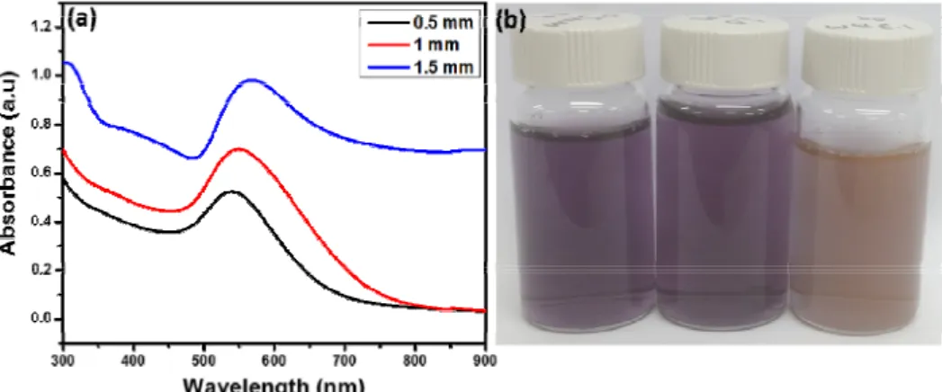
The color change of the reaction solution from yellow to violet after 30 min of reaction indicated the formation of AuNPs. This color originates due to the collective oscillation of the free electrons of the AuNPs interacting with light. UV-Vis spectroscopy is the principal characterization technique for metal nanoparticles. To determine the effect of the concentration of HAuCl4 on the synthesis, the UV-Vis spectra of AuNPs synthesized from the reaction of different concentrations of HAuCl4 (0.5, 1.0, or 1.5 mM) with 500 µL of Schisandra chenesis fruit extract were recorded after 45 min of reaction. Figure 1 shows the absorption peaks of AuNPs synthesized using different concentrations of HAuCl4 at 541, 548, and 566 nm. The increased intensity of the peaks with increasing concentration of HAuCl4
can clearly be observed. Since the increased absorbance can be attributed to the increase of the concentration or the size of NPs, it can be validated that high concentration of HAuCl4 produced more amount of Au atoms that were converted into NPs.
Moreover, the absorbance peak shifted from 541 ~ 566 nm with the increase in HAuCl4 concentration. Earlier studies demonstrated that the characteristic surface plasmon absorption peak of AuNPs occurs in the range of 520 ~ 550 nm depending on the size of particles. AuNPs synthesized using Rhus chinensis were found to have a particle size of 20 ~ 40 nm; their
Figure 1. (a) UV-Vis spectra of AuNPs synthesized by Schisandra chenesis fruit extract with different concentrations of HAuCl4 (0.5, 1.0, or 1.5 mM). (b) Photos of the synthesized AuNPs solutions.
corresponding surface plasmon absorbance peak was observed at 532 nm, and the intensity of the peak increased with increasing concentration of HAuCl4[13]. In this study using Schisandra chenesis fruit extract, the intensity of the peak increased and the absorbance peak was red-shifted. In general, as the size of NPs increases, the oscillation of the free electrons in the metal NPs becomes retarded, which in turn shifts the plasmonic absorbance toward longer wavelength direction. In this sense, the red-shifted absorbance may indicate the synthesis of larger AuNPs under high concentration of HAuCl4.
3.2. TEM analysis
Although UV-Vis spectra of AuNPs showed the increased and red-shifted absorbance with the concentration of HAuCl4, more detailed structural information could not be obtained from the UV-Vis spectra. Therefore, TEM analysis was performed to study the morphology of the AuNPs synthesized using Schisandra chenesis fruit extract in Figure 2(a ~ c). The AuNPs synthesized with an HAuCl4 concentration of 0.5 mM had mostly spherical and small plate structures with sizes of 10 ~ 20 nm, while the AuNPs synthesized with 1 mM HAuCl4 had spherical and plate structures with sizes ranging from 20 ~ 40 nm. In contrast, the AuNPs synthesized with 1.5 mM HAuCl4 were mostly triangular and hexagonal plates having sizes greater than 250 nm and small spherical and irregular nanoparticles with sizes of 50 ~ 100 nm.
This means that the size and shape of AuNPs became polydisperse under the high concentration of HAuCl4 (1.5 mM), i.e., larger triangular, hexagonal, and quasi-spherical AuNPs were formed. It has previously been reported that the use of plant
extracts with appropriate capping and stabilizing capacity leads to the formation of spherical AuNPs [23]. When the concentration of the gold precursor was lower, the resulting AuNPs were more spherical due to the sufficient capping and stabilizing ability of the Schisandra chenesis fruit extract at this concentration. As the concentration of gold precursor increased, triangularly shaped Au nanoparticles dominated, due to the chloride ions donated by HAuCl4.
3.3. DLS Analysis
The PSA results further supported the TEM analysis of the particle size of the AuNPs in Figure 2. The charge on the nanoparticles indicates their stability, which was measured as the zeta potential. The zeta potential value of the AuNPs synthesized using Schisandra chenesis fruit extract was determined to be -30.1 mV in Figure 3. The negative value of zeta potential indicated that the surface of AuNPs, which would be coated by Schisandra chenesis fruit extract, had negative charges. Since the Figure 2. TEM images (a ~ c) and PSA analyses (d ~ f) of AuNPs synthesized by Schisandra chenesis fruit extract with different concentrations
of HAuCl4 (0.5, 1.0, or 1.5 mM).
Figure 3. The zeta potential of the AuNPs synthesized with 1.5 mM of HAuCl4.
zeta potential value of ± 30 mV can be considered as the criteria for the good stabilization, it can be validated that the synthesized AuNPs have sufficient colloidal stability without noticeable aggregation.
3.4. XRD Analysis
Figure 4 depicts the XRD pattern of the AuNPs synthesized using Schisandra chenesis fruit extract. The XRD pattern shows Bragg reflection peaks at 38.2°, 44.3°, 64.6°, and 77.6°, which can be assigned to the (111), (200), (220), and (311) planes of the FCC structure of gold, respectively. We found the obtained XRD results were in a good agreement with the standard diffraction pattern of gold metal (Joint Committee on Powder Diffraction Standards-JCPDS no: 04-0784). The intense peak at 38.2° reveals the predominant growth of the (111) plane of FCC gold. Since XRD patterns provide the detailed crystalline information of given nanoparticles, the absence of other peaks in Figure 4 confirmed the purity of the crystalline phase of AuNPs.
3.5. EDX analysis
The EDX spectrum in Figure 5 shows a strong peak near 2 KeV, which is characteristic of elemental gold. Previously reported AuNPs showed similar EDX analysis results [26]. The elements Cl, C, O, and N were also observed, which might be due to Schisandra chenesis fruit extract attached to the AuNPs during the synthesis. The weight percentage of Au found from the EDX analysis was 80%.
3.6. Catalytic activity for reduction of 4-nitrophenol
After the morphological studies and characterization of the AuNPs synthesized using Schisandra chenesis fruit extract, we then carried out the reduction of 4-nitrophenol to 4-aminophenol using AuNPs to investigate their catalytic activity. An aqueous solution of 4-nitrophenol showed an absorption peak at 320 nm.Upon the addition of freshly prepared NaBH4, the 4-nitrophenol solution became bright yellow and its absorbance peak shifted to 400 nm; this color change and the peak shift were due to 4-nitrophenolate anions. It needs to be noted that the reduction of 4-nitrophenol to 4-aminophenol is thermodynamically favorable, but kinetically inhibited by the high activation energy [24].
Therefore, the reduction did not occur in the absence of AuNPs. Upon the addition of AuNPs, the yellow color of 4-nitrophenolate anions began to fade. The reduction of the 4-nitrophenolate anions to 4-aminophenol was monitored using UV-Vis spectroscopy. In Figure 6, the characteristic peak of 4-nitrophenol at 400 nm diminished within 27 min. In addition, new absorption peak at 300 nm, which corresponds to the absorbance of 4-aminophenol, newly appeared and increased in
the intensity with the decreased absorbance of 4-nitrophenol at 400 nm. These results indicate that the AuNPs synthesized with Schisandra chinensis fruit extract show good catalytic activity.
4. Conclusions
In summary, we developed a simple, facile, and green synthetic method for the synthesis of AuNPs, using Schisandra chinensis fruit extract. The shape and size of the AuNPs could be controlled by the concentration of HAuCl4 and Schisandra chinensis fruit extract. Their morphology and size distribution were analyzed using TEM and PSA analysis. The study revealed that the HAuCl4 concentration used during the AuNP synthesis the shape and size of the AuNPs could be controlled by the concentration of HAuCl4 and Schisandra chinensis fruit extract as a surfactant. In the low HAuCl4 concentration (0.5 mM), the synthesized Au NPs had mostly spherical and small plate structures with sizes of 10 ~ 20 nm. By increasing the HAuCl4
concentration up to 1.5 mM, Au NPs with triangular and hexagonal plates having sizes greater than 250 nm became dominant. The crystalline nature of the AuNPs was confirmed using XRD analysis, which revealed the predominant growth of the (111) plane of FCC gold. When the synthesized Au NPs were applied to a model catalytic reaction, they efficiently catalyzed the reduction of 4-nitrophenol to 4-aminophenol, revealing their practical usefulness.
Acknowledgement
This work was supported by a Research Grant of Pukyong National University (2019).
References
1. Zhang, Q., Li, W., Moran, C., Zeng, J., Chen, J., Wen, L-P., and Xia, Y., “Seed-Mediated Synthesis of Ag Nanocubes with Controllable Edge Lengths in the Range of 30-200 nm and Comparison of Their Optical Properties,” J. Am. Chem. Soc., 132, 11372-11378 (2010).
2. Pietrobon, B., and Kitaev, V., “Photochemical Synthesis of Monodisperse Size-Controlled Silver Decahedral Nanoparticles and Their Remarkable Optical Properties,” Chem. Mater, 20(16), 5186-5190 (2008).
3. Zhang, J., Langille, M. R., and Mirkin, C. A., “Photomediated Synthesis of Silver Triangular Bipyramids and Prisms: The Effect of pH and BSPP,” J. Am. Chem. Soc, 132(35), 12502-12510 (2010).
4. Crespo-Biel, O., Ravoo, B. J., Huskens, J., and Reinhoudt, D. N., “Writing with Molecules on Molecular Printboards,”
Dalton. Trans, 23, 2737-2741 (2006).
5. Gupta, K., Jana, P. C., and Meikap, A. K., “Optical and Electrical Transport Properties of Polyaniline-Silver Nanocomposite,” Synth. Met. 160(13), 1566-1573 (2010).
6. Liz-Marzan, L. M., “Tailoring Surface Plasmons through the Morphology and Assembly of Metal Nanoparticles,” Langmuir, 22(1), 32-41 (2006).
7. Manivasagan, P., Bharathiraja, S., Moorthy, M. S., Oh, Y-O., Song, K., Seo, H., and Oh, J., “Anti-EGFR Antibody Conjugation of Fucoidan-Coated Gold Nanorods as Novel Photothermal Ablation Agents for Cancer Therapy,” ACS Appl. Interfaces, 9(17), 14633-14646 (2017).
8. Manivasagan, P., Bharathiraja, S., Bui, N. Q., Jang, B., Oh, Y-O., Lim, I. G., and Oh, J., “Doxorubicin-Loaded Fucoidan Capped Gold Nanoparticles for Drug Delivery and Photoacoustic Imaging,” Int. J. Biol. Macromol, 91, 578-588 (2016).
9. Sun, H., Yuan, Q., Zhang, B., Ai, K., Zhang, P., and Lu, L.,
“Gd Functionalized Gold Nanorods for Multimodal Imaging Application,” Nanoscale, 3, 1990-1996 (2011).
10. Xia, Y., Xiong, Y., Lim, B., and Skrabalak, S. E.,
“Shape-Controlled Synthesis of Metal Nanocrystals: Simple Chemistry Meets Complex Physics?” Angew. Chem. Int. Ed., 48(1), 60-103 (2009).
11. Varma, R. S., “Greener Approach to Nanomaterials and Their Sustainable Applications,” Current Opinion in Chem. Eng., 1(2), 123-128 (2012).
12. Rokade, A. A., Kim, J. H., Lim, S. R., Yoo, S. I., Jin, Y.
E., and Park, S. S., “A Novel Green Synthesis of Silver Nanoparticles Using Rubus crataegifolius Bge Fruit Extract,”
J. Clus. Sci., 28(4), 2017-2026 (2017).
13. Patil, M. P., Ngabire, D., Thi, H. H. P., Kim, M-D., and Kim, G-D., “Eco-friendly Synthesis of Gold Nanoparticles and Evaluation of Their Cytotoxic on Cancer Cells,” J. Clus.
Sci., 28(1), 119-132 (2017).
14. Patil, M. P., Rokade, A. A., Ngabire, D., and Kim, G-D.,
“Green Synthesis of Silver Nanoparticles Using Water Extract from Galls of Rhus Chinensis and Its Antibacterial Activity,”
J. Clus. Sci., 27(5), 1737-1750 (2016).
15. Sharma, R. K., Gulati, S., and Mehata, S., “Preparation of Gold Nanoparticles Using Tea: A Green Chemistry
Experiment,” J. Chem. Educ., 89(10), 1316-1318 (2012).
16. Shankar, S. S., Rai, A., Ahmad, A., and Sastry, M.,
“Controlling the Optical Properties of Lemongrass Extract Synthesized Gold Nanotriangles and Potential Application in Infrared-Absorbing Optical Coatings,” Chem. Mater., 17(3), 566-572 (2005).
17. Xie, J., Lee, J. Y., and Wang, D. I. C., “Synthesis of Single-Crystalline Gold Nanoplates in Aqueous Solutions through Biomineralization by Serum Albumin Protein,” J.
Phys. Chem. C, 111(28), 10226-10232 (2007).
18. El-Seedi, H. R., El-Shabasy, R. M., Khalifa, S. A. M., Saeed, A., Shah, A., Shah, R., Iftikhar, F. J., Abdel-Daim, M. M., Omri, A., Hajrahand, N. H., Sabir, J. S. M., Zou, X., Halabi, M. F., Sarhan, W., and Guo, W., “Metal Nanoparticles Fabricated by Green Chemistry Using Natural Extracts:
Biosynthesis, Mechanisms, and Applications,” RSC Adv., 9, 24539-24559 (2019).
19. Yong, C., Jinbao, T., Xiaoke, W., Fengxiang, S., and Shujuan, L., “An Immunostimulatory Polsaccharide (SCP-IIa) from the Fruit of Schisandra (Turcz.) Brazil,” Inter. J. Biol. Macromol, 50(3), 844-848 (2012).
20. Tafesh, A. M., and Weiguny, J., “A Riview of the Selective Catalytic Reduction of Aromatic Nitro Compounds into Arometic Amines, Isocyanates, and Ureas Using CO,” Chem.
Rev., 96(6), 2035-2052 (1996).
21. Esumi, K., Isono, R., and Yoshimura, T., “Preparation of PAMAN- and PPI-Metal (Silver, Platinum, and Palladium) Nanocomposites and Their Catalytic Activities for Reduction of 4-Nitrophenol,” Langmuir, 20(1), 237-243 (2004).
22. Pradhan, N., Pal, A., and Pal, T., “Silver Nanoparticle Catalyzed Reduction of Aromatic Nito compounds,” Colloids Surf. A, 196(2), 247-257 (2002).
23. Philip, D., “Rapid Green Synthesis of Spherical Gold Nanoparticles Using Mangifera Indica Leaf,” Spectrochimica Acta Part A, 77(4), 807-810 (2010).
24. Liang, M., Su, R., Huang, R., Qi, W., Yu, Y., Wang, L., and He, Z., “Facile in Situ Synthesis of silver Nanoparticles on Procyanidin-Grafted Eggshell Membrane and Their Catalytic Properties,” ACS Appl. Mater. Interfaces, 6(7), 4638-4649 (2014).