INTRODUCTION
Bicuspid aortic valve (BAV) is the most common congenital heart valve disease, with an overall incidence of approximate- ly 1% in the general population.1,2 Subjects with BAV often pres- ent with aortic dilatation and may exhibit mechanical function- al alterations in vasculatures.3,4 In addition, BAV is a highly heritable trait, often associated with other congenital heart defects or genetic syndromes.5,6
Regarding myocardial characteristics in subjects with BAV, most previous studies have focused on subclinical left ven-
Prevalence, Characteristics, and Clinical Significance of Concomitant Cardiomyopathies in Subjects with Bicuspid Aortic Valves
Hyeonju Jeong1, Chi Young Shim2, Darae Kim3, Jah Yeon Choi2, Kang-Un Choi2, Soo Youn Lee4, Geu-Ru Hong2, and Jong-Won Ha2
1Division of Cardiology, Department of Internal Medicine, Myongji Hospital, Hanyang University College of Medicine, Goyang;
2Division of Cardiology, Department of Internal Medicine, Severance Cardiovascular Hospital, Yonsei University College of Medicine, Seoul;
3Department of Medicine, Heart Vascular Stroke Institute, Samsung Medical Center, Sungkyunkwan University School of Medicine, Seoul;
4Division of Cardiology, Department of Internal Medicine, Sejong General Hospital, Bucheon, Korea.
Purpose: The present study aimed to investigate the prevalence, characteristics, and clinical significance of concomitant specific cardiomyopathies in subjects with bicuspid aortic valves (BAVs).
Materials and Methods: A total of 1186 adults with BAV (850 males, mean age 56±14 years) at a single tertiary center were com- prehensively reviewed. Left ventricular non-compaction, hypertrophic cardiomyopathy, and idiopathic dilated cardiomyopathy were confirmed when patients fulfilled current clinical and echocardiographic criteria. Clinical and echocardiographic charac- teristics, including comorbidities, heart failure presentation, BAV morphology, function, and aorta phenotypes, in BAV subjects with or without specific cardiomyopathies were compared.
Results: Overall, 67 subjects (5.6%) had concomitant cardiomyopathies: 40 (3.4%) patients with left ventricular non-compaction, 17 (1.4%) with hypertrophic cardiomyopathy, and 10 (0.8%) with dilated cardiomyopathy. BAV subjects with hypertrophic car- diomyopathy had higher prevalences of diabetes mellitus and heart failure with preserved ejection fraction, and tended to have type 0 phenotype, while BAV subjects with dilated cardiomyopathy showed higher prevalences of chronic kidney disease and heart failure with reduced ejection fraction. BAV subjects with left ventricular non-compaction were significantly younger and predominantly male, and had greater BAV dysfunction and a higher prevalence of normal aorta shape. In multiple regression analysis, cardiomyopathy was independently associated with heart failure (odds ratio 2.795, 95% confidential interval 1.603–
4.873, p<0.001) after controlling for confounding factors.
Conclusion: Concomitant cardiomyopathies were observed in 5.6% of subjects with BAV. A few different clinical and echocardio- graphic characteristics were found. The presence of cardiomyopathy was independently associated with heart failure.
Key Words: Bicuspid aortic valve, hypertrophic cardiomyopathy, non-compaction, dilated cardiomyopathy
pISSN: 0513-5796 · eISSN: 1976-2437
Received: March 25, 2019 Revised: June 28, 2019 Accepted: July 15, 2019
Corresponding author: Chi Young Shim, MD, PhD, Division of Cardiology, De- partment of Internal Medicine, Severance Cardiovascular Hospital, Yonsei Univer- sity College of Medicine, 50-1 Yonsei-ro, Seodaemun-gu, Seoul 03722, Korea.
Tel: 82-2-2228-8453, Fax: 82-2-2227-7742, E-mail: [email protected]
•The authors have no potential conflicts of interest to disclose.
© Copyright: Yonsei University College of Medicine 2019
This is an Open Access article distributed under the terms of the Creative Com- mons Attribution Non-Commercial License (https://creativecommons.org/licenses/
by-nc/4.0) which permits unrestricted non-commercial use, distribution, and repro- duction in any medium, provided the original work is properly cited.
Yonsei Med J 2019 Sep;60(9):816-823 https://doi.org/10.3349/ymj.2019.60.9.816
tricular dysfunction associated with increased aortic stiff- ness.7-9 Although a possible association between BAV and spe- cific cardiomyopathies (CMs) based on common genetic traits has been proposed in several case reports,10,11 data on the prev- alence of coexisting specific CMs in subjects with BAV are lim- ited. In a previous study, an incidence of 11% for left ventricular non-compaction (LVNC) in 109 patients with BAV was report- ed.12 In a recent large population study, a prevalence of only 0.4% for hypertrophic CM (HCM) was reported in 5430 pa- tients with BAV, similar to the general population.13 However, the prevalence of concomitant specific CMs might be differ- ent based on ethnicity. Moreover, data are lacking regarding clinical and echocardiographic characteristics based on the type of concomitant CMs. Therefore, in the present study, the prevalence, characteristics, and clinical significance of concomi- tant CMs, including LVNC, HCM, and idiopathic dilated CM (DCM), were determined using a large Korean BAV registry.
MATERIALS AND METHODS
Study population
We retrospectively reviewed subjects diagnosed with BAV us- ing transthoracic echocardiography in Severance Cardiovas- cular Hospital from January 2003 to December 2017. A total of 1186 subjects (850 males, mean age 56±14 years) were includ- ed in this study. All echocardiographic studies in subjects with BAV were manually reviewed for confirmation. Patient medi- cal data as recorded by the physicians were carefully reviewed by two experienced observers who were blinded to echocar- diography results. Patients who presented with acute heart fail- ure were not included in the present study. The institutional review board of Severance Hospital approved the present study (IRB No. 2015-0403-001), which was conducted in compli- ance with the Declaration of Helsinki. The subjects were clas- sified into four groups based on the presence of specific CMs.
Standard two-dimensional and Doppler measurements were performed following the American Society of Echocar- diography guidelines.14 BAV was diagnosed based on anatom- ic evaluation of the aortic valve, when only two cusps were un- equivocally identified in systole and diastole in the short-axis view and with a clear “fish mouth” appearance during systo- le.15 BAV morphology was classified into four types based on position and pattern of raphe and cusps: Type 1 exhibited fu- sion of the left coronary and right coronary cusps, type 2 indi- cated fusion of the right coronary and noncoronary cusps, and type 3 comprised fusion of the left coronary and noncoronary cusps. Type 0 was recorded when there were two developed cusps and no raphe (true type).15,16 The severity of aortic ste- nosis or aortic regurgitation was assessed using integrated ap- proaches.17,18 The dimensions of the sinus of Valsalva, sinotu- bular junction, and ascending aorta were measured as previously described.7,15 The presence of aortopathy was defined as an
ascending aorta dimension ≥40 mm, as previously defined.
HCM was clinically diagnosed based on the presence of un- explained myocardial hypertrophy (wall thickness ≥15 mm) in the absence of local or systemic etiologies capable of pro- ducing the extent of hypertrophy evident.13 Mild systemic hy- pertension was not an exclusion criterion in the diagnosis of HCM. Subjects with coexisting HCM were subdivided into three morphologically obstructive HCM subgroups, non-ob- structive HCM, non-apical HCM, or non-obstructive apical HCM, based on either the presence of left ventricular outflow track obstruction or the predominant hypertrophy at the left ventricular apex. LVNC was diagnosed based on previously suggested echocardiographic criteria19-21 and/or ratios between noncompacted and compacted layers of the left ventricular wall on cardiac magnetic resonance imaging.22 If the diagno- sis of LVNC was suspected but not confirmed using echocar- diography, cardiac magnetic resonance imaging was performed.
DCM was defined as an ejection fraction <40% in the presence of increased left ventricular dimension. Subjects with ischemic heart disease, uncorrected or corrected severe aortic stenosis or aortic regurgitation, and other reversible causes were ex- cluded from diagnosis of idiopathic DCM in this study. Echo- cardiographic data were gathered and analyzed by experienced sonographers blinded to each patient’s clinical data. Heart fail- ure was diagnosed using current diagnostic criteria23 and was categorized based on left ventricular ejection fraction (LVEF) as follows: heart failure with preserved ejection fraction (LVEF
≥50%) and heart failure with reduced ejection fraction (LVEF
<50%).
Continuous variables are expressed as a mean±standard de- viation. Categorical variables are expressed as a number (per- centage). Comparisons between groups were performed us- ing standard λ2 tests for categorical variables and paired t-tests for continuous variables. Univariate and multivariate logistic regression analyses were performed. All statistical analyses were performed using SPSS Statistics, version 23.0 (IBM, Ar- monk, NY, USA). p values <0.05 were considered statistically significant.
RESULTS
Prevalence of coexistent CMs in BAV subjects
Overall, 67 subjects (5.6%) had concomitant CMs: 10 (0.8%) subjects with DCM, 17 (1.4%) with HCM, and 40 (3.4%) with LVNC. Among the subjects with coexistent HCM, five had ob- structive HCM, six presented with non-obstructive HCM, and six with apical HCM (Fig. 1).
Characteristics of BAV subjects with HCM, LVNC, or DCM
Baseline characteristics of the subjects with or without specif- ic CMs are shown in Table 1. Subjects with DCM had a higher
prevalence of chronic kidney disease and heart failure with reduced ejection fraction, compared with those without CM.
Subjects with HCM showed higher prevalences of diabetes mellitus and heart failure with preserved ejection fraction than those without CM. Subjects with LVNC were younger and pre- dominantly male, compared with those without CM. In addi- tion, subjects with LVNC exhibited a lower prevalence of heart failure than patients with DCM or HCM.
Table 2 shows the structural and functional characteristics of the left ventricle in each group. The echocardiographic vari- ables, including left ventricle dimension, wall thickness, and LVEF, were significantly different among the groups because of their own disease characteristics. LA volume index values in BAV subgroups with specific CMs were significantly greater
than those in the BAV group without CMs. Early diastolic mi- tral annular tissue (e’) velocity and ratio of early diastolic mi- tral inflow velocity to e’ velocity (E/e’) in BAV subgroups with HCM and DCM were also significantly greater than those in BAV subgroups without CMs. However, right ventricular systol- ic pressure was not significantly different among the groups.
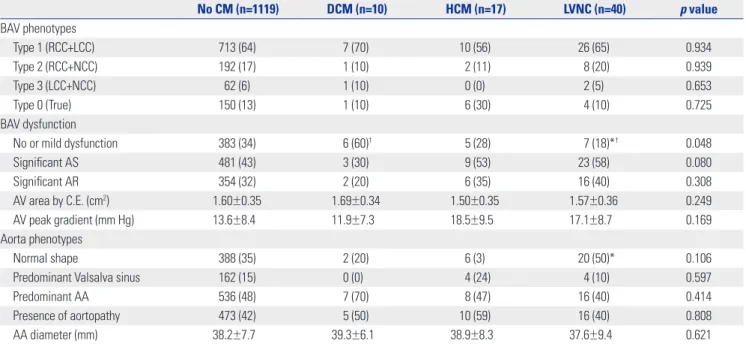
The BAV characteristics in Table 3 and Fig. 2 show that the type 1 BAV phenotype (fusion of right and left coronary cusps) was the most prevalent morphology in all groups. Although subjects with HCM tended to have type 0 phenotype, there was no statistically significant differences in BAV phenotypes among the groups. Subjects with DCM comprised a higher prevalence of no or mild dysfunction, while subjects with LVNC exhibited a lower prevalence of no or mild dysfunction. Regarding aorta Table 1. Baseline Characteristics according to the Presence of Specific Cardiomyopathy
No CM (n=1119) DCM (n=10) HCM (n=17) LVNC (n=40) p value
Age (yr) 56±15 56±5 60±13 51±16*‡ 0.097
Male 793 (71) 8 (80) 13 (77) 35 (88)* 0.135
Body mass index (kg/m2) 23.9±3.6 23.7±3.5 24.7±2.2 23.4±3.2 0.666
Hypertension 462 (41) 3 (30) 10 (59) 17 (43) 0.444
Diabetes mellitus 209 (19) 3 (30) 9 (52)* 4 (10)‡ 0.001
Dyslipidemia 231 (21) 1 (10) 3 (18) 10 (25) 0.742
CAD 182 (16) 0 (0) 2 (12) 5 (13) 0.464
CKD 97 (9) 3 (30)* 2 (12) 4 (10) 0.124
Heart failure 178 (16) 6 (60)* 8 (47) 8 (20)†‡ <0.001
Preserved EF 129 (12) 0 (0) 8 (47)*† 6 (15)‡ <0.001
Reduced EF 49 (4) 6 (60)* 0 (0)† 2 (5)† <0.001
CM, cardiomyopathy; DCM, dilated cardiomyopathy; HCM, hypertrophic cardiomyopathy; LVNC, left ventricular non-compaction; CAD, coronary artery disease;
CKD, chronic kidney disease; EF, ejection fraction.
Values are presented as mean±standard deviation or n (%) unless otherwise indicated.
*p<0.05 compared to the group with no CM; †p<0.05 compared to the DCM group; ‡p<0.05 compared to the HCM group.
BAV subjects (n=1186)
BAV with CM (n=67, 5.6%) Obstructive
(n=5, 7.4%)
Non-obstructive (n=6, 9.0%)
Apical (n=6, 9.0%)
BAV without CM
(n=1119, 94.4%) LVNC
(n=40, 59.7%) HCM (n=17, 25.4%) DCM
(n=10, 14.9%) DCM (n=10, 0.8%)
LVNC (n=40, 3.4%) HCM (n=17, 1.4%)
BAV subjects with CM (n=67)
A B
DCM HCM LVNC
DCM HCM LVNC
Fig. 1. Prevalence of specific cardiomyopathies in bicuspid aortic valves (BAV) subjects. (A) A total of 1186 BAV subjects. (B) Sixty-seven BAV subjects with cardiomyopathy (CM). LVNC, left ventricular non-compaction; DCM, dilated cardiomyopathy; HCM, hypertrophic cardiomyopathy.
phenotypes, subjects with LVNC revealed a higher prevalence of normal shape than those without CMs. Although statistical significance was not observed, more DCM patients had pre- dominant AA phenotype, and none showed predominant si- nus of Valsalva. Fig. 3 illustrates the representative cases of co- existing CMs in subjects with BAV.
Clinical significance of concomitant CMs in BAV Logistic regression analysis was performed to investigate fac- tors associated with heart failure in subjects with BAV. In uni- variate analysis, increased age was significantly associated with
heart failure [odds ratio (OR) 1.035, p<0.001], while gender was not. Comorbidities, such as hypertension (OR 1.793, p<0.001), diabetes mellitus (OR 1.704, p=0.003), and chronic kidney dis- ease (OR 2.226, p<0.001), were significantly associated with heart failure. Aortic valve dysfunction was not a significant as- sociating factor for heart failure. However, an increased aorta diameter (OR 1.020, p=0.046) was significantly associated with heart failure. Moreover, presence of CM was the strongest factor associated with heart failure (OR 2.582, p<0.001). In mul- tivariate analysis, age, hypertension, chronic kidney disease, and presence of concomitant CM were independently associ-
Table 2. Left Ventricle Characteristics according to the Presence of Specific Cardiomyopathy
No CM (n=1119) DCM (n=10) HCM (n=17) LVNC (n=40) p value
LVEDD (mm) 53.5±9.1 68.7±10.3* 50.7±9.9† 59.0±10.2*†‡ <0.001
LVESD (mm) 36.3±8.7 59.2±9.8* 31.9±8.1*† 41.9±9.5*†‡ <0.001
IVS thickness (mm) 10.7±2.2 10.1±2.6 14.7±4.2*† 10.7±2.3‡ <0.001
PW thickness (mm) 10.5±2.0 10.3±2.2 12.2±2.3*† 10.7±2.0‡ 0.005
LV ejection fraction (%) 63±12 29±11* 70±8*† 58±12*†‡ <0.001
LA volume index (mL/m2) 32.0±16.4 46.9±21.4* 36.0±14.4 37.7±16.4* 0.004
e’ velocity (cm/s) 6.1±2.5 3.5±1.4* 4.1±1.6* 6.7±3.0†‡ <0.001
E/e’ 13.0±5.9 20.0±6.6* 17.5±7.3* 12.8±5.4†‡ <0.001
RVSP (mm Hg) 29±11 33±12 25±5 29±10 0.526
CM, cardiomyopathy; DCM, dilated cardiomyopathy; HCM, hypertrophic cardiomyopathy; LVNC, left ventricular non-compaction; LVEDD, left ventricular end-dia- stolic dimension; LVESD, left ventricular end-systolic dimension; IVS, interventricular septum; PW, posterior wall; LV, left ventricle; LA, left atrium; e’, early dia- stolic mitral annular tissue; E/e’, ratio of early diastolic mitral inflow velocity to early diastolic mitral annular tissue velocity; RVSP, right ventricular systolic pres- sure.
Values are presented as mean±standard deviation unless otherwise indicated.
*p<0.05 compared to the group with no CM; †p<0.05 compared to the DCM group; ‡p<0.05 compared to the HCM group.
Table 3. BAV and Aorta Characteristics according to the Presence of Specific Cardiomyopathy
No CM (n=1119) DCM (n=10) HCM (n=17) LVNC (n=40) p value
BAV phenotypes
Type 1 (RCC+LCC) 713 (64) 7 (70) 10 (56) 26 (65) 0.934
Type 2 (RCC+NCC) 192 (17) 1 (10) 2 (11) 8 (20) 0.939
Type 3 (LCC+NCC) 62 (6) 1 (10) 0 (0) 2 (5) 0.653
Type 0 (True) 150 (13) 1 (10) 6 (30) 4 (10) 0.725
BAV dysfunction
No or mild dysfunction 383 (34) 6 (60)† 5 (28) 7 (18)*† 0.048
Significant AS 481 (43) 3 (30) 9 (53) 23 (58) 0.080
Significant AR 354 (32) 2 (20) 6 (35) 16 (40) 0.308
AV area by C.E. (cm2) 1.60±0.35 1.69±0.34 1.50±0.35 1.57±0.36 0.249
AV peak gradient (mm Hg) 13.6±8.4 11.9±7.3 18.5±9.5 17.1±8.7 0.169
Aorta phenotypes
Normal shape 388 (35) 2 (20) 6 (3) 20 (50)* 0.106
Predominant Valsalva sinus 162 (15) 0 (0) 4 (24) 4 (10) 0.597
Predominant AA 536 (48) 7 (70) 8 (47) 16 (40) 0.414
Presence of aortopathy 473 (42) 5 (50) 10 (59) 16 (40) 0.808
AA diameter (mm) 38.2±7.7 39.3±6.1 38.9±8.3 37.6±9.4 0.621
BAV, bicuspid aortic valve; CM, cardiomyopathy; DCM, dilated cardiomyopathy; HCM, hypertrophic cardiomyopathy; LVNC, left ventricular noncompaction; RCC, right coronary cusp; LCC, left coronary cusp; NCC, non-coronary cusp; AS, aortic stenosis; AR, aortic regurgitation; AV, aortic valve; C.E., continuity equation; AA, ascending aorta.
Values are presented as mean±standard deviation or n (%) unless otherwise indicated.
*p<0.05 compared to the group with no CM; †p<0.05 compared to the DCM group.
100
80
60
40
20
0
100
80
60
40
20
Without CM With DCM With HCM With LVNC 0 Without CM With DCM With HCM With LVNC
% %
BAV phenotypes Aorta phenotypes
A B
Type 2
Type 1 Type 3 Type 0 Normal shape Predominant Valsalva sinus Predominant ascending aorta Fig. 2. (A) Bicuspid aortic valves (BAV) phenotypes according to the presence of specific cardiomyopathies (CMs). (B) Aorta phenotypes according to the presence of specific CMs. DCM, dilated cardiomyopathy; HCM, hypertrophic cardiomyopathy; LVNC, left ventricular non-compaction.
63.9 70
52.9
65
35.7 30 35.3
50
5.5 10
0
5
17.2 10
11.8
20
14.9
0
23.5
10
13.4 10
35.3
10
49.4
70
41.2 40
A
B
C
Fig. 3. Representative cases of coexisting cardiomyopathies in bicuspid aortic valves subjects. (A) Hypertrophic cardiomyopathy, (B) left ventricular non- compaction, and (C) dilated cardiomyopathy.
ated with heart failure (Table 4). Neither the presence of aor- topathy nor increased aorta diameter was independently as- sociated with heart failure in multivariate analysis.
DISCUSSION
The principal findings of the present study are as follows. First, approximately 6% of BAV subjects had concomitant CMs. LVNC was the most prevalent; however, its prevalence in BAV sub- jects was only 3.4% lower than that previously reported. Sec- ond, BAV subjects with specific CMs had distinct clinical and echocardiographic features. Third, age, hypertension, and pres- ence of CM were significantly associated with heart failure. To the best of our knowledge, this is the first study in which the prevalence, characteristics, and clinical implications of con- comitant specific CMs were identified in adults with BAV using data from a large Korean registry.
Case reports of specific CMs in BAV subjects have been spo- radic, and studies on their prevalences are limited. Agarwal, et al.12 reported the incidence of LVNC in patients with BAV. In their retrospective observational study, 12 of 109 BAV patients (11.0%) were diagnosed with LVNC based on echocardiograph- ic criteria. The mean age at diagnosis was 33±17 years. The in- cidence was greater and mean age at diagnosis was signifi- cantly lower values thereof than in our study. Chandra, et al.24 researched the incidence of LVNC in Caucasians and African- Americans, but not Asians, and revealed inter-racial differenc- es in the frequency thereof in BAV patients. Therefore, the dif- ferences in incidences between the present study and Agarwal, et al.12 may likely be due to population characteristics, includ- ing age and race. Padang, et al.13 reported the incidence of HCM in patients with BAV. In their retrospective cohort, 23 patients were diagnosed with HCM and BAV based on echocardio- graphic criteria. The mean age at diagnosis was 52±16 years.
Their study showed 0.9% of patients with HCM had coexistent BAV and 0.4% of patients with BAV had coexistent HCM. The incidence of BAV in the general population is usually approxi-
mately 1%; therefore, the incidence of BAV was not deemed dif- ferent between HCM and general populations. In our study, 1.4% of patients had concomitant BAV and HCM, and these results are similar to a previous study.13 In the present study, id- iopathic DCM was identified in 0.8% of BAV subjects, similar to the results found in the general population. To date, studies on the prevalence of DCM in BAV subjects have not been pub- lished, probably because a certain degree of aortic valve dysfunc- tion can result in chamber dilatation and myocardial dysfunc- tion. To eliminate confounding factors, patients with significant aortic valve dysfunction were excluded when diagnosing idio- pathic DCM. Therefore, the actual prevalence of coexisting DCM in BAV subjects might be underestimated in this study. In a pre- vious study, genetic factors were suggested as a possible cause of coexistent BAV and HCM.25 However, few studies exist on genes in patients with BAV and specific CMs. In the future, ge- netic analysis will be important in studying the relationship be- tween BAV and CM.
Recently, research into BAV has focused not only in the valve itself, but also in relation to the left ventricle and aorta.7-9 Since BAV patients with concomitant CMs have prominent charac- teristics of their own myocardial disease, there were no signif- icantly different characteristics in BAV morphology, aortic valve dysfunction, or aorta phenotype in overall group comparison.
Although type 0 BAV phenotype and presence of aortopathy tended to be more prevalent in BAV patients with HCM, these tendencies did not show statistical significance.
Predictably, the presence of specific CMs may influence a patient’s clinical course, especially in regards to heart failure in BAV subjects. Heart failure was associated with 16.9% of all BAV subjects and 32.8% of BAV subjects with specific CMs. In univariate analysis, age and well-known comorbidities were correlated with heart failure, as expected. Regarding several specific characteristics in BAV subjects, increased aortic di- ameter was positively associated with heart failure, while sig- nificant aortic valve dysfunction was not. These findings sup- port a possible ventricular vascular interaction in BAV subjects, which has been suggested in previous studies.3,7-9 However, the
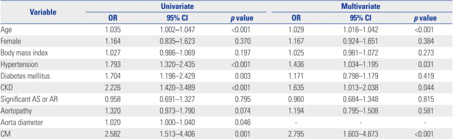
Table 4. Factors associated with Heart Failure in BAV Subjects
Variable Univariate Multivariate
OR 95% CI p value OR 95% CI p value
Age 1.035 1.002–1.047 <0.001 1.029 1.016–1.042 <0.001
Female 1.164 0.835–1.623 0.370 1.167 0.924–1.651 0.384
Body mass index 1.027 0.986–1.069 0.197 1.025 0.981–1.072 0.273
Hypertension 1.793 1.320–2.435 <0.001 1.436 1.034–1.195 0.031
Diabetes mellitus 1.704 1.196–2.429 0.003 1.171 0.798–1.179 0.419
CKD 2.226 1.420–3.489 <0.001 1.635 1.013–2.038 0.044
Significant AS or AR 0.958 0.691–1.327 0.795 0.960 0.684–1.348 0.815
Aortopathy 1.320 0.973–1.790 0.074 1.194 0.795–1.508 0.581
Aorta diameter 1.020 1.000–1.040 0.046 - - -
CM 2.582 1.513–4.406 0.001 2.795 1.603–4.873 <0.001
BAV, bicuspid aortic valve; OR, odds ratio; CI, confidence interval; CKD, chronic kidney disease; AS, aortic stenosis; AR, aortic regurgitation; CM, cardiomyopathy.
strongest factor associated with heart failure was the presence of CM. Consequently, in multivariate analysis, age, hyperten- sion, and presence of CM were independently associated with heart failure.
The present study had several limitations. First, this study included only Korean BAV subjects from a single tertiary re- ferral center, which may result in bias. Therefore, multinational studies including various ethnic groups are needed to evaluate the prevalence of CMs in BAV subjects. However, we believe this study was the first to report on the prevalence of concom- itant CMs in a large Korean registry using comprehensive re- views. Second, data were lacking regarding common genetic backgrounds in BAV patients with CMs. The results of this study may be a basis for future genetic research. Third, chronic he- modynamic effects of significant AS resulting in acquired LV hypertrophy or noncompaction cannot be fully excluded, al- though patients with severe AS were not included in this study.
Forth, aortic diameters were measured based on echocardio- graphic imaging alone because only some BAV subjects under- went computed tomography or cardiac magnetic resonance imaging.
In conclusion, concomitant BAV with CMs was observed in 5.6% of our BAV population. Several clinical and echocardio- graphic characteristics, including comorbidities, heart failure presentation, BAV phenotypes, valve function, and presence of aortopathy were found in these patients. The presence of CM was independently associated with heart failure.
ACKNOWLEDGEMENTS
This study was supported in part by a faculty research grant of Yonsei University College of Medicine (6-2012-0206).
AUTHOR CONTRIBUTIONS
Conceptualization: Chi Young Shim. Data curation: Soo Youn Lee and Hyeonju Jeong. Formal analysis: Hyeonju Jeong and Chi Young Shim. Investigation: Hyeonju Jeong. Methodology: Hyeonju Jeong and Chi Young Shim. Project administration: Chi Young Shim. Re- sources: Hyeonju Jeong, Chi Young Shim, Darae Kim, Jah Yeon Choi, Kang-Un Choi, Soo Youn Lee, Geu-Ru Hong, and Jong-Won Ha. Soft- ware: Hyeonju Jeong and Chi Young Shim.
ORCID iDs
Hyeonju Jeong https://orcid.org/0000-0002-7916-3624 Chi Young Shim https://orcid.org/0000-0002-6136-0136 Darae Kim https://orcid.org/0000-0003-3284-0904 Jah Yeon Choi https://orcid.org/0000-0002-6793-4137 Kang-Un Choi https://orcid.org/0000-0002-3385-3152 Soo Youn Lee https://orcid.org/0000-0002-1241-891X Geu-Ru Hong https://orcid.org/0000-0003-4981-3304 Jong-Won Ha https://orcid.org/0000-0002-8260-2958
REFERENCES
1. Ward C. Clinical significance of the bicuspid aortic valve. Heart 2000;83:81-5.
2. Prakash SK, Bossé Y, Muehlschlegel JD, Michelena HI, Limongelli G, Della Corte A, et al. A roadmap to investigate the genetic basis of bicuspid aortic valve and its complications: insights from the international BAVCon (Bicuspid Aortic Valve Consortium). J Am Coll Cardiol 2014;64:832-9.
3. Shim CY, Cho IJ, Yang WI, Kang MK, Park S, Ha JW, et al. Central aortic stiffness and its association with ascending aorta dilation in subjects with a bicuspid aortic valve. J Am Soc Echocardiogr 2011;24:847-52.
4. Kim M, Shim CY, You SC, Cho IJ, Hong GR, Ha JW, et al. Charac- teristics of carotid artery structure and mechanical function and their relationships with aortopathy in patients with bicuspid aor- tic valves. Front Physiol 2017;8:622.
5. Niaz T, Poterucha JT, Johnson JN, Craviari C, Nienaber T, Palfree- man J, et al. Incidence, morphology, and progression of bicuspid aortic valve in pediatric and young adult subjects with coexisting congenital heart defects. Congenit Heart Dis 2017;12:261-9.
6. Niaz T, Poterucha JT, Olson TM, Johnson JN, Craviari C, Nienaber T, et al. Characteristic morphologies of the bicuspid aortic valve in patients with genetic syndromes. J Am Soc Echocardiogr 2018;31:194-200.
7. Lee SY, Shim CY, Hong GR, Seo J, Cho I, Cho IJ, et al. Association of aortic phenotypes and mechanical function with left ventricu- lar diastolic function in subjects with normally functioning bicus- pid aortic valves and comparison to subjects with tricuspid aortic valves. Am J Cardiol 2015;116:1547-54.
8. Weismann CG, Lombardi KC, Grell BS, Northrup V, Sugeng L.
Aortic stiffness and left ventricular diastolic function in children with well-functioning bicuspid aortic valves. Eur Heart J Cardio- vasc Imaging 2016;17:225-30.
9. Lee SY, Shim CY, Hong GR, Cho IJ, Chang HJ, Ha JW, et al. Deter- minants and prognostic significance of symptomatic status in pa- tients with moderately dysfunctional bicuspid aortic valves. PLoS One 2017;12:e0169285.
10. Feizi O, Farrer Brown G, Emanuel R. Familial study of hypertro- phic cardiomyopathy and congenital aortic valve disease. Am J Cardiol 1978;41:956-64.
11. Brown PS Jr, Roberts CS, McIntosh CL, Roberts WC, Clark RE.
Combined obstructive hypertrophic cardiomyopathy and stenot- ic congenitally bicuspid aortic valve. Am J Cardiol 1990;66:1273-5.
12. Agarwal A, Khandheria BK, Paterick TE, Treiber SC, Bush M, Ta- jik AJ. Left ventricular noncompaction in patients with bicuspid aortic valve. J Am Soc Echocardiogr 2013;26:1306-13.
13. Padang R, Gersh BJ, Ommen SR, Geske JB. Prevalence and im- pact of coexistent bicuspid aortic valve in hypertrophic cardiomy- opathy. Heart Lung Circ 2018;27:33-40.
14. Lang RM, Badano LP, Mor-Avi V, Afilalo J, Armstrong A, Ernande L, et al. Recommendations for cardiac chamber quantification by echocardiography in adults: an update from the American Soci- ety of Echocardiography and the European Association of Cardio- vascular Imaging. Eur Heart J Cardiovasc Imaging 2015;16:233-70.
15. Lee SY, Shim CY, Kim D, Cho I, Hong GR, Ha JW, et al. Factors de- termining aortic valve dysfunction in Korean subjects with a bi- cuspid aortic valve. Am J Cardiol 2017;119:2049-55.
16. Roberts WC, Ko JM. Frequency by decades of unicuspid, bicus- pid, and tricuspid aortic valves in adults having isolated aortic valve replacement for aortic stenosis, with or without associated aortic regurgitation. Circulation 2005;111:920-5.
17. Zoghbi WA, Enriquez-Sarano M, Foster E, Grayburn PA, Kraft CD, Levine RA, et al. Recommendations for evaluation of the severity of native valvular regurgitation with two-dimensional and Dop- pler echocardiography. J Am Soc Echocardiogr 2003;16:777-802.
18. Baumgartner H, Hung J, Bermejo J, Chambers JB, Evangelista A, Griffin BP, et al. Echocardiographic assessment of valve stenosis:
EAE/ASE recommendations for clinical practice. J Am Soc Echo- cardiogr 2009;22:1-23.
19. Jenni R, Oechslin E, Schneider J, Attenhofer Jost C, Kaufmann PA.
Echocardiographic and pathoanatomical characteristics of isolat- ed left ventricular non-compaction: a step towards classification as a distinct cardiomyopathy. Heart 2001;86:666-71.
20. Chin TK, Perloff JK, Williams RG, Jue K, Mohrmann R. Isolated noncompaction of left ventricular myocardium. A study of eight cases. Circulation 1990;82:507-13.
21. Stöllberger C, Finsterer J, Blazek G. Left ventricular hypertrabecu- lation/noncompaction and association with additional cardiac
abnormalities and neuromuscular disorders. Am J Cardiol 2002;
90:899-902.
22. Petersen SE, Timperley J, Neubauer S. Left ventricular thrombi in a patient with left ventricular non-compaction in visualisation of the rationale for anticoagulation. Heart 2005;91:e4.
23. Yancy CW, Jessup M, Bozkurt B, Butler J, Casey DE Jr, Colvin MM, et al. 2017 ACC/AHA/HFSA focused update of the 2013 ACCF/
AHA Guideline for the management of heart failure: a report of the American College of Cardiology/American Heart Association Task Force on Clinical Practice Guidelines and the Heart Failure Society of America. J Am Coll Cardiol 2017;70:776-803.
24. Chandra S, Lang RM, Nicolarsen J, Gayat E, Spencer KT, Mor-Avi V, et al. Bicuspid aortic valve: Inter-racial difference in frequency and aortic dimensions. JACC Cardiovasc Imaging 2012;5:981-9.
25. Somerville J, McDonald L. Congenital anomalies in the heart with hypertrophic cardiomyopathy. Br Heart J 1968;30:713-22.