http://dx.doi.org/10.5468/ogs.2016.59.1.17 pISSN 2287-8572 · eISSN 2287-8580
Introduction
Elective prophylactic cervical cerclage is usually performed in women who have a history of spontaneous second-trimester miscarriages and preterm delivery or those who underwent cervical procedures such as conization that may cause cervical incompetence [1]. However, not all cerclage operations re- sulted in full term delivery; some patients delivered as preterm even after a cerclage operation. A recent study showed that women who underwent the procedure, regardless of whether
Post-cerclage ultrasonographic cervical length can predict preterm delivery in elective cervical cerclage patients
Rok Katerina Song
1, Hyun-Hwa Cha
2, Mi-Young Shin
1, Suk-Joo Choi
1, Soo-young Oh
1, Jong-Hwa Kim
1, Cheong-Rae Roh
1Department of Obstetrics and Gynecology, 1Samsung Medical Center, Sungkyunkwan University School of Medicine, Seoul; 2Kyungpook National University Hospital, Kyungpook National University School of Medicine, Daegu, Korea
Objective
To evaluate the usefulness of transvaginal ultrasound measurements of cervical length before and after elective prophylactic cervical cerclage in predicting preterm delivery before 32 weeks of gestation.
Methods
Women who underwent an elective cerclage operation at 14 to 19 weeks of gestation and who delivered between January 2004 and December 2009 were enrolled in this study (n=52). Ultrasonography was performed to measure cervical length before and after cerclage. The primary outcome was defined as preterm delivery before 32 weeks of gestation. A receiver operating characteristic curve was used to determine the most discriminating cut-off values of ultrasonographic cervical parameters predictive of preterm delivery before 32 weeks of gestation.
Results
Among the 52 patients studied, ten delivered before 32 weeks of gestation. Among the ultrasonographic cervical parameters compared, only the cervical length after cerclage was significantly different (shorter) in patients who delivered before 32 weeks of gestation (P=0.037) compared to that of those who delivered after 32 weeks of gestation in univariate and multivariate analyses (odds ratio, 0.402; 95% confidence interval, 0.174 to 0.925; P=0.021).
The receiver operating characteristic curve showed that a cervical length of 25 mm or less after cerclage was predictive of preterm delivery before 32 weeks of gestation (area under curve, 0.71; 95% confidence interval, 0.56 to 0.87;
P=0.029) with a sensitivity of 91.0% and a specificity of 30.0%.
Conclusion
Patients with a cervical length less than 25 mm after elective cerclage may be at higher risk of preterm delivery before 32 weeks of gestation.
Keywords: Cerclage; Cervical insufficiency; Cervical length; Premature birth
Received: 2015.2.25. Revised: 2015.7.25. Accepted: 2015.9.23.
Corresponding author: Cheong-Rae Roh
Department of Obstetrics and Gynecology, Samsung Medical Center, Sungkyunkwan University School of Medicine, 81 Irwon-ro, Gangnam- gu, Seoul 06351, Korea
Tel: +82-2-3410-3516 Fax: +82-2-3410-0630 E-mail: [email protected]
http://orcid.org/0000-0002-5266-6168
Articles published in Obstet Gynecol Sci are open-access, distributed under the terms of the Creative Commons Attribution Non-Commercial License (http://creativecommons.
org/licenses/by-nc/3.0/) which permits unrestricted non-commercial use, distribution, and reproduction in any medium, provided the original work is properly cited.
Copyright © 2016 Korean Society of Obstetrics and Gynecology
it was elective or emergent, had a higher rate of preterm birth compared to that of the general population [2]. The results from studies of the effectiveness of elective cerclage have been mixed [3-6], and few studies have attempted to deter- mine parameters that can predict preterm delivery after elec- tive cerclage [7-12].
Cervical length measurement is the most powerful method to predict preterm delivery in women with a history of prior preterm birth [13]. Studies of post-elective cerclage have shown that cervical length is correlated with eventual preg- nancy outcome [7,14]. Some studies showed that prophylactic cerclage procedures resulted in measurable cervical lengthen- ing [8,10,15]. It is not known whether this relative restoration of cervical anatomy after prophylactic or urgent cerclage pre- dicts pregnancy outcome [8].
Our aims in this study are therefore to determine 1) ultraso- nographic cervical parameters predictive of preterm delivery before 32 weeks of gestation after elective cerclage and 2) cut-off values for these cervical parameters predictive of pre- term delivery before 32 weeks of gestation [7].
Materials and methods
In this retrospective study, the records of women who under- went elective cerclage and who subsequently delivered at a tertiary referral hospital in Seoul, Korea from January 2004 to December 2009 were examined. Patients were included based on their prior obstetric histories. Twin pregnancies (n=4), the transferred after cerclage (n=13) and, re-cerclage operation re- ceivers (n=2) in one pregnancy were excluded from this study.
This study was approved by the institutional review board for Clinical Research in our institute. Among a total of 72 elective cerclage cases performed during the study period, 52 patients fulfilling the inclusion criteria were investigated. The indica- tions for elective cerclage were patients with (1) a prior ob- stetric history of advanced cervical dilation and/or membrane bulging to the external os in the second trimester (n=29), (2) a history of conization (n=22), (3) a history of cervical pregnancy, and (4) a history of preterm delivery at 28 weeks of gestation.
One patient underwent cerclage for (1) and (2) concurrently.
The presence of funneling and the overall cervical length from the internal os to the external os, regardless of the cer- clage knot, was evaluated before and after cerclage using a 4–9 MHz transvaginal ultrasound probe (Accuvix V10, Medi-
son, Seoul, Korea). Cervical length was measured in the op- erating room at least three times and the shortest value was recorded. In all patients, the McDonald cerclage procedure was performed using 5 mm Mersilene tape at 14 to 19 weeks of gestation. None of the patients had uterine contractions, vaginal bleeding, or a watery vaginal discharge at the time of cerclage. After cerclage, we used tocolytics such as nifedipine, ritodrine, or atosiban when preterm labor was diagnosed. An- tibiotics were prescribed when premature preterm rupture of membranes was confirmed. Steroids were used when delivery was pending before 34 weeks of gestation.
The study subjects were stratified into two groups to assess the predictive values of cervical parameters after cerclage for preterm delivery: those who delivered before 32 weeks of gestation (n=10, group 1) and those who delivered after 32 weeks of gestation (n=42, group 2). The cutoff of 32 weeks of gestation was chosen based on the findings of the French The etude epidémiologique sur les petits ages gestationnels cohort study [16], namely that delivery before 32 weeks of gestation was associated with a significant increase in neona- tal mortality and morbidity [13].
Demographic characteristics including prior obstetric history and a history of conization, cervical length before and after cerclage, measured cervical length change before and after cerclage, and pregnancy outcomes were compared between the two groups. Prior obstetric history was defined as a history of a previous cerclage operation and/or a history of cervical in- competence defined as advanced cervical dilation and/or fetal membrane bulging to the external os in the second trimester.
Categorical data were analyzed with Fisher’s exact tests, and continuous data were analyzed with Mann-Whitney U-tests.
The significance of the change in the cervical length before
and after cerclage was analyzed using Wilcoxon’s signed rank
test. Multivariable analysis was used to adjust for known con-
founders identified in previous studies and for variables associ-
ated with preterm delivery before 32 weeks in the univariate
analysis with P<0.2, namely a history of cerclage, history of
conization, gestational week at which the cerclage operation
was performed, cervical length before and after cerclage, and
change in cervical length before and after cerclage. Multivari-
able logistic was used to assess the effects of confounding
variables. Receiver operating characteristic (ROC) curves were
used to identify the best cut-off values of cervical parameters
for predicting preterm birth before 32 weeks of gestation af-
ter elective cerclage [13]. Statistical analyses were performed
using PASW ver. 18 (SPSS Inc., Chicago, IL, USA), and a P-
value less than 0.05 was considered significant.Results
The mean maternal age was 32.6±3.7 years. A cerclage operation was performed at an average gestational age of 15.3±1.5 weeks. The mean cervical length before cerclage was 29.4±9.2 mm (range, 10.0 to 52.5 mm) compared to 35.6±9.4 mm (range, 13.0 to 55.0 mm) after cerclage. Gesta- tional age at delivery was 36.5±5.8 weeks. The mean cerclage to delivery duration was 147.7±40.1 days. The relationships between ultrasonographic parameters and early preterm deliv- ery before 32 weeks are shown in Table 1. Cervical lengths be-
fore cerclage were not significantly different between the two groups. In contrast, cervical lengths after cerclage were signifi- cantly different between the two groups (P=0.037); the mean cervical length after cerclage was longer in group 2. Neverthe- less, there was no significant difference in the change in cervi- cal length before and after cerclage between the two groups.
There was a significant increase in overall cervical length after cerclage in both groups, as shown in Table 2.
In additional analysis with the patients who have a prior ob- stetric history of advanced cervical dilation and/or membrane bulging to the external os in the second trimester (n=29), the history of definite cervical insufficiency, cervical lengths before and after cerclage were not significantly different between the two groups (Table 3). There was a significant increase in overall cervical length after cerclage in both groups, as shown
Table 1. Demographic characteristics and pregnancy outcome of elective cerclage cases
GA at delivery <32 wk (n=10) ≥32 wk (n=42) P-valuea)
Age (yr) 32 (26–39) 33 (26–43) 0.543
Parity, history of
Full term delivery 3 (30.0) 15 (35.7) 1.000
Preterm delivery 4 (40.0) 22 (52.4) 0.482
Abortion 6 (60.0) 22 (52.4) 0.736
History of cervical incompetence 5 (50.0) 24 (57.1) 0.734
History of previous cerclage 4 (40.0) 13 (31.0) 0.711
History of conization 6 (60.0) 16 (38.1) 0.290
GA at cerclage (wk) 14 (12–16) 15 (13–19) 0.371
CL before cerclage (mm) 26.4 (10.0–36.6) 31.3 (12.9–52.5) 0.060
CL after cerclage (mm) 31.8 (13.0–38.7) 37.7 (14.0–55.0) 0.037
CL change (mm) 3.3 (0.8–16.1) 6.7 (-4.7–16.0) 0.377
GA at delivery (wk) 27 (16–32) 38 (33–41) <0.001
Cerclage to delivery duration (day) 87 (17–120) 167 (110–190) <0.001
Birth weight (kg) 1.02 (0.33–1.83) 3.06 (2.02–3.98) <0.001
Data represented as median (minimum–maximum) or number (%) unless otherwise specified.
GA, gestational age; CL, cervical length.
a)Significance at P<0.05.
Table 2. Change of cervical length before and after cerclage
Patients CL before cerclage (mm) CL after cerclage (mm) P-valuea)
Elective cerclage (n=52) 30.2 (10.0–52.5) 35.8 (13.0–55.0) <0.001
GAD <32 wk (n=10) 26.4 (10.0–36.6) 31.8 (13.0–38.7) 0.006
GAD ≥32 wk (n=42) 31.3 (12.9–52.5) 37.7 (14.0–55.0) <0.001
Data represented as median (minimum–maximum).
CL, cervical length; GAD, gestational age at delivery.
a)Significance at P<0.05 by Wilcoxon signed rank test.
in Table 4.
In multivariable logistic with multiple confounding variables accounting for preterm birth, only cervical length after cer- clage showed a significant association with delivery before 32 weeks of gestation; in other words, preterm delivery before 32 weeks of gestation decreased as the cervical length after cerclage increased (odds ratio, 0.402; 95% confidence inter- val, 0.174 to 0.925; P=0.021).
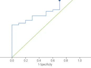
ROC curve analysis (Fig. 1) revealed that cervical length mea- sured after cerclage was a moderate predictor of early preterm delivery before 32 weeks of gestation (area under curve, 0.71;
95% confidence interval, 0.56 to 0.87; P=0.029). The best cut-off value of cervical length after cerclage for the prediction of preterm delivery before 32 weeks of gestation was 25 mm, with a sensitivity of 91.0% and a specificity of 30.0%.
Discussion
In this study, we found an association between cervical length after cerclage and preterm delivery before 32 weeks of gestation. As the post-operative cervical length got lon- ger, the probability of preterm delivery before 32 weeks of gestation was reduced. The ROC curve had an area under the curve of 0.71 with a 95 percent confidence interval of 0.56–0.87. Therefore, a cervical length of 25 mm measured immediately after cerclage could be used as a moderately confident baseline reference for early preterm delivery be- fore 32 weeks of gestation. However, the sample size in this study was fairly small; additional studies are required to confirm our reference cut-off value of post-cerclage cervical length.
Table 3. Demographic characteristics and pregnancy outcome of cervical insufficiency cases
GA at delivery <32 wk (n=5) ≥32 wk (n=24) P-valuea)
Age (yr) 34 (27–37) 33 (26–43) 0.899
Parity, history of
Full term delivery 0 (0) 11 (45.8) 0.126
Preterm delivery
4 (80.0) 21 (87.5) 0.553Abortion
3 (60.0) 11 (45.8) 0.651History of previous cerclage
4 (80.0) 12 (50.0) 0.343History of conization
1 (20.0) 0 (0) 0.172GA at cerclage (wk)
14 (13–17) 15 (13–19) 0.295CL before cerclage (mm)
30.0 (10.0–37.0) 33.0 (13.0–53.0) 0.222CL after cerclage (mm)
36.0 (13.0–39.0) 42.0 (19.0–55.0) 0.101CL change (mm)
3.0 (2.0–7.0) 8.0 (–5.0–16.0) 0.129GA at delivery (wk)
25 (17–32) 39 (34–42) <0.001Cerclage to delivery duration (day)
85 (17–120) 167 (110–189) <0.001Birth weight (kg)
0.82 (0.36–1.55) 3.06 (2.09–3.98)<0.001
Data represented as median (minimum–maximum) or number (%).
GA, gestational age; CL, cervical length.
a)Significance at P<0.05.
Table 4. Change of cervical length before and after cerclage
Patients CL before cerclage (mm) CL after cerclage (mm) P-valuea)
Cervical insufficiency (n=29) 33.0 (10.0–52.0) 39.0 (13.0–55.0) <0.001
GAD <32 wk (n=5) 30.0 (10.0–37.0) 36.0 (13.0–39.0) 0.043
GAD ≥32 wk (n=24) 33.0 (13.0–53.0) 42.0 (19.0–55.0) <0.001
Data represented as median (minimum–maximum).
CL, cervical length; GAD, gestational age at delivery.
a )Significance at P<0.05 by Wilcoxon signed rank test.
The relative risk of preterm delivery has been shown to increase as cervical length after cerclage becomes shorter. It is reasonable to hypothesize that prophylactic cerclage may reduce the risk of preterm delivery by restoring the cervical anatomy, which can act as a barrier to ascending infections [8].
Several studies have reported an increased cervical length af- ter cerclage. Dijkstra et al. reported that both prophylactic and urgent cerclage resulted in a statistically significant increase in cervical length, although the degree of cervical lengthening after cerclage did not contribute to the success of the proce- dure based on the primary outcome of delivery after 37 weeks of gestation [8]. One study evaluated whether a successful cerclage depended on suture placement as close as possible to the internal cervical os, which can be represented by cer- clage height [17]. Another study reported a trend toward an increased incidence of preterm delivery as cerclage height decreased, although this trend was not statistically significant [18]. A greater cerclage height results in a greater cervical length after cerclage because the overall cervical length is the sum of the cervical height (cervical length below the knot) and the cervical length above the knot. We therefore measured the overall cervical length in this study rather than cervical height.
During a cerclage operation, surgeons attempt to make the cervix as long as possible. Nevertheless, a surgeon may not be able to achieve the cervical length s/he intended, especially
in cases with a very short cervical length prior to cerclage.
Because these patients are at high risk for preterm delivery ac- cording to the results of this study, serial cervical length mea- surements after a cerclage operation are important to predict preterm birth, which can be expected if progressive cervical shortening is noted in the late second or early third trimester [19]. Serial measurements of cervical length after cerclage can help the physician to predict and prepare for preterm delivery by, for example, administering steroids to the mother to foster fetal lung maturation.
As the study being retrospective, we carefully selected and limited the patients enrolled to include only elective cerclage patients.
Limitations of our study include confounding factors such as the use of tocolytics or antibiotics. These factors might have affected the primary outcome of preterm delivery before 32 weeks. However, we argue that these factors are unlikely to have had a significant impact on the primary outcome of this study because we adopted standard management strategies for preterm labor and premature preterm rupture of mem- branes. Also, most of the pre and post operational cervical lengths used in the study were measured immediately before and after the surgery, although some immediate measure- ments were missing in patient records and were replaced by those measured in the nearest follow ups.
Practice Bulletin No. 142 “Cerclage for the management of cervical insufficiency” from the American College of Ob- stetrics and Gynecologists does not recommend prophylactic cerclage after conization [20]. Along with past reports in which cerclage after conization had no benefit on pregnancy outcomes [21-23] a previous study in our center also showed that cervical cerclage after electrosurgical conization did not convincingly reduce the rate of spontaneous preterm delivery [24]. Present study includes the cerclage cases after conization reflecting the past practice. Cerclage cases with the definite history of cervical insufficiency need to be further evaluated with a larger population.
In summary, we found that cervical length measurements after elective cerclage operations are important for predict- ing preterm delivery. Patients with a post-cerclage cervical length shorter than 25 mm need to be followed-up with serial measurements. A prospective randomized study with a larger number of subjects is required to confirm our findings.
Fig. 1. Cervical length measured after cerclage was closely as- sociated with early preterm delivery before 32 weeks of gestation (area under curve, 0.71; 95% confidence interval, 0.56 to 0.87;
P=0.029). The best cut-off value of cervical length after cerclage for the prediction of preterm delivery before 32 weeks of gestation was 25 mm (sensitivity, 91.0%; specificity, 30%).
1.0
0.8
0.6
0.4
0.2
0.0
1-Specificity 25 mm
0.0 0.2 0.4 0.6 0.8 1.0
Sensitivity