676
Increased Arterial Stiffness in Behcet’s Disease Patients
Moo-Yong Rhee, MD1, Sang-Hoon Na, MD1, Young-Kwon Kim, MD1, Myoung-Mook Lee, MD1, Seong-Kyu Kim, MD2 and Wan Kim, MD3
1Division of Cardiology, Department of Internal Medicine, Dongguk University College of Medicine, Gyeongju,
2Division of Rheumatology, Department of Internal Medicine, Dankook University College of Medicine, Cheonan,
3Department of Internal Medicine, Gwangju Veterans Hospital, Gwangju, Korea
ABSTRACT
Background and Objectives:Pulse wave velocity (PWV) is an ideal indicator of arterial stiffness. This study investigated arterial stiffness of different vascular regions in patients suffering with Behcet’s disease (BD), and we assessed whether arterial stiffness was affected by the clinical parameters of BD. Subjects and Methods:This study included 53 BD patients (mean age: 38±8 years) and 65 healthy controls (mean age: 38±8 years) who were without any known cardiovascular diseases. After recording the clinical parameters of the BD patients, pulse wave velocity was measured with an automated device in the heart-femoral, heart-carotid, heart-brachial and femoral-ankle segments. Results:Patients with BD had significantly higher PWV values than did the controls in all the regional arterial segments. The PWV values were not correlated with the duration of the disease, corticosteroid use or the presence of active disease at the time of examination. The clinical variables related to severe BD manifestations, which included severe disease, male gender, vascular lesions or immunosuppressant use, were partly associated with increased PWV on the univariate analysis, but any statistical significance for these clinical variables was lost in all the regional arterial segments on multivariate analysis. In addition, mul- tivariate regression analysis revealed that age and the mean arterial pressure were independently associated with increased PWV in most regional arterial segments for BD patients. Conclusion:The patients with BD had significantly increased arterial stiffness in all the regional arterial segments when compared with the healthy controls. Longitudinal studies that employ a large population are required to determine the pathophysiologic and prognostic implications of increased arterial stiffness in BD. (Korean Circulation J 2006;36:676-682) KEY WORDS:Behcet’s disease;Arteries.
Introduction
Behcet’s disease(BD) is a chronic inflammatory dis- order that involves multiple organs. Although the exact pathogenesis for BD is not completely understood, it has been suggested that the disease is triggered in ge- netically susceptible individuals by environmental fa- ctors such as infectious agents. The histological hallmark of the disease is noted to be a vasculitis. A variety of vascular lesions, including venous or arterial occlusive lesions and arterial aneurysm, can occur in approxi- mately one-third of BD patients. These venous and ar-
terial lesions can be infiltrated by different types of in- flammatory cells, including lymphocytes, monocytes, plasma cells and neutrophils.1-3) In general, those BD pa- tients with major vessel disease exhibit a poor prognosis.4)
Although the definite pathogenic mechanism for the vascular lesions in BD remains unclear, endothelial dysfunction is thought to play an important role in the development of these lesions.5)6) It has been recently demonstrated that brachial artery flow-mediated dila- tation is impaired in BD.6) Because flow-mediated di- lataion is endothelium-dependent and it is largely con- trolled by the release of endothelial nitric oxide(NO),7) an impairment of endothelium-dependent flow-medi- ated dilatation suggests that there is decreased endo- thelial NO activity. This lack of activity may contribute to the vascular lesions that are often seen in BD. Fur- thermore, endothelial NO has been found to directly regulate large artery stiffness in vivo.8) We and other investigators have shown that potentially functional
Received:April 5, 2006 Accepted:June 22, 2006
Correspondence:Moo-Yong Rhee, MD,Division of Cardiology, Depart- ment of Internal Medicine, Dongguk University College of Medicine, 814 Siksa-dong, Ilsandong-gu, Goyang 410-773, Korea
Tel: 82-31-961-7125, Fax: 82-31-961-7785 E-mail: [email protected]
DNA polymorphism in the endothelial NO synthetase gene, which could cause reduced NO activity, may be associated with susceptibility to BD.9)10)
Arterial stiffness is a reliable and strong indepen- dent predictor of subsequent cardiovascular events and mortality, and it may be closely related to the process of atherosclerosis.11-14) Since stiffened arteries transmit pulse waves faster than do the more elastic blood ve- ssels, pulse wave velocity(PWV) is an ideal indicator of arterial stiffness. In addition, arterial abnormalities may be attributed to functional changes like endothe- lial dysfunction and also structural alterations such as atherosclerosis.15)16) Acute systemic inflammation and chronic systemic vasculitis are noted to be associated with endothelial dysfunction.17)18) Moreover, inflamm- ation is known to be an important risk factor for fu- ture cardiovascular events.19) These findings have led to the hypothesis that the acute and chronic inflamma- tory processes associated with BD may cause endothe- lial dysfunction. This can then lead to subsequent vas- cular damage and the increased arterial stiffness that are closely related to the clinical course of BD patients.
However, any information has been rather limited re- garding arterial stiffness in BD. Thus, this study in- vestigated the arterial stiffness of different arterial regi- ons in Korean BD patients, and we then assessed whe- ther arterial stiffness was affected by the clinical para- meters of BD.
Subjects and Methods
SubjectsThis study included 53 BD patients who fulfilled the International Study Group(ISG) criteria,20) and 65 healthy controls who were matched to the BD pa- tients for their age, gender, blood pressure, heart rate, height, cholesterol and glucose levels. The frequency of smokers, if any, was also taken into account. Those su- bjects who had hypertension, diabetes mellitus or a previous history of coronary artery disease or myocar- dial infarction were excluded.
At the time of examination, the presence of more than two of the following Behcet’s clinical features was considered as active disease: oral ulceration, genital ul- ceration, skin lesions, ocular lesions, active major vessel disease and active major organ involvement, including active gastrointestinal or neurological lesions. During the course of the disease, the presence of one or more of the following clinical features was regarded as severe disease:9) posterior uveitis or retinal vasculitis, gastroin- testinal ulcerations with bleeding or perforation, major organ involvement and major vessel involvement. In addition, those BD patients with venous or arterial oc- clusive diseases or arterial aneurysm were considered to have vascular lesions, but those patients with super-
ficial thrombophlebitis were not considered to have vascular lesions. The duration of the disease in the BD group was calculated from the time when the patients fulfilled the ISG criteria to the time of their examina- tions. The mean of this time period was 4.7±4.8 years.
Patient information was obtained on any corticost- eroids treatment and immunosuppressants use for the three months before the start of this study. Using sta- ndard laboratory methods, the levels of total cholest- erol, triglyceride and glucose were measured from the fasting blood samples that were taken from all the su- bjects. The study was approved by the Hospital Ethics Committee. An informed, written consent was obtained from each subject.
Measurements of PWV
PWV was measured in the morning with the patient in a supine position after 15 minutes of bed rest in a quite room, following 12-hour abstinence from smok- ing, alcohol and coffee consumption. A single trained observer performed all the measurements. PWV meas- urements were taken using an automated device(VP- 2000, Colin Co. Ltd., Japan).21) This device simultan- eously records the PWV, the brachial and tibial arterial blood pressures, and the ECG and heart sounds. In brief, ECG electrodes were placed on both wrists, and a heart sound microphone was placed on the left st- ernal border. Occlusion cuffs, which were connected to both plethysmographic and oscillometric sensors, were wrapped around both upper arms and the ankles.
The brachial and tibial arterial blood pressures were measured simultaneously. The brachial and tibial ar- terial pulse waves were acquired using oscillometric sen- sors. The carotid and femoral arterial waves were ac- quired using tonometric sensors that were placed at the left carotid artery and the left femoral artery.
The heart-carotid pulse transit time was measured from the second heart sound to the notch of the car- otid pulse wave. The heart-brachial pulse transit time was taken from the second heart sound to the notch of the brachial pulse waves. The heart-femoral pulse tra- nsit time(from the aortic orifice to the femoral artery) was calculated by summing the carotid-femoral pulse transit time and the heart-carotid pulse transit time.
The femoral-ankle pulse transit time was measured by the foot-to-foot method, from the foot of the femoral arterial pulse waves to the tibial arterial pulse waves.
Path lengths of the heart-femoral, heart-carotid, heart- brachial and femoral-ankle segments were calculated automatically with using the patient’s height. The heart-femoral(hfPWV), heart-carotid(hcPWV), fem- oral-ankle(faPWV), and heart-brachial(hbPWV) PWV levels were calculated for each arterial segment by di- viding the path lengths by the corresponding pulse transit times, and they were expressed as m/sec.
Statistical analysis
The data were analyzed using the SPSS statistical package program version 11.5 for Windows(SPSS Inc., Chicago, IL, USA). Differences of the continuous va- riables between groups were compared using indepen- dent t tests, and the results we obtained were re-ev- aluated using multiple regression analysis to exclude the confounding effects of the clinical variables of BD and also for excluding the confounding effects of the other risk factors on the PWV values. The bivariate correlations between two continuous variables were ev- aluated using Pearson’s correlation coefficient when indicated. P less than 0.05 were considered to be sta- tistically significant.
Results
Table 1 summarizes the clinical features of the 53 BD patients. Major vascular lesions were detected in nine patients: inferior vena cava occlusion was found in three patients, superior vena cava occlusion was fo- und in one patient, deep vein thrombosis at the lower extremities was found in three patients, cavernous si- nus thrombosis was found in one patient and femoral arterial aneurysm was found in one patient. Immuno- suppressive agents and corticosteroids were used by 15(azathioprine in 10 and cyclosporine in 5) and 38 patients, respectively. No differences between the BD patients and the controls were found in terms of age, the gender ratio and the other parameters that are known to affect the PWV, including height, blood pre- ssure, heart rate and the total cholesterol levels. The
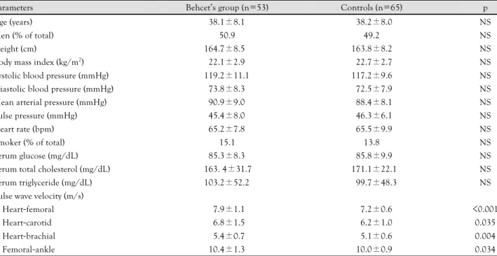
BD patients had significantly higher PWV values did the controls in all the regional arterial segments, inclu- ding the heart-femoral, heart-carotid, heart-brachial and femoral-ankle segments(Table 2).
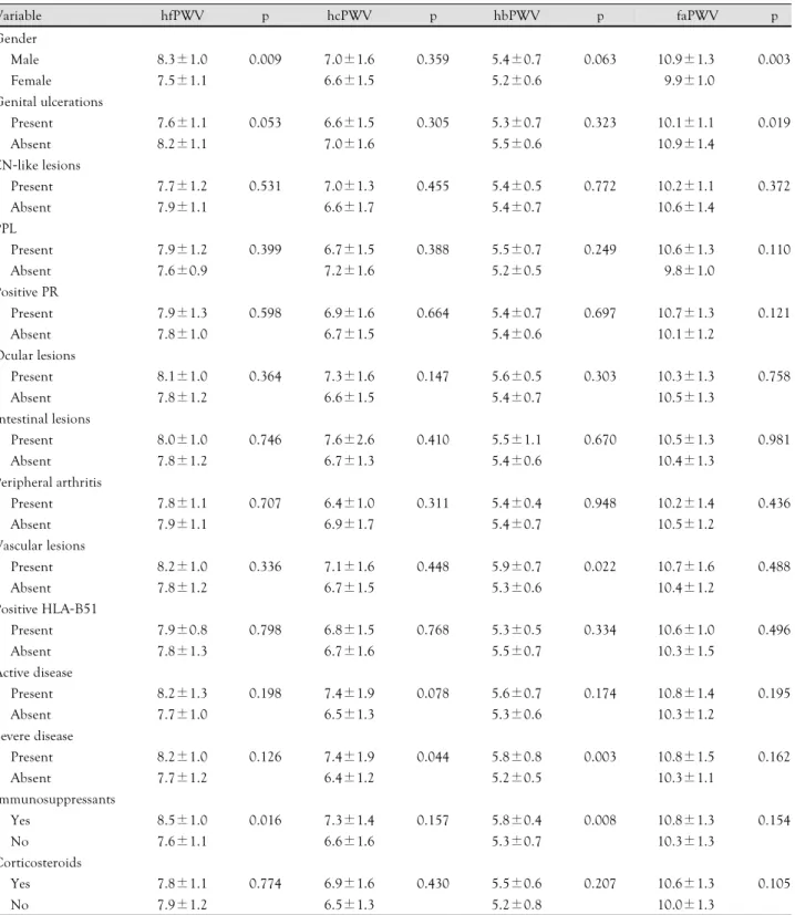
Table 3 shows the relationship between the PWV values and the clinical parameters of BD in the differ- ent regions. Male BD patients exhibited a tendency towards elevated PWV values in all the four regions when compared to the female patients. However, signi- ficantly higher levels of PWV were observed in only the heart-femoral and femoral-ankle segments. It’s in- teresting that the patients without genital ulcerations had a tendency to have higher PWV levels than did
Table 1. Clinical features of 53 patients with Behcet’s disease Clinical features Number of patients (%)
Oral ulcerations 53 (100)
Genital ulcerations 32 (60.4)
Erythema nodosum-like lesions 21 (39.4)
Papulopustular lesions 44 (83.0)
Positive pathergy reaction 28 (52.8)
Ocular lesions 12 (22.6)
Intestinal lesions 06 (11.3)
Peripheral arthritis 14 (26.4)
Vascular lesions 09 (17.0)
Central nervous system lesions 02 (03.8)
Positive HLA-B51 22 (42.3)
Active disease 16 (30.2)
Severe disease 19 (35.8)
Immunosuppressant use 15 (28.3)
Corticosteroid use 38 (71.7)
Table 2. Comparisons of the demographic data, laboratory values and cardiovascular parameters between patients with Behcet’s disease and the controls
Parameters Behcet’s group (n=53) Controls (n=65) p
Age (years) 038.1±8.1 038.2±8.0 NS
Men (% of total) 50.9 49.2 NS
Height (cm) 164.7±8.5 163.8±8.2 NS
Body mass index (kg/m2) 022.1±2.9 022.7±2.7 NS
Systolic blood pressure (mmHg) 119.2±11.1 117.2±9.6 NS
Diastolic blood pressure (mmHg) 073.8±8.3 072.5±7.9 NS
Mean arterial pressure (mmHg) 090.9±9.0 088.4±8.1 NS
Pulse pressure (mmHg) 045.4±8.0 046.3±6.1 NS
Heart rate (bpm) 065.2±7.8 065.5±9.9 NS
Smoker (% of total) 15.1 13.8 NS
Serum glucose (mg/dL) 085.3±8.3 085.8±9.9 NS
Serum total cholesterol (mg/dL) 163. 4±31.7 171.1±22.1 NS
Serum triglyceride (mg/dL) 103.2±52.2 099.7±48.3 NS
Pulse wave velocity (m/s)
Heart-femoral 007.9±1.1 007.2±0.6 <0.001
Heart-carotid 006.8±1.5 006.2±1.0 0.035
Heart-brachial 005.4±0.7 005.1±0.6 0.004
Femoral-ankle 010.4±1.3 010.0±0.9 0.034
Data are expressed as means±SD
the patients with genital ulcerations. Significantly hi- gher levels were observed only in the femoral-ankle segment for the patients without genital ulcerations.
In the case of the other clinical features of BD, no sig- nificant differences were found between the patients
with and without the individual features of Behcet’s disease, with the exception that the patients with va- scular lesions had significantly higher PWV values in the heart-brachial segment than did the patients wi- thout vascular lesions. In addition, the PWV levels did
Table 3. Analysis of pulse wave velocity (mean±SD, m/s) in the four regions according to the clinical parameters of Behcet’s disease
Variable hfPWV p hcPWV p hbPWV p faPWV p
Gender
Male 8.3±1.0 0.009 7.0±1.6 0.359 5.4±0.7 0.063 10.9±1.3 0.003
Female 7.5±1.1 6.6±1.5 5.2±0.6 09.9±1.0
Genital ulcerations
Present 7.6±1.1 0.053 6.6±1.5 0.305 5.3±0.7 0.323 10.1±1.1 0.019
Absent 8.2±1.1 7.0±1.6 5.5±0.6 10.9±1.4
EN-like lesions
Present 7.7±1.2 0.531 7.0±1.3 0.455 5.4±0.5 0.772 10.2±1.1 0.372
Absent 7.9±1.1 6.6±1.7 5.4±0.7 10.6±1.4
PPL
Present 7.9±1.2 0.399 6.7±1.5 0.388 5.5±0.7 0.249 10.6±1.3 0.110
Absent 7.6±0.9 7.2±1.6 5.2±0.5 09.8±1.0
Positive PR
Present 7.9±1.3 0.598 6.9±1.6 0.664 5.4±0.7 0.697 10.7±1.3 0.121
Absent 7.8±1.0 6.7±1.5 5.4±0.6 10.1±1.2
Ocular lesions
Present 8.1±1.0 0.364 7.3±1.6 0.147 5.6±0.5 0.303 10.3±1.3 0.758
Absent 7.8±1.2 6.6±1.5 5.4±0.7 10.5±1.3
Intestinal lesions
Present 8.0±1.0 0.746 7.6±2.6 0.410 5.5±1.1 0.670 10.5±1.3 0.981
Absent 7.8±1.2 6.7±1.3 5.4±0.6 10.4±1.3
Peripheral arthritis
Present 7.8±1.1 0.707 6.4±1.0 0.311 5.4±0.4 0.948 10.2±1.4 0.436
Absent 7.9±1.1 6.9±1.7 5.4±0.7 10.5±1.2
Vascular lesions
Present 8.2±1.0 0.336 7.1±1.6 0.448 5.9±0.7 0.022 10.7±1.6 0.488
Absent 7.8±1.2 6.7±1.5 5.3±0.6 10.4±1.2
Positive HLA-B51
Present 7.9±0.8 0.798 6.8±1.5 0.768 5.3±0.5 0.334 10.6±1.0 0.496
Absent 7.8±1.3 6.7±1.6 5.5±0.7 10.3±1.5
Active disease
Present 8.2±1.3 0.198 7.4±1.9 0.078 5.6±0.7 0.174 10.8±1.4 0.195
Absent 7.7±1.0 6.5±1.3 5.3±0.6 10.3±1.2
Severe disease
Present 8.2±1.0 0.126 7.4±1.9 0.044 5.8±0.8 0.003 10.8±1.5 0.162
Absent 7.7±1.2 6.4±1.2 5.2±0.5 10.3±1.1
Immunosuppressants
Yes 8.5±1.0 0.016 7.3±1.4 0.157 5.8±0.4 0.008 10.8±1.3 0.154
No 7.6±1.1 6.6±1.6 5.3±0.7 10.3±1.3
Corticosteroids
Yes 7.8±1.1 0.774 6.9±1.6 0.430 5.5±0.6 0.207 10.6±1.3 0.105
No 7.9±1.2 6.5±1.3 5.2±0.8 10.0±1.3
Data are expressed as means±SD. EN: erythema nodosum, PPL: papulopustular lesions, PR: pathergy reaction, hcPWV: heart-carotid pulse wave velocity, hbPWV: heart-brachial pulse wave velocity, hfPWV: heart-femoral pulse wave velocity, faPWV: femoral-ankle pulse wave velocity
not significantly differ between the patients with and without active disease at the time of examination. In contrast, the BD patients with severe disease or those BD patients who had been taking immunosuppressive agents showed a tendency towards increased PWV va- lues in comparison with the patients without severe disease or those BD patients who had been taking im- munosuppressants. Significant differences were obser- ved in the heart-carotid and heart-brachial segments when severe disease was present; significant differences were also found in the heart-femoral and heart-bra- chial segments in the patients who had been exposed to immunosuppressive agents. The patients who had been treated with corticosteroids had similar PWV le- vels to the patients who had not received corticosteroids.
On the bivariate correlation analyses of BD patients with using Pearson’s correlation coefficient, all the re- gional PWV levels except the femoral-ankle segment correlated well with increasing patient age. In parti- cular, the degree of correlation was greater in the cen- tral arteries(hfPWV and hcPWV) than in the periph- eral arteries(hbPWV and faPWV). The PWV values
in all the regional arterial segments were positively co- rrelated with the mean blood pressure. However, all the regional PWV values did not correlate with the dura- tion of disease(Table 4). In addition, the regional PWV values did not correlate with height and the levels of cholesterol, glucose and triglyceride(data not shown).
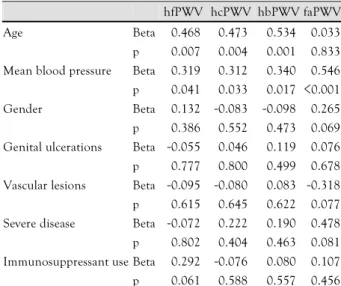
Multiple regression analysis was used to adjust for any potential confounding influences of age, gender, the mean blood pressure, the duration of the disease and the clinical variables of BD. Only age and the mean blood pressure appeared to be significant factors asso- ciated with increased PWV in most of the regional ar- terial segments for the BD patients, except for the fe- moral-ankle segment in regards to age. In addition, the statistical significance was lost upon univariate analysis for some of the clinical variables associated with incre- ased PWV in all the regional arterial segments(Table 5).
Discussion
Behcet’s disease(BD) is a chronic inflammatory dis- order that’s characterized by recurrent oral and genital ulcerations, ocular lesions and skin lesions. Although the exact pathogenesis of this disease remains unclear, small vessel vasculitis accounts for a considerable por- tion of the pathogenic process in BD. In addition, large venous or arterial lesions can occur in up to one-third of BD patients.1)3) Although the precise pathogenic mechanism for the vascular lesions in BD remains murky at best, endothelial dysfunction is thought to play an central role in the development of these le- sions.5)6) Acute systemic inflammation and chronic systemic vasculitis are also known to be associated with endothelial dysfunction.17)18) In the present study, we measured the regional PWVs of four arterial segments in BD patients and the healthy controls. The two groups were well matched for the factors known to affect arterial stiffness. The BD patients had significantly hi- gher PWV values in all the four arterial regions than did the controls, indicating the presence of increased arterial stiffness in the patients with BD. Such incre- ased arterial stiffness in the BD patients may be attri- buted to endothelial dysfunction and the acute or ch- ronic inflammatory processes associated with BD.
Univariate analysis showed that the BD patients with severe disease or those BD patients that had taken immunosuppressive agents exhibited a tendency to- wards increased PWVs. In addition, the male BD pa- tients, who are known to have a more severe disease course as compared to female patients,4) also had hi- gher PWV levels than did the female patients. In the case of the patients with major vessel lesions, which are a severe manifestation of BD, significantly incre- ased PWV values were found only in the heart-br- achial segment. Any statistical significance for the cli-
Table 4. Correlations between the regional PWV values and age, mean blood pressure and disease duration
Age Mean blood
pressure
Disease duration
r p r p r p
hfPWV 0.483 <0.001 0.472 <0.001 -0.080 0.567 hcPWV 0.531 <0.001 0.547 <0.001 -0.065 0.646 hbPWV 0.423 <0.002 0.560 <0.001 -0.069 0.622 faPWV 0.208 <0.134 0.609 <0.001 -0.224 0.106 hcPWV: heart-carotid pulse wave velocity, hbPWV: heart-bra-chial pulse wave velocity, hfPWV: heart-femoral pulse wave velocity, faPWV: femoral-ankle pulse wave velocity
Table 5. Multiple regression analysis for the co-factors that affect regional PWV in Behcet’s disease
hfPWV hcPWV hbPWV faPWV
Age Beta 0.468 0.473 0.534 0.033
p 0.007 0.004 0.001 0.833 Mean blood pressure Beta 0.319 0.312 0.340 0.546 p 0.041 0.033 0.017 <0.001 Gender Beta 0.132 -0.083 -0.098 0.265 p 0.386 0.552 0.473 0.069 Genital ulcerations Beta -0.055 0.046 0.119 0.076 p 0.777 0.800 0.499 0.678 Vascular lesions Beta -0.095 -0.080 0.083 -0.318 p 0.615 0.645 0.622 0.077 Severe disease Beta -0.072 0.222 0.190 0.478 p 0.802 0.404 0.463 0.081 Immunosuppressant use Beta 0.292 -0.076 0.080 0.107 p 0.061 0.588 0.557 0.456 hcPWV: heart-carotid pulse wave velocity, hbPWV: heart-brachial pulse wave velocity, hfPWV: heart-femoral pulse wave velocity, faPWV:
femoral-ankle pulse wave velocity
nical parameters that were noted to be related to the severe BD manifestations, which included severe disease, immunosuppressives use, male gender and vascular le- sions, was lost upon multiple regression analysis for all the regional arterial segments, so further studies with a larger number of patients are needed to elucidate the relationship between the regional PWV values and th- ose clinical parameters of BD. On the other hand, si- milar to the well-known effects of age and mean blood pressure on PWV,13)22) we found on multiple regre- ssion analysis that age and the mean blood pressure were the independent significant factors associated with increased PWV in the BD patients. With respect to age, the effect on the PWV values in BD patients was greater in the central arteries than in the periph- eral arteries.
The use of corticosteroid as a risk factor for cardio- vascular disease is controversial.23-26) In the current study, the patients who had been treated with cortico- steroids had PWV values that were similar to the patients who had not received these medications. This may be explained by the double-edged nature of corti- costeroids, i.e., that while patients treated with corti- costeroids usually have severe disease, and these drugs can reduce arterial wall inflammation. On the other hand, the fact that the PWV values did not correlate with the duration of the disease in the present study may reflect the previous observation that the overall activity and severity of BD tend to abate with time.27)
It has been noted that prolonged exposure to low levels of acute-phase reactants, including C-reactive protein and amyloid A protein, may be associated with subsequent vascular injury.19) However, C-reactive pro- tein is not likely to be a suitable marker for disease activity in BD patients. In the current study, no differ- ence in the PWV values was observed between the BD patients with and without active disease, outwity de- termined by the presence of active lesions at the time of examination. Since the disease activity usually fluc- tuates in BD patients during the course of the disease, only the presence of active lesions at a single point would not result in the increased PWV levels.
Information has been limited regarding the arterial stiffness in chronic inflammatory disorders that dis- play systemic vasculitis, such as BD. Booth et al.28) re- ported that antineutrophil cytoplasmic antibody-ass- ociated systemic vasculitis is associated with increased arterial stiffness, and that the stiffness correlated with the degree of active inflammation. Kurum et al.29) de- scribed that the PWV levels in 14 BD patients did not significantly differ from the PWV levels in healthy co- ntrols. When compared with our study, their study po- pulation was small and they excluded subjects with active BD. More recently, Ikonomidis et al.30) reported that BD patients had an increased aortic diameter,
lower mean aortic strain and distensibility, and also a higher mean aortic stiffness index on echocardiogra- phic study. However, all these investigations, including our study, employed a cross-sectional study design;thus, causality can not be established.
In conclusion, the present study showed that BD patients have significantly increased central and peri- pheral arterial stiffness when compared with healthy controls. Age and the mean blood pressure were the independent significant factors associated with incre- ased PWV. Longitudinal studies with a large subject population are required to determine the pathophysio- logic and prognostic implications of increased arterial stiffness in BD.
REFERENCES
1) Sakane T, Takeno M, Suzuki N, Inaba G. Behcet’s disease. N Engl J Med 1999;341:1284-91.
2) Gul A. Behcet’s disease: an update on the pathogenesis. Clin Exp Rheumatol 2001;19(Suppl 24):S6-12.
3) Koc Y, Gullu I, Akpek G, et al. Vascular involvement in Behcet’s disease. J Rheumatol 1992;19:402-10.
4) Kural-Seyahi E, Fresko I, Seyahi N, et al. The long-term mor- tality and morbidity of Behcet syndrome: a 2-decade outcome survey of 387 patients followed at a dedicated center. Medicine 2003;82:60-76.
5) Schmitz-Huebner U, Knop J. Evidence for an endothelial cell dysfunction in association with Behcet’s disease. Thromb Res 1984;34:277-85.
6) Chambers JC, Haskard DO, Kooner JS. Vascular endothelial function and oxidative stress mechanisms in patients with Be- hcet’s syndrome. J Am Coll Cardiol 2001;37:517-20.
7) Joannides R, Haefeli WE, Linder L, et al. Nitric oxide is respon- sible for flow-dependent dilatation of human peripheral conduit arteries in vivo. Circulation 1995;91:1314-9.
8) Wilkinson IB, Qasem A, McEniery CM, Webb DJ, Avolio AP, Cockcroft JR. Nitric oxide regulates local arterial distensibility in vivo. Circulation 2002;105:213-7.
9) Kim JU, Chang HK, Lee SS, et al. Endothelial nitric oxide sy- nthase gene polymorphisms in Behcet’s disease and rheumatic diseases with vasculitis. Ann Rheum Dis 2003;62:1083-7.
10) Salvarani C, Boiardi L, Casali B, et al. Endothelial nitric oxide synthase gene polymorphisms in Behcet’s disease. J Rheumatol 2002;29:535-40.
11) Laurent S, Boutouyrie P, Asmar R, et al. Aortic stiffness is an independent predictor of all-cause and cardiovascular mortality in hypertensive patients. Hypertension 2001;37:1236-41.
12) Mattace-Raso FU, van der Cammen TJ, Hofman A, et al. Art- erial stiffness and risk of coronary heart disease and stroke: the Rotterdam Study. Circulation 2006;113:657-63.
13) Zieman SJ, Melenovsky V, Kass DA. Mechanisms, pathophysio- logy, and therapy of arterial stiffness. Arterioscler Thromb Vasc Biol 2005;25:932-43.
14) Kim YK, Kim D. The relation of pulse wave velocity with Fra- mingham Risk Score and SCORE Risk Score. Korean Circ J 2005;35:22-9.
15) Booth AD, Wallace S, McEniery CM, et al. Inflammation and arterial stiffness in systemic vasculitis: a model of vascular in- flammation. Arthritis Rheum 2004;50:581-8.
16) Klocke R, Cockcroft JR, Taylor GJ, Hall IR, Blake DR. Arterial stiffness and central blood pressure, as determined by pulse wave
analysis, in rheumatoid arthritis. Ann Rheum Dis 2003;62:414-8.
17) Hingorani AD, Cross J, Kharbanda RK, et al. Acute systemic in- flammation impairs endothelium-dependent dilatation in humans.
Circulation 2000;102:994-9.
18) Raza K, Thambyrajah J, Townend JN, et al. Suppression of infla- mmation in primary systemic vasculitis restores vascular endoth- elial function: lessons for atherosclerotic disease? Circulation 2000;102:1470-2.
19) Danesh J, Whincup P, Walker M, et al. Low grade inflammation and coronary heart disease: prospective study and updated meta- analyses. BMJ 2000;321:199-204.
20) International Study Group for Behcet’s Disease (ISGBD). Cri- teria for diagnosis of Behcet’s disease. Lancet 1990;335:1078-80.
21) Rhee MY. Acute and chronic effects of smoking on the arterial wall properties and the hemodynamics in smokers with hyperten- sion. Korean Circ J 2005;35:493-9.
22) Park JS, Seo JJ, Chung JW, et al. Association of the invasively measured aortic stiffness and coronary artery disease. Korean Circ J 2005;35:766-72.
23) Wolfe F, Mitchell DM, Sibley JT, et al. The mortality of rheu-- matoid arthritis. Arthritis Rheum 1994;37:481-94.
24) Petri M, Perez-Gutthann S, Spence D, Hochberg MC. Risk factors for coronary artery disease in patients with systemic lupus ery-
thematosus. Am J Med 1992;93:513-9.
25) Manzi S, Meilahn EN, Rairie JE, et al. Age-specific incidence rates of myocardial infarction and angina in women with syste- mic lupus erythematosus: comparison with the Framingham Study.
Am J Epidemiol 1997;145:408-15.
26) Rasker JJ, Cosh JA. Cause and age at death in a prospective study of 100 patients with rheumatoid arthritis. Ann Rheum Dis 1981;
40:115-20.
27) Yazici H, Tuzun Y, Pazarli H, et al. Influence of age of onset and patient’s sex on the prevalence and severity of manifestations of Behcet’s syndrome. Ann Rheum Dis 1984;43:783-9.
28) Booth AD, Wallace S, McEniery CM, et al. Inflammation and arterial stiffness in systemic vasculitis: a model of vascular infla- mmation. Arthritis Rheum 2004;50:581-8.
29) Kurum T, Yildiz M, Soy M, Ozbay G, Alimgil L, Tuzun B. Art- erial distensibility as determined by carotid-femoral pulse wave velocity in patients with Behcet's disease. Clin Rheumatol 2005;
24:134-8.
30) Ikonomidis I, Lekakis J, Stamatelopoulos K, Markomihelakis N, Kaklamanis PG, Mavrikakis M. Aortic elastic properties and left ventricular diastolic function in patients with Adamantiades- Behcet's disease. J Am Coll Cardiol 2004;43:1075-81.