pISSN 1598-9992 eISSN 2233-6869
REVIEW ARTICLE
췌장액체고임의 치료
윤승배, 장재혁, 이인석
가톨릭대학교 의과대학 내과학교실
Treatment of Pancreatic Fluid Collections
Seung Bae Yoon, Jae Hyuck Chang and In Seok Lee
Department of Internal Medicine, College of Medicine, The Catholic University of Korea, Seoul, Korea
Pancreatic Fluid Collection (PFC) develops as a result of acute pancreatitis, chronic pancreatitis, trauma, and postoperation. Although percutaneous drainage, surgery and Endoscopic Retrograde Panceatogram are used as conventional treatments in complicated PFC, the clinical course of PFC is unsatisfactory due to its clinical success rate and the risk of procedure-related complications. Endoscopic ultrasonography-guided transmural drainage of PFC is a safe and effective modality for the management of PFC, particularly in pa- tients with pancreas necrosis. A range of techniques and stents have been introduced and a newly designed metal stent is now available. (Korean J Gastroenterol 2018;72:97-103)
Key Words: Pancreatic pseudocyst; Necrosis; Endosonography; Pancreatitis
Received August 23, 2018. Revised September 13, 2018. Accepted September 14, 2018.
CC This is an open access article distributed under the terms of the Creative Commons Attribution Non-Commercial License (http://creativecommons.org/licenses/
by-nc/4.0) which permits unrestricted non-commercial use, distribution, and reproduction in any medium, provided the original work is properly cited.
Copyright © 2018. Korean Society of Gastroenterology.
교신저자: 이인석, 06591, 서울시 서초구 반포대로 222, 가톨릭대학교 서울성모병원 내과
Correspondence to: In Seok Lee, Department of Internal Medicine, Seoul St. Mary’s Hospital, The Catholic University of Korea, 222 Banpo-daero, Seocho-gu, Seoul 06591, Korea. Tel: +82-2-2258-6022, Fax: +82-2-2258-2055, E-mail: [email protected], ORCID: https://orcid.org/0000-0002-1127-1522
Financial support: None. Conflict of interest: None.
서 론
췌장액체고임(pancreatic fluid collection, PFC)은 췌장 염, 외상 또는 수술 후 발생한다. 개정된 Atlanta 분류1에서는 PFC를 급성과 만성으로 나누고 급성 PFC는 췌장괴사의 유무 에 따라 acute peripancreatic fluid collections와 acute ne- crotic collections로, 만성 PFC는 가성낭종과 walled-off pancreatic necrosis로 구분하였다. PFC의 치료는 보존적 치 료, 수술, 경피적 배액술 및 역행성 내시경 치료가 사용되어 왔으나 치료 효과에 비하여 재원 기간이 길고 시술 연관 합병 증이 높아 임상에서 췌장액체고임의 효과적 치료가 어려웠 다.2,3 PFC의 내시경 치료는 경유두적 접근(transpapillary approach)과 경벽적 접근(transmural approach) 방법이 있다.
경벽적 배액술은 돌출된 1975년 Rogers 등4에 의하여 처음 이루어졌고, 초음파 내시경에 의한 경벽적 배액술은 1992년
Grimm 등5에 의하여 시도된 이후 내시경 치료는 성공률이 높고 효과가 좋으며 안전한 시술로 PFC의 기본 치료로 인정 받고 있다. 이번 원고는 PFC의 초음파 내시경을 이용한 치료 의 방법과 효과에 대해 고찰하고자 한다.
본 론
1. PFC 종류 및 특징
급성 가성낭종은 급성 췌장염에서 발생하고 최소 4주 이상 소요되며 액화로 인하여 내부에 고형조직이 없다. 만성 가성 낭종은 만성 췌장염에서 발생하며 췌관 협착이나 췌석에 의하 여 췌관이 막히면 췌액이 누출되면서 발생하며 내부에 고형괴 사조직이 관찰되지 않는다.6
감염된 가성낭종(infected pancreatic pseudocysts)은 내 부에 고형조직이 없는 경우 감염된 괴사조직과 달리 배액이
Fig. 1. Display of different types of drain tubes. (A) Plastic Stent (Cook Medical, Bloomington, IN, USA), (B) Solus stent (Cook Medical), (C) Axios stent (Boston Scientific, Marlborough, MA, USA), (D) Spaxus stent (Taewoong Medical, Gimpo, Korea), (E) Nagi stent (Taewoong Medical).
가능하므로 췌장농양으로 진단하기도 한다. 췌장괴사는 췌장 실질이 파괴된 것으로, 췌장염의 초기에는 CT에서 실질의 조 영증강이 되지 않는 것으로 진단한다. 췌장괴사로 인하여 주 췌관의 단절이 발생하며, 괴사가 수주 지속되면 액체고임의 형태가 되며 내부는 액체 및 고형의 괴사조직이 혼재한다. 이 경우 내시경 배액술을 시행하면 고형조직이 제거되지 않아 흔 히 감염이 발생할 수 있다.
2. PFC drain의 적응증
가성낭종에서 전형적인 배액 적응증은 증상이 발생하거나 감염이 된 경우이다. 과거에는 크기 6 cm 이상의 경우 배액이 나 수술을 하였으나 시술 연관 합병증의 위험이 있어 현재는 크기를 기준으로 시술을 결정하지 않는다. 가성낭종이 크기가 계속 커지는 경우에도 배액을 시도할 수 있지만 무증상이면 경과 관찰도 가능하다.7 무균성 췌장괴사(sterile pancreatic necrosis)는 무증상인 경우 배액술이 필요 없지만 반복되는 복통, 위장관 폐쇄 증상, 식욕부진, 체중 감소, 전신쇠약 등의 증상이 지속되면 배액의 적응이 된다. 내시경 치료는 합병증 의 위험이 높고 대상 환자가 이미 중한 상태에서 시술되므로 감염이 없는 췌장괴사에서 내시경 배액은 신중하여야 한다.8 감염된 췌장괴사(infected pancreatic necrosis)는 배액의 적 응증이 된다. 이 경우 무균성 췌장괴사에서도 발열이나 백혈 구 증가 소견을 동반할 수 있으므로 임상 소견과 세침흡인술
을 통한 세균배양 검사 등으로 정확한 진단이 중요하다.
3. 내시경 시술 전 점검 사항
시술 전 췌장액체고임에 대한 정확한 진단이 필수적이다.
특히 cystic pancreatic neoplasm, duplication cyst, true pancreatic cyst, pseudoaneurysm, solid necrotic neo- plasm (eg, retroperitoneal sarcoma), lymphocele 등과 같 은 췌장 낭종성 질환에 대한 감별을 요한다. 사전 문진에서 환자에게 췌장염의 과거력이 있는지 꼭 확인하여야 하며, 정 확한 진단을 위하여 초음파 내시경이나 조영증강 CT, MRI 등의 검사를 시행하여 정확한 진단 및 주변 혈관이나 장기 등 해부학적 구조을 확인하고, 특히 췌장액체고임 내부에 췌 장궤사를 동반하고 있는지 확인한다. 췌장액체고임의 원인이 불명한 경우 초음파 내시경 흡입술 및 체액분석 검사를 시도 하여 정확한 진단이 이루어진 후 시술하도록 한다.9 시술 전 혈액응고 장애가 있는지 혈액 검사를 통하여 확인하고 시술의 합병증에 대한 대비를 하고 동의서를 받도록 한다.
4. 초음파 내시경 유도 가성낭종 배액술 시술 방법
환자를 진정 유도 후, 겸자공 직경이 3.7 mm 이상의 종주 형 초음파 내시경(linear echoendoscope)을 삽입하여 위, 식 도 또는 십이지장 주변의 가성낭종을 찾는다. 초음파 내시경 의 컬러 도플러로 천자하려는 장관벽과 가성낭종 사이에 혈관
A
A BB
CC DD EE
A
A BB CC DD
E
E FF GG HH
I
I JJ KK LL
Fig. 2. Procedure of EUS-guided pseudocyst drainage. (A) Infected cyst found at tail, the cyst wall protruded at the posterior side of the stomach on gastroscopy (B) and EUS (C). Transmural puncture was done under EUS-guided technique (D-F), the guidewire was introduced into the cyst (G, H) and then balloon dilatation of the cystic wall was performed (I, J), finally 2 double pigtail plastic stents were inserted (K, L). EUS, endoscopic ultrasonography.
이 있는지 확인하고 19-gauge의 흡인 바늘을 이용하여 장관 벽을 통하여 가성낭종을 천자한다. 천자 후 흡인 바늘을 통하 여 소량의 조영제를 주입하면서 영상기기를 통하여 천자가 잘 되었는지 확인하고 가이드와이어를 흡인 바늘 내부를 통하여 가성낭종으로 삽입하여 가성낭종 내부에서 2-3바퀴 감기도록 충분히 삽입하고 흡인 바늘을 제거한다. 이후 가이드와이어로 절개도(cystotome)를 삽입하고 전류를 통전하면서 가성낭종 내로 진입시켜 입구를 확장한다. 이후 충분한 장관벽 천자공 을 확보하기 위하여 가이드와이어를 통하여 부우지나 풍선확 장술을 시행할 수 있다. 확장이 끝나면 배액관(플라스틱 또는 금속 스텐트)을 삽입한다(Fig. 1). 특히 배액관을 삽입하는 동 안 초음파 내시경이나 방사선 조영하에서 배액관이 정확하게 위치하도록 주의하여야 한다. 시술이 완료되면 종주형 초음파 내시경 및 배액관을 제외한 부속 기구들을 환자의 몸에서 제
거한다(Fig. 2).
5. PFC의 내시경 치료의 효과
PFC의 치료 효과는 액체고임의 위치, 모양 및 양상에 따라 다르다. 특히 액체고임 내 액체의 밀도와 괴사조직의 유무에 따라 배액 효과의 차이가 있다. 전형적인 가성낭종은 대부분 의 연구에서 90% 이상에서 치유가 되는 반면, 췌장괴사에서 는 50-60% 정도에서만 내시경 치료의 효과를 보인다.10
가성낭종은 액체의 밀도가 낮아 수개 이내의 플라스틱 스 텐트로도 배액이 가능한 반면, 액체 밀도가 높고 내부에 괴사 조직을 동반하는 췌장괴사는 수개의 스텐트로는 충분한 배액 이 되지 않으며 시술 후 감염의 위험이 증가한다. 따라서 췌장 괴사에서 배액하는 경우 경벽누공의 직경이나 배액관이 굵을 필요가 있고 괴사 부위가 광범위하면 수술이나 경피적 배액이
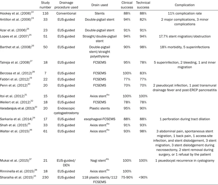
Table 1. Endoscopic Treatment of a Pancreas Pseudocyst Study
number
Drainage
procedure used Drain used Clinical success
Technical
success Complication
Hookey et al. (2006)22 116 Conventional Stents 88% 88% 11% complication rate
Antillon et al. (2006)23 33 EUS-guided Double-pigtail stent 94% 82% 2 major complications, 3 minor complications Azar et al. (2006)24 23 EUS-guided Double-pigtail stent 91% 91%
Lopes et al. (2007)25 51 EUS-guided Straight/double-pigtail stent
94% 94% 17.7% stent migration/obstruction
Barthet et al. (2008)26 50 EUS-guided Double-pigtail stent/straight polyethylene
90% 98% 18% morbidity, 5 superinfections
Talreja et al. (2008)27 18 EUS-guided FCSEMS 95% 78% 5 superinfection, 2 bleeding, 1 and inner migration
Berzosa et al. (2012)28 7 EUS-guided FCSEMS 100% 83%
Fabbri et al. (2012)29 22 EUS-guided FCSEMS 77% 77%
Penn et al. (2012)30 20 EUS-guided FCSEMS 70% 70% 2 pseudocyst infection, 1 post transmural drainage fever and post-ERCP pancreatitis Itoi et al. (2012)31 15 EUS-guided Axios stent®a 100% 100%
Weilert et al. (2012)32 18 EUS-guided FCSEMS 78% 78%
Varadarajulu et al. (2013)33 20 Endoscopic cystogastrostomy
Plastic stents 95% 90%
Sarkaria et al. (2014)34 17 EUS-guided esophageal-FCSEMS 88% 88% 1 perforation during tract dilation
Shah et al. (2015)35 33 EUS-guided Axios stent®a 91% 93%
Walter et al. (2015)36 61 EUS-guided Axios stent®a 93% 98% 3 abdominal pain, spontaneous stent migration, 1 back pain, 1 access-site infection, and stent dislodgement, 3 stent
migration, 3 stent dislodgement during necrosectomy, 2 stent removal during
surgery, or 1 refusal by the patient Mukai et al. (2015)37 21 EUS-guided/
DEN
Nagi stent®b 100% 100% 1 psuedocyst recurrence in cystogramy
Rinninella et al. (2015)38 18 EUS-guided Axios stent®a 100%
Sharaiha et al. (2015)15 230 EUS-guided 118 plastic stents/112 FCSEMS
75-90% <90%
EUS, endoscopic ultrasonography; FCSEMS, full covered self expandable metal stent; ERCP, endoscopic retrograde cholangiopancreatography;
DEN, direct endoscopic necrosectomy.
aAxios Stent® (Boston Scientific, Marlborough, MA, USA); bNagi stent® (Taewoong Medical, Gimpo, Korea).
추가로 필요한 경우가 있다. 췌장괴사에서 치료 효과를 높이 는 방법으로는 경벽 배액관과 경비적 배액관를 같이 삽입하여 경비 배액관으로 주기적 세척을 하는 방법, 여러 부위에 천공 및 배액관을 삽입하는 방법(multiple transluminal gateway technique),11 직경이 굵은 금속 스텐트를 삽입하는 방법 및 내시경 췌장괴사 제거술 등의 방법이 있다.12
금속 스텐트는 플라스틱 스텐트보다 상대적으로 큰 직경으 로 스텐트의 막힘이 없고 배액 효과가 커서 췌장액체고임의 치료에 더 유리할 것으로 생각되었다. 이전의 금속 스텐트를 이용한 비교 연구들은 치료 성공률, 부작용 및 재발률에서 차 이가 없었지만,13,14 이후 Sharaiha 등15의 연구에서 플라스틱 스텐트군에서 합병증 발생이 2.5배 높고 관해율이 89%로 금
속 스텐트군 98%보다 낮았다. 최근 Yoon 등16의 메타분석에 서도 금속 스텐트가 플라스틱 스텐트에 비하여 높은 임상적 성공률(OR 3.39, 95% CI 2.05-5.60)과 낮은 부작용(OR 0.37, 95% CI 0.21-0.66)을 보였으며, 하부집단 분석에서도 가성낭 종(OR 5.35, 95% CI 1.35-21.19) 및 췌장궤사(OR 3.37, 95%
CI 1.89-5.99)에서 높은 치료 성공률을 보여 금속 스텐트가 유용하였다. 금속 스텐트 역시 삽입 후 복통, 감염 및 스텐트 이탈, 출혈, 천공 등의 합병증이 발생할 수 있으므로 시술 후 신중한 경과 관찰이 필요하다.17,18 가성낭종(Table 1) 및 췌장 괴사에서 내시경 배액술(Table 2)의 치료 성적 및 합병증에 대하여 정리하였다.19
경벽적 배액술 후 적절한 배액관의 거치 기간에 대한 기준
Table 2. Endoscopic Treatment of Wall-off Necrosis Study
number
Drainage procedure
used
Drain used Clinical success
Technical
success Complication
Seewald et al. (2005)39 13 DEN Double-pigtail stent
91% 91% 4 minor bleeding after balloon dilation, necrosectomy
Charnley et al. (2006)40 13 DEN Double-pigtail stents
92.3% 92.3%
Voermans et al. (2007)41 25 DEN Double-pigtail stents
93% 93% 2 surgery, 1 hemorrhage, 1 perforation of cyst wall
Papachristou et al.
(2007)42
53 DEN Double-pigtail stents
81% 81% 12 required operation after initial endoscopic drainage/debridement, due to 3 persistence of WOPN, 2 recurrence of a PFCs, 2 cutaneous fistula
formation, or 1 technical failure, persistence of pancreatic pain, colonic obstruction, perforation,
and flank abscess Escourrou et al. (2008)43 13 DEN Double-pigtail
stents
100% 100% 3 bleeding, 3 transient aggravation of sepsis
Seifert et al. (2009)44 93 DEN Multiple stents 80% 80%
Gardner et al. (2009)45 45 DEN and EUS-guided
drainage
Multiple stents 45% DEN 88%, standard endoscopy drainage 45%
13 bleeding, 5 perforations of the necrosis, 2 fistular formation, 2 air embolism, 2 complications
at ohter organs
Gardner et al. (2011)46 104 DEN Multiple stents 91% 91% 14%; included 5 retrogastric perforations/pneumoperitoneum Attam et al. (2014)47 10 DEN and
transmural drain
Novel large-bore esophageal
FCSEMS
90% 100%
Smoczyński et al.
(2014)18
112 Endoscopic drainage
Multiple stents 84% 93% 19 stoma bleeding, 4 GI perforation, 2 collection perforation, 1 sepsis, 3 stent migration Sarkaria et al. (2014)34 17 EUS-guided Esohageal
FCSEMS
83% 83%
Mukai et al. (2015)37 8 DEN Nagi stent®a 100% 100%
Rinninella et al. (2015)38 52 EUS guided
Axios stent®b 90.4% 100% 3 surgery due to infection, 1 perforated wall
Walter et al. (2015)36 46 EUS guided
Axios stent®b 81% 81% 9%
DEN, direct endoscopic necrosectomy; WOPN, walled-off pancreatic necrosis; PFC, pancreatic fluid collection; EUS, endoscopic ultrasonography;
FCSEMS, full covered self expandable metal stent; GI, gastrointestinal.
aNagi stent® (Taewoong Medical, Gimpo, Korea); bAxios Stent® (Boston Scientific, Marlborough, MA, USA).
은 아직 결정되어 있지 않다. 배액관은 췌장액체고임이 소실 되었을 때 시행하는 것이 원칙이다. 췌장액체고임의 재발은 췌장실질이나 췌관의 손상이 주된 원인으로 알려져 있다. 췌 관의 손상이 없는 단순 가성낭종의 경우 매달 영상 검사를 시행하여 췌장액체고임이 소실되는 것이 확인되면 제거하도 록 한다.20반면 감염된 가성낭종이나 췌장괴사를 동반하는 경 우 액체의 점도가 높고 괴사조직 등으로 가성낭종에 비하여 신속한 배액이 되지 않아 장기간의 거치 기간을 필요로 한다.
그러나 거치 기간이 길어지면 배액관의 장관이나 복강 내 이 탈이 발생할 수 있으므로 정기적인 영상 검사를 통하여 배액
관의 상태나 췌장액체고임의 소실을 확인하고 제거 시점을 결 정하여야 한다.
최근에는 금속 스텐트의 양끝을 확장시키고 직경이 굵은 형태의 Lumen-Apposing Metal Stent (LAMS)가 개발되어 시술에 사용되고 있으며 Axios StentⓇ (Boston Scientific, Marlborough, MA, USA)와 Nagi stentⓇ (Taewoong Medical, Gimpo, Korea), Spaxus stentⓇ (Taewoong Medical)가 있다.
LAMS는 일체형 구조로 플라스틱 스텐트에 비하여 시술이 간편 하며 장벽과 가성낭종벽을 밀착시켜 시술 시 천공이나 출혈을 예방하는 효과가 있고, 양선단의 특징적인 모양으로 스텐트
이탈을 예방하며, 직경이 굵어 내시경이 가성낭종 내로 진입할 수 있으므로 괴사된 조직을 제거할 수 있다는 장점이 있다.
반면, Bang 등21이 시행한 전향적 연구에서 3건의 가성동맥류에 서의 출혈, 2건의 장관점막이 증식하여 LAMS 선단을 덮어버리 거나 1건의 담관의 협착 등이 발생하였으며, 이러한 합병증은 시술 3주 후 발생함을 보고하여 시술 환자들은 시술 3주 후 복부 CT를 시행하여 가성낭종이 소실되면 LAMS 제거를 고려 해야 한다고 보고하였다.
결 론
췌장액체고임은 초음파 내시경 유도 경벽 배액술이 치료에 도입되면서 안전하고 효과적인 치료법으로 시술되고 있다. 그 러나 내시경 시술은 췌장액체고임이 발생하고 4주 이상의 시 간 경과 후 시행하는 것이 원칙이다. 시술에 사용되는 배액관 은 췌장액체의 양상에 따라 배액관의 종류 및 개수 선택이 필요하다. 최근 국내외에서 다양한 금속 스텐트가 배액관으로 사용되고 있으며 치료 효과가 좋다는 것이 입증되고 있다. 특 히 동반된 췌장괴사가 광범위한 경우 다수의 배액관 삽입이나 내시경 췌장괴사 제거술 등이 필요할 수 있고, 이런 환자들은 시술 후 감염이나 출혈 등의 합병증이 발생할 확률이 높다는 것을 고려해야 한다. 따라서 췌장괴사에 의한 액체고임 치료 는 내과, 영상의학과 및 외과 간의 다학제 접근으로 환자 상태 에 적합한 치료 전략을 수립하는 것이 바람직하겠다.
우리나라에서는 초음파 내시경 유도 가성낭종 배액술은 2013년 신 의료기술로 인정받았다. 그러나 시술에 사용되는 부우지 확장기, 확장용 풍선, 침형절개도, 플라스틱 스텐트 및 금속 스텐트 등 시술에 사용되는 치료 재료들이 식약청의 사 용 허가를 받지 못하여 급여권에서 시술을 시행할 수 없었으 나 최근 국내에서 개발된 유도선, 절개도(cystotome) 및 배액 관(금속 스텐트)이 식약청 허가 임상 연구 후 허가를 취득하여 향후 보험 적용도 가능해질 것으로 생각된다.
REFERENCES
1. Banks PA, Bollen TL, Dervenis C, et al. Classification of acute pan- creatitis--2012: revision of the Atlanta classification and defi- nitions by international consensus. Gut 2013;62:102-111.
2. Shin DH, Shin JH, Kang YW, et al. Management of pancreatic pseudocysts. Korean J Gastroenterol 1999;33:831-836.
3. Kwon SB, Kim IY, Bae KS, et al. Treatment of pancreatic pseudocysts. Korean J Gastroenterol 2000;36:529-535.
4. Rogers BH, Cicurel NJ, Seed RW. Transgastric needle aspiration of pancreatic pseudocyst through an endoscope. Gastrointest Endosc 1975;21:133-134.
5. Grimm H, Binmoeller KF, Soehendra N. Endosonography-guided drainage of a pancreatic pseudocyst. Gastrointest Endosc
1992;38:170-171.
6. Andrén-Sandberg A, Dervenis C. Pancreatic pseudocysts in the 21st century. Part I: classification, pathophysiology, anatomic considerations and treatment. JOP 2004;5:8-24.
7. Baron TH, Treatment of pancreatic pseudocysts, pancreatic ne- crosis, and pancreatic duct leaks. Gastrointest Endosc Clin N Am 2007;17:559-579, vii.
8. Kozarek RA. Endoscopic management of pancreatic necrosis:
not for the uncommitted. Gastrointest Endosc 2005;62:
101-104.
9. Levy MJ, Smyrk TC, Reddy RP, et al. Endoscopic ultra- sound-guided trucut biopsy of the cyst wall for diagnosing cystic pancreatic tumors. Clin Gastroenterol Hepatol 2005;3:974-979.
10. Varadarajulu S, Bang JY, Phadnis MA, Christein JD, Wilcox CM.
Endoscopic transmural drainage of peripancreatic fluid collec- tions: outcomes and predictors of treatment success in 211 con- secutive patients. J Gastrointest Surg 2011;15:2080-2088.
11. Varadarajulu S, Phadnis MA, Christein JD, Wilcox CM. Multiple transluminal gateway technique for EUS-guided drainage of symptomatic walled-off pancreatic necrosis. Gastrointest Endosc 2011;74:74-80.
12. Varadarajulu S, Wilcox CM. Endoscopic placement of permanent indwelling transmural stents in disconnected pancreatic duct syndrome: does benefit outweigh the risks? Gastrointest Endosc 2011;74:1408-1412.
13. Melman L, Azar R, Beddow K, et al. Primary and overall success rates for clinical outcomes after laparoscopic, endoscopic, and open pancreatic cystgastrostomy for pancreatic pseudocysts.
Surg Endosc 2009;23:267-271.
14. Navaneethan U, Njei B, Sanaka MR. 734 Endoscopic transmural drainage of pancreatic pseudocysts: multiple plastic stents ver- sus metal stents- a systematic review and meta-analysis.
Gastrointest Endosc 2014;79:AB167-AB168.
15. Sharaiha RZ, DeFilippis EM, Kedia P, et al. Metal versus plastic for pancreatic pseudocyst drainage: clinical outcomes and success. Gastrointest Endosc 2015;82:822-827.
16. Yoon SB, Lee IS, Choi MG. Metal versus plastic stents for drainage of pancreatic fluid collection: a meta-analysis. United European Gastroenterol J 2018;6:729-738.
17. Shah RJ, Shah JN, Waxman I, et al. Safety and efficacy of endo- scopic ultrasound-guided drainage of pancreatic fluid collec- tions with lumen-apposing covered self-expanding metal stents.
Clin Gastroenterol Hepatol 2015;13:747-752.
18. Smoczyński M, Marek I, Dubowik M, et al. Endoscopic drain- age/debridement of walled-off pancreatic necrosis--single cen- ter experience of 112 cases. Pancreatology 2014;14:137-142.
19. Tyberg A, Karia K, Gabr M, et al. Management of pancreatic fluid collections: a comprehensive review of the literature. World J Gastroenterol 2016;22:2256-2270.
20. Singhal S, Rotman SR, Gaidhane M, Hahaleh M. Pancreatic fluid collection drainage by endoscopic ultrasound: an update. Clin Endosc 2013;46:506-514.
21. Bang JY, Hasan M, Navaneethan U, Hawes R, Varadarajulu S.
Lumen-apposing metal stents (LAMS) for pancreatic fluid collec- tion (PFC) drainage: may not be business as usual. Gut 2017;66:2054-2056.
22. Hookey LC, Debroux S, Delhaye M, Arvanitakis M, Le Moine O, Devière J. Endoscopic drainage of pancreatic-fluid collection in 116 patients: a comparison of etiologies, drainage techniques, and outcomes. Gastrointest Endosc 2006;63:635-643.
23. Antillon MR, Shah RJ, Stiegmann G, Chen YK. Single-step EUS-guided transmural drainage of simple and complicated pan- creatic pseudocysts. Gastrointest Endosc 2006;63:797-803.
24. Azar RR, Oh YS, Janec EM, Early DS, Jonnalagadda SS, Edmundowicz SA. Wire-guided pancreatic pseudocyst drainage by using a modified needle knife and therapeutic echoendoscope.
Gastrointest Endosc 2006;63:688-692.
25. Lopes CV, Pesenti C, Bories E, Caillol F, Giovannini M.
Endoscopic-ultrasound-guided endoscopic transmural drain- age of pancreatic pseudocysts and abscesses. Scand J Gastroenterol 2007;42:524-529.
26. Barthet M, Lamblin G, Gasmi M, Vitton V, Desjeux A, Grimaud JC.
Clinical usefulness of a treatment algorithm for pancreatic pseudocysts. Gastrointest Endosc 2008;67:245-252.
27. Talreja JP, Shami VM, Ku J, Morris TD, Ellen K, Kahaleh M.
Transenteric drainage of pancreatic-fluid collections with fully covered self-expanding metallic stents (with video). Gastrointest Endosc 2008;68:1199-1203.
28. Berzosa M, Maheshwari S, Patel KK, Shaib YH. Single-step endo- scopic ultrasonography-guided drainage of peripancreatic fluid collections with a single self-expandable metal stent and stand- ard linear echoendoscope. Endoscopy 2012;44:543-547.
29. Fabbri C, Luigiano C, Cennamo V, et al. Endoscopic ultra- sound-guided transmural drainage of infected pancreatic fluid collections with placement of covered self-expanding metal stents: a case series. Endoscopy 2012;44:429-433.
30. Penn DE, Draganov PV, Wagh MS, Forsmark CE, Gupte AR, Chauhan SS. Prospective evaluation of the use of fully covered self-expanding metal stents for EUS-guided transmural drainage of pancreatic pseudocysts. Gastrointest Endosc 2012;76:679-684.
31. Itoi T, Binmoeller KF, Shah J, et al. Clinical evaluation of a novel lumen-apposing metal stent for endosonography-guided pan- creatic pseudocyst and gallbladder drainage (with videos).
Gastrointest Endosc 2012;75:870-876.
32. Weilert F, Binmoeller KF, Shah JN, Bhat YM, Kane S. Endoscopic ultrasound-guided drainage of pancreatic fluid collections with indeterminate adherence using temporary covered metal stents. Endoscopy 2012;44:780-783.
33. Varadarajulu S, Bang JY, Sutton BS, Trevino JM, Christein JD, Wilcox CM. Equal efficacy of endoscopic and surgical cystogas- trostomy for pancreatic pseudocyst drainage in a randomized trial. Gastroenterology 2013;145:583-590.
34. Sarkaria S, Sethi A, Rondon C, et al. Pancreatic necrosectomy us- ing covered esophageal stents: a novel approach. J Clin Gastroenterol 2014;48:145-152.
35. Shah RJ, Shah JN, Waxman I, et al. Safety and efficacy of endo-
scopic ultrasound-guided drainage of pancreatic fluid collec- tions with lumen-apposing covered self-expanding metal stents.
Clin Gastroenterol Hepatol 2015;13:747-752.
36. Walter D, Will U, Sanchez-Yague A, et al. A novel lumen-apposing metal stent for endoscopic ultrasound-guided drainage of pan- creatic fluid collections: a prospective cohort study. Endoscopy 2015;47:63-67.
37. Mukai S, Itoi T, Sofuni A, Tsuchiya T, Gotoda T, Moriyasu F. Clinical evaluation of endoscopic ultrasonography-guided drainage us- ing a novel flared-type biflanged metal stent for pancreatic fluid collection. Endosc Ultrasound 2015;4:120-125.
38. Rinninella E, Kunda R, Dollhopf M, et al. EUS-guided drainage of pancreatic fluid collections using a novel lumen-apposing metal stent on an electrocautery-enhanced delivery system: a large ret- rospective study (with video). Gastrointest Endosc 2015;82:
1039-1046.
39. Seewald S, Groth S, Omar S, et al. Aggressive endoscopic therapy for pancreatic necrosis and pancreatic abscess: a new safe and effective treatment algorithm (videos). Gastrointest Endosc 2005;62:92-100.
40. Charnley RM, Lochan R, Gray H, O’Sullivan CB, Scott J, Oppong KE. Endoscopic necrosectomy as primary therapy in the manage- ment of infected pancreatic necrosis. Endoscopy 2006;38:
925-928.
41. Voermans RP, Veldkamp MC, Rauws EA, Bruno MJ, Fockens P.
Endoscopic transmural debridement of symptomatic organized pancreatic necrosis (with videos). Gastrointest Endosc 2007;
66:909-916.
42. Papachristou GI, Takahashi N, Chahal P, Sarr MG, Baron TH.
Peroral endoscopic drainage/debridement of walled-off pancre- atic necrosis. Ann Surg 2007;245:943-951.
43. Escourrou J, Shehab H, Buscail L, et al. Peroral transgastric/trans- duodenal necrosectomy: success in the treatment of infected pancreatic necrosis. Ann Surg 2008;248:1074-1080.
44. Seifert H, Biermer M, Schmitt W, et al. Transluminal endoscopic necrosectomy after acute pancreatitis: a multicentre study with long-term follow-up (the GEPARD study). Gut 2009;58:1260-1266.
45. Gardner TB, Chahal P, Papachristou GI, et al. A comparison of di- rect endoscopic necrosectomy with transmural endoscopic drainage for the treatment of walled-off pancreatic necrosis.
Gastrointest Endosc 2009;69:1085-1094.
46. Gardner TB, Coelho-Prabhu N, Gordon SR, et al. Direct endo- scopic necrosectomy for the treatment of walled-off pancreatic necrosis: results from a multicenter U.S. series. Gastrointest Endosc 2011;73:718-726.
47. Attam R, Trikudanathan G, Arain M, et al. Endoscopic transluminal drainage and necrosectomy by using a novel, through-the-scope, fully covered, large-bore esophageal metal stent: preliminary ex- perience in 10 patients. Gastrointest Endosc 2014;80:312-318.