Multicenter clinical study on the hydrodynamic piezoelectric internal sinus elevation (HPISE) technique
Hyung-Ju Lee1, Jee-Won Moon1, Ju-Hyoung Lee1, In-Sook Park1, Nam-Ho Kim2, Dong-Seok Sohn1
1Department of Dentistry and Oral and Maxillofacial Surgery, Daegu Catholic University Medical Center, Daegu,
2Department of Oral and Maxillofacial Surgery, Chung-Ang University College of Medicine, Seoul, Korea
Abstract(J Korean Assoc Oral Maxillofac Surg 2012;38:85-9)
Objectives: This study was to evaluate the effect of vertical bone gain and success rate and analyze the failure cases using the hydrodynamic piezoelectric internal sinus elevation (HPISE) technique.
Materials and Methods: Patients who had been operated in the three centers including Daegu Catholic University Medical Center were selected for this study. The mucoperiosteal flap was elevated, and the sinus floor was then broken by specially designed piezoelectric insert, with hydraulic pressure applied to the sinus membrane for even elevation. Afterward, implants were placed. Panoramic radiogram or computed tomogram was taken before and after surgery and at the second operation and prosthesis placement. Later, changes in vertical height were measured and compared. The survival rate was based on the criteria of Buser et al. and Cochran et al.
Results: In this study, 8 implants failed out of a total of 169 implants, resulting a success rate of 95.3%. These failure cases were due to insufficient initial stability or sinus membrane perforation. The mean of radiographic vertical height change at prosthesis placement was 5.7 mm (0.5-10.5 mm).
Conclusion: In this study, HPISE technique was found to be a predictable treatment for atrophic maxilla and an alternative technique to the lateral approach.
Key words: Hydrodynamic piezoelectric internal sinus elevation, Hydraulic pressure, Crestal approach, Piezosurgery
[paper submitted 2011. 6. 22 / revised 2011. 11. 18 / accepted 2012. 3. 6]
bone graft through osteotomy on the sinus lateral walls, sinus bone graft has been investigated aggressively for its clinical skill and results, reporting many successful clinical outcomes and high predictability5-7. Osteotome-mediated sinus floor elevation (OMSFE), developed by many clinicians since it was suggested by Summers, has been widely used because there were less injuries and symptoms after the surgery such as pain and edema compared to bone graft through osteotomy of the sinus lateral walls8-10. However, this technique can also cause sinus membrane perforation due to the initial bone preparation and the pressure when pushing of bone graft material and many complications such as benign paroxysmal positional vertigo caused by internal ear damage when using osteotome and mallet were reported11,12. Moreover, since bone graft material must be used for this technique, there is high chance of infection by bone graft material on the perforated sinus membrane.
The hydrodynamic piezoelectric internal sinus elevation (HPISE) technique using ultrasonic device with a specially designed ultrasonic wave surgery insert causes neither benign
I. Introduction
Recently, dental implant placement is widely used from the restoration of a single tooth to the full-mouth reconstruction.
However, it has been limited by the absorption of the alveolar bone and location of important anatomical structures. In particular, placing implants in the maxillary posterior region is very difficult due to the pneumatization of sinus and fast absorption of alveolar bone. To address such problems, various techniques have been used for atrophic alveolar bone, with sinus bone graft using various bone graft materials1-3.
Since Boyne and James4 reported the clinical result of sinus Dong-Seok Sohn
Department of Dentistry and Oral and Maxillofacial Surgery, Daegu Catholic University Medical Center, 3056-6, Daemyung 4-dong, Nam-gu, Daegu 705- 718, Korea
TEL: +82-53-650-4291 FAX: +82-53-622-7067 E-mail: dssohn@cu.ac.kr
This is an open-access article distributed under the terms of the Creative Commons Attribution Non-Commercial License (http://creativecommons.org/licenses/by-nc/3.0/), which permits unrestricted non-commercial use, distribution, and reproduction in any medium, provided the original work is properly cited.
CC
Catholic University Medical Center from January 2008 to May 2009. For 103 patients, a total of 169 implants were placed in 114 sinuses. Male patients were 53, and female patients were 50; average age was 52 (20-90). Smokers and patients of accommodative systemic disease were not excluded in this study.
2. Methods
Patients’ personal information such as gender and age as well as the time of HPISE surgery, the time of implant placement, complications, and vertical bone height before and after surgery were investigated based on the patients' medical records, panoramic radiogram, and computed tomogram (CT).
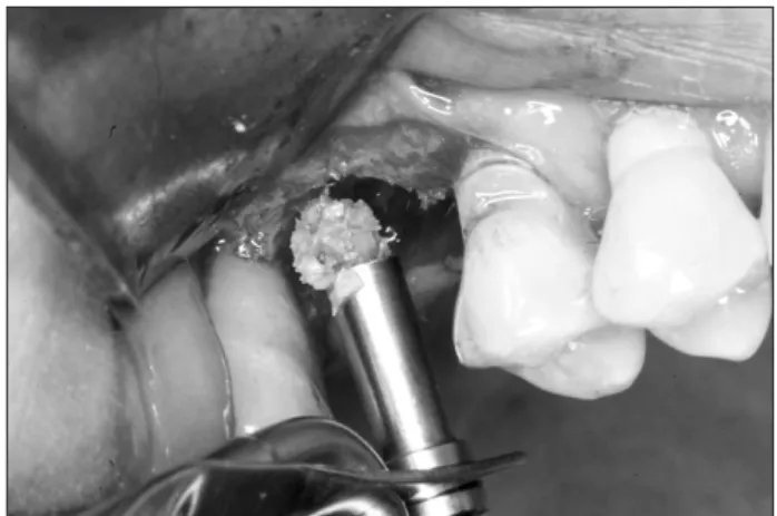
1) Surgical operation (Figs. 1-3)
All surgeries were performed under local anesthesia. After crestal incision, the mucoperiosteal flap was elevated, and the cortical bone of the sinus floor was then perforated with specially designed insert (S028I; BukBu Dental Co., Daegu, Korea). And sterilized saline solution was injected into the sinus membrane for 10-12 seconds with strong hydraulic paroxysmal positional vertigo nor membrane perforation
caused by malleting by directly reducing bones to the sinus floor in sinus bone graft. The greater elevation can be gained than the existing crestal approaches because removal of the sinus floor and elevating the sinus membrane with hydraulic pressure simultaneously, it can minimize the risk of sinus membrane perforation.
This study was to evaluate the effect and clinical appli- cation of HPISE technique by investigating the success rate by conducting post-operative measurement of vertical bone gain in the patients who had surgery using HPISE technique due to the atrophic maxilla among the patients who visited the hospital for the implant placement in the maxillary posterior region, and to improve the predictability by analyzing the failure cases.
II. Materials and Methods
1. Materials (Table 1)
This study examined patients who had been operated using the HPISE technique for the placement of implants in their maxillary posterior region in three centers including Daegu
Fig. 1. The sinus floor was perforated with the hydrodynamic piezoelectric internal sinus elevation insert, and hydraulic pressure was applied for a few seconds.
Hyung-Ju Lee et al: Multicenter clinical study on the hydrodynamic piezoelectric internal sinus elevation (HPISE) technique. J Korean Assoc Oral Maxillofac Surg 2012
Fig. 2. Bone graft material was inserted under the elevated sinus membrane using amalgam carrier.
Hyung-Ju Lee et al: Multicenter clinical study on the hydrodynamic piezoelectric internal sinus elevation (HPISE) technique. J Korean Assoc Oral Maxillofac Surg 2012 Table 1. Patients’ descriptive data
Center No. of patients Gender
(male/female) Mean age
(year) No. of sinuses No. of implants Mean residual bone height (mm) A
B C Total
32 40 31 103
15/17 16/24 22/9 53/50
52 50 54 52
33 49 32 114
48 72 49 169
7.27 5.83 5.54 618 Hyung-Ju Lee et al: Multicenter clinical study on the hydrodynamic piezoelectric internal sinus elevation (HPISE) technique. J Korean Assoc Oral Maxillofac Surg 2012
before the operation, on the day of operation, after the second operation, and at the initial functional loading. One operator performed three measurements of the vertical bone amount in the buccolingual section of CT image, the average measurements were taken, and the measurements of increased bone level were calculated.
3) Data analysis
To objectify data, a unified clinical examination chart was created after reviewing the medical records of each center.
Data was collected from clinical examination chart based on the medical records and clinical examination. The clinical examination chart includes mainly (1) sex and age, (2) location of operation, (3) type of the bone material, (4) type of the implant, (5) implant width and length, (6) location of implant placement, (7) vertical bone height at each stage, and (8) failure of implant and the estimated reason. The success rate and vertical bone height were analyzed using the data from the clinical examination chart; the success rate was measured according to the criteria of Cochran et al.13 and Buser et al.14.
III. Results
1. Vertical bone gain after HPISE surgery (Table 2) Vertical bone gain was calculated by comparing the value pressure to elevate the sinus membrane. Once the sinus
membrane was elevated, S028I was rotated in every direction for even elevation. In some cases, bone graft material was used after the sinus membrane was elevated; in other cases, either bone graft material was not used, or only concentrated growth factor and/or collagen sponge were inserted. After implants were placed, the flap was relocated, and the operator sutured. The sutures were removed after 2 weeks.
2) Radiological evaluation and measurement (Fig. 4) Radiological bone change was measured by examining the digital panoramic radiographs and CT images taken
Fig. 3. Implant was placed.
Hyung-Ju Lee et al: Multicenter clinical study on the hydrodynamic piezoelectric internal sinus elevation (HPISE) technique. J Korean Assoc Oral Maxillofac Surg 2012
Fig. 4. Measurement of vertical bone height. A. Before surgery. B. After surgery. C. Second operation. D. Prosthesis placement.
Hyung-Ju Lee et al: Multicenter clinical study on the hydrodynamic piezoelectric internal sinus elevation (HPISE) technique. J Korean Assoc Oral Maxillofac Surg 2012
Table 2. Patients’ findings Mean healing time
(week) Mean residual bone height
(mm) Mean vertical bone gain
(mm) Mean loading period
(week) Success rate
(%)
33.6 6.18 5.7 100.1 95.3
Hyung-Ju Lee et al: Multicenter clinical study on the hydrodynamic piezoelectric internal sinus elevation (HPISE) technique. J Korean Assoc Oral Maxillofac Surg 2012
and Krieger15, who used the OMSFE technique, reported an average success rate of 92.1%; Wallace and Froum16 reported an average success rate of 94%, and Del Fabbro et al.17, 91.49%. Likewise, Toffler et al.18 reported high success rate of 97.8%. This study also showed similar success rate of 95.3%.
In this study, the mean vertical bone gain was 5.7 mm (0.5-10.5 mm); in most cases, however, it was maintained at the apex of the implant over time regardless of the amount of bone graft and level of elevation in the sinus. It is considered to be caused by the pneumatization of maxillary sinus, and it was maintained regardless of the height of the initial residual bone. Therefore, more vertical bone gain could be obtained if the height of the initial residual bone is low. And the perforation of the sinus membrane during operation was found to be important not only for the success rate but also for vertical bone gain because the latter was insufficient or very minimal in cases with perforation. Cases with perforation were caused by excessive force applied to the sinus membrane when handling instruments. Therefore, when braking the sinus floor, instruments should be operated carefully because excessive force may cause perforation even though ultrasonic wave instruments are not working on soft tissues. If the sinus membrane is perforated during the surgery and the operator fails to control it properly, the surgery is likely to fail. Therefore, closing the perforated part by inserting collagen membrane is recommended rather than using bone graft material; otherwise, carry out a lateral approach to control the perforated part19,20.
Initial stability in implant placement is important for successful osseointegration21. Among the failed implants in this study, 2 were caused by insufficient initial stability due to insufficient residual bone or improper bone preparation.
To overcome this, it is considered that use the implant with tapered design or place the implant that one size lager. In addition, recent studies reported the formation of new bone by only the elevation of the sinus membrane without bone graft before the surgery and the value at prosthesis placement, and
the mean of vertical height change was 5.7 mm (0.5-10.5 mm). Prosthesis placement was performed 33.6 weeks (14- 94 weeks) after the surgery on the average. In most cases, the sinus membrane was elevated to above of the implant right after the surgery, but it fell to the apex of implant over time.
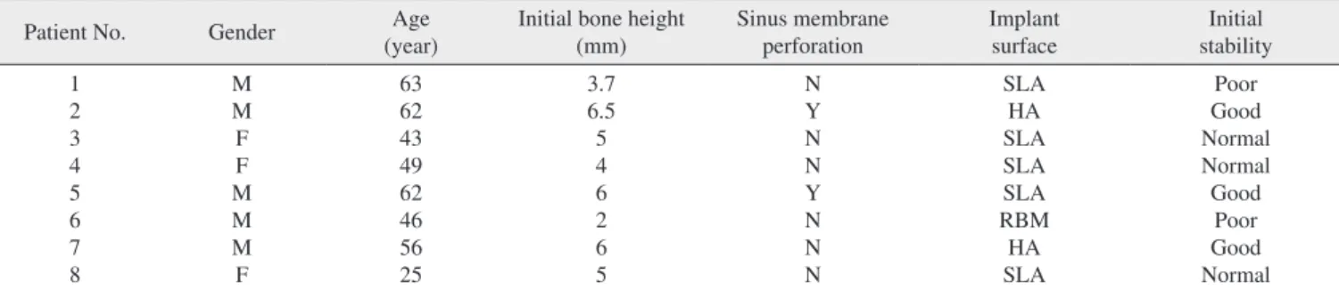
2. Success rate and failure (Table 3)
Out of a total of 169 implants placed in 114 sinuses of 103 patients, 8 implants failed, resulting a success rate of 95.3%.
The reason for failed implants was sinus membrane perforation in sinus elevation or insufficient initial stability caused by insufficient residual bone. The failed implants were 5 of sand blasted, large grit, acid-etched implant, 2 of hydroxiapatite coated and 1 of resorbable blasting media. In 4 cases, they failed during the second operation after the implant placement or impression procedure and in the other 4 cases, they failed during the functional loading.
IV. Discussion
The HPISE technique was developed to elevate the sinus membrane with hydraulic pressure after removal the sinus floor directly with micro-vibration of ultrasonic waves using specially designed insert that supports internal irrigation.
Compared with the OMSFE technique, it causes less sinus membrane perforation, the sinus membrane can be divided from the sinus floor more easily, and it is safe from benign paroxysmal positional vertigo. When using HPISE technique, bone graft can be performed easily without resistance to the sinus membrane because it elevates the sinus membrane with hydraulic pressure while braking the sinus floor, and this can shorten the operation time.
Among the studies on the success rate of implants placed in the maxillary posterior region using sinus elevation, Coatoam
Table 3. Failure list
Patient No. Gender Age
(year) Initial bone height
(mm) Sinus membrane
perforation Implant
surface Initial
stability 1
2 3 4 5 6 7 8
M M F F M M M F
63 62 43 49 62 46 56 25
3.7 6.5 5 4 6 2 6 5
N Y N N Y N N N
SLA HA SLA SLA SLA RBM HA SLA
Poor Good Normal Normal Good
Poor Good Normal (M: male, F: female, N: no, Y: yes, SLA: sand blasted, large grit, acid-etched implant, HA: hydroxiapatite coated, RBM: resorbable blasting media.) Hyung-Ju Lee et al: Multicenter clinical study on the hydrodynamic piezoelectric internal sinus elevation (HPISE) technique. J Korean Assoc Oral Maxillofac Surg 2012
4. Boyne PJ, James RA. Grafting of the maxillary sinus floor with autogenous marrow and bone. J Oral Surg 1980;38:613-6.
5. Adell R, Lekholm U, Gröndahl K, Brånemark PI, Lindström J, Jacobsson M. Reconstruction of severely resorbed edentulous maxillae using osseointegrated fixtures in immediate autogenous bone grafts. Int J Oral Maxillofac Implants 1990;5:233-46.
6. Raghoebar GM, Vissink A, Reintsema H, Batenburg RH. Bone grafting of the floor of the maxillary sinus for the placement of endosseous implants. Br J Oral Maxillofac Surg 1997;35:119-25.
7. Aghaloo TL, Moy PK. Which hard tissue augmentation techniques are the most successful in furnishing bony support for implant placement? Int J Oral Maxillofac Implants 2007;22 Suppl:49-70.
8. Summers RB. A new concept in maxillary implant surgery: the osteotome technique. Compendium 1994;15:152, 154-6, 158 passim; quiz 162.
9. Summers RB. The osteotome technique: Part 2--The ridge expansion osteotomy (REO) procedure. Compendium 1994;15:422, 424, 426, passim; quiz 436.
10. Summers RB. The osteotome technique: Part 3--Less invasive methods of elevating the sinus floor. Compendium 1994;15:698, 700, 702-4 passim; quiz 710.
11. Di Girolamo M, Napolitano B, Arullani CA, Bruno E, Di Girolamo S. Paroxysmal positional vertigo as a complication of osteotome sinus floor elevation. Eur Arch Otorhinolaryngol 2005;262:631-3.
12. Peñarrocha M, Garcia A. Benign paroxysmal positional vertigo as a complication of interventions with osteotome and mallet. J Oral Maxillofac Surg 2006;64:1324.
13. Cochran DL, Buser D, ten Bruggenkate CM, Weingart D, Taylor TM, Bernard JP, et al. The use of reduced healing times on ITI implants with a sandblasted and acid-etched (SLA) surface: early results from clinical trials on ITI SLA implants. Clin Oral Implants Res 2002;13:144-53.
14. Buser D, Mericske-Stern R, Bernard JP, Behneke A, Behneke N, Hirt HP, et al. Long-term evaluation of non-submerged ITI implants.
Part 1: 8-year life table analysis of a prospective multi-center study with 2359 implants. Clin Oral Implants Res 1997;8:161-72.
15. Coatoam GW, Krieger JT. A four-year study examining the results of indirect sinus augmentation procedures. J Oral Implantol 1997;23:117-27.
16. Wallace SS, Froum SJ. Effect of maxillary sinus augmentation on the survival of endosseous dental implants. A systematic review.
Ann Periodontol 2003;8:328-43.
17. Del Fabbro M, Testori T, Francetti L, Weinstein R. Systematic review of survival rates for implants placed in the grafted maxillary sinus. Int J Periodontics Restorative Dent 2004;24:565-77.
18. Toffler M, Toscano N, Holtzclaw D. Osteotome-mediated sinus floor elevation using only platelet-rich fibrin: an early report on 110 patients. Implant Dent 2010;19:447-56.
19. Sohn DS, Moon JW, Moon KN, Cho SC, Kang PS. New bone formation in the maxillary sinus using only absorbable gelatin sponge. J Oral Maxillofac Surg 2010;68:1327-33.
20. Shin HI, Sohn DS. A method of sealing perforated sinus membrane and histologic finding of bone substitutes: a case report. Implant Dent 2005;14:328-33.
21. Le Gall MG. Localized sinus elevation and osteocompression with single-stage tapered dental implants: technical note. Int J Oral Maxillofac Implants 2004;19:431-7.
22. Lundgren S, Andersson S, Gualini F, Sennerby L. Bone refor- mation with sinus membrane elevation: a new surgical technique for maxillary sinus floor augmentation. Clin Implant Dent Relat Res 2004;6:165-73.
23. Palma VC, Magro-Filho O, de Oliveria JA, Lundgren S, Salata LA, Sennerby L. Bone reformation and implant integration following maxillary sinus membrane elevation: an experimental study in primates. Clin Implant Dent Relat Res 2006;8:11-24.
24. Sohn DS, Lee JS, Ahn MR, Shin HI. New bone formation in the maxillary sinus without bone grafts. Implant Dent 2008;17:321-31.
material during sinus graft22-24. In the case of the OMSFE technique, sinus bone graft without bone graft material is impossible because it elevates the sinus membrane by filling the bone graft material. However, the HPISE technique elevates the sinus membrane with hydraulic pressure, and can acquire sufficient elevation of sinus membrane similar to the lateral approach technique, enabling the implant placement only by the elevation of the sinus membrane without bone graft material.
This study evaluated the usefulness and prognosis of the HPISE technique used in various centers based on retrospective clinical evaluation and obtained satisfactory clinical results even in cases for the parts where unfavorable anatomical structure to place an implant. Even though many clinical applications and long-term examination are required, the HPISE technique can be considered as an alternative technique to the lateral approach technique.
V. Conclusion
This study examined the 169 implants of 103 patients who had been operated using the HPISE technique for the placement of implants in their maxillary posterior region in three centers including Department of Dentistry and Oral and Maxillofacial Surgery, Daegu Catholic University Medical Center from January 2008 to May 2009. After investigating the vertical bone change by period and factor, the following conclusions were drawn:
1. The mean of vertical bone gain was 5.7 mm (0.5-10.5 mm).
2. Among 169 implants, 8 implants failed; the success rate of HPISE was 95.3%.
3. Instruments should be handled carefully to prevent the perforation of the sinus membrane. In case of per foration, the operator should take the necessary measures.
4. Implant with tapered design or implant that is one size larger is considered because insufficient initial stability may cause failure.
References
1. Atwood DA. Bone loss of edentulous alveolar ridges. J Periodontol 1979;50:11-21.
2. Misch CE. Maxillary sinus augmentation for endosteal implants:
organized alternative treatment plans. Int J Oral Implantol 1987;
4:49-58.
3. Smiler DG, Johnson PW, Lozada JL, Misch C, Rosenlicht JL, Tatum OH Jr, et al. Sinus lift grafts and endosseous implants.
Treatment of the atrophic posterior maxilla. Dent Clin North Am 1992;36:151-86.