Synovial chondromatosis is a benign nodular cartilaginous pro- liferation arising in the synovium of joints. Usually the major weight bearing joint is involved and approximately 70% of cases are the knee joint. The radiolographic features of this condition are variable. Classically, radiographs reveal multiple intraarticular calcifications of similar size and shape, distributed throughout the joint, with typical “ring-and-arc” chondroid mineralization. The classic appearance is not often encountered. Rarely, it would be confused with malignancy such as chondrosarcoma, osteosar- coma or synovial sarcoma.
We report a case of primary synovial chondromatosis origi- nating from the intra-articular tissues of the knee, mimicking a parosteal osteoarcoma with the clinical, radiographic and com- puted tomography (CT) findings.
Case Report
A 24-year-old man was referred to Orthopedic Clinic complain- ing of intermittent pain in the posterior aspect of the knee. Dur-
Case Report
J Korean Bone Joint Tumor Soc 2010; 16: 95-98 • doi:10.5292/jkbjts.2010.16.2.95 www.kbjts.or.krGiant Synovial Chondromatosis of the Knee Mimicking a Parosteal Osteosarcoma: A Case Report
Chang Ho Kang, M.D.*, Jong Hoon Park, M.D., Dae Hee Lee, M.D., Chul Hwan Kim, M.D.
†, Jeong Mi Park, M.D.
‡, and Won Seok Lee, M.D.
Departments of Orthopedic Surgery, *Radiology,
†Pathology, Korea University Anam Hospital,
‡Department of Radiology, Catholic University St. Mary's Hospital, Seoul, Korea
ing physical examination, the flexion of his left knee joint was limited to 110o and fixed hard mass was palpated in the popliteal fossa. Neither tenderness nor local heating was detected. He did not give any trauma or previous specific medical history.
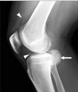
Plain radiographs showed a densely ossified mass which seemed to abut on the posterior aspect of the proximal tibia (Fig.
Received September 30, 2010 Revised November 5, 2010 Accepted November 30, 2010
Correspondence to: Jong Hoon Park, M.D.
Department of Orthopedic Surgery, Korea University Anam Hospital, 126-1, Anam- dong 5-ga, Sungbuk-gu, Seoul 136-705, Korea
TEL: +82-2-920-6609 FAX: +82-2-924-2471 E-mail: [email protected]
Synovial chondromatosis is a benign nodular cartilaginous proliferation arising in the synovium of joints. The radiolographic features of this condition are variable. Rarely, it would be confused with malignancy such as chondrosarcoma, osteosarcoma or synovial sarcoma. We re- port a case of primary synovial chondromatosis of the posterior aspect of the proximal tibia mimicking a parosteal osteoarcoma on the radi- ography, which showed a homogeneously radiopaque juxtacortical mass. However, subsequent computed tomography (CT) showed multiple intra-articular masses containing chondroid mineralization, suggesting synovial chondromatosis.
Key words: synovial chondromatosis, parosteal osteosarcoma, diagnostic imaging
Copyrights © 2010 by The Korean Bone and Joint Tumor Society
“This is an Open Access article distributed under the terms of the Creative Commons Attribution Non-Commercial License (http://creativecommons.org/licenses/by-nc/3.0/) which permits unrestricted non-commercial use, distribution, and reproduction in any medium, provided the original work is properly cited.”
대한골관절종양학회지:제16권 제2호 2010
Figure 1. Lateral radiograph shows a soft tissue mass (arrow) containing dense mineralization extending into the popliteal fossa. Partial attachment to the cortex as well as radiolucent cleavage plane was evident. Two small radiodense soft tissue masses (arrowheads) were initially missed on the radiograph.
96
Chang Ho Kang, et al.
1) and two small radiodense masses in the suprapatellar pouch and the Hoffa’s fat pad were initially missed on the radiograph.
A radiolucent cleft separating the part of the ossified large mass from the posterior cortex of the proximal tibia was detected ra- diographically. The combined radiological and clinical differen- tial diagnosis at this point included a parosteal osteosarcoma as well as myositis ossificans and tumoral calcinosis, with parosteal osteosarcoma being preferentially considered in the differential diagnosis. CT scan was performed to further evaluate, which showed a large, well-circumscribed calcified mass extending into the popliteal fossa with another small mineralized masses (Fig.
2). According to the findings of CT scan, a radiologist reported that radiologic diagnosis was compatible with a synovial chon- dromatosis. We thought that limitation of the knee joint motion was occurred due to the mechanical block of the huge posterior mass. The operation for mass excision was performed. The large mass in the popliteal fossa, as well as another small sclerotic nodular masses were excised through direct posterior approach.
The intra-articular mass showed only thin, tenuous attachments to the synovial membrane and could easily be removed. Because the synovium and the capsule of the joint appeared grossly unre- markable, a partial or complete synovectomy was not performed.
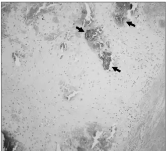
Histologically, the mass was composed of hyaline cartilage with focal areas of acellular bone, diagnostic of primary synovial os-
teochondromatosis. On microscopic examination, variably cel- lular hyaline cartilage covered by a fine fibrous layer and some myxoid change was reported (Fig. 3). After operation, the range of knee joint motion was markedly improved with normal range and no evidence of recurrence was observed postoperatively 14 months.
Figure 3. Photomicrograph (original magnification, H&E stain ×200) shows the typical hyaline cartilaginous nodule with calcification (arrows).
Figure 2. CT scan demonstrates the calcified small masses with target appearance occurring in the synovium more clearly (arrowheads), as well as large mass showing (arrows) extensive ring-and-arc mineralization that represents enchondral ossification of the multiple chondral bodies.
97
Giant Synovial Chondromatosis of the Knee Mimicking a Parosteal Osteosarcoma: A Case Report
Discussion
Synovial chondromatosis is a benign arthropathy character- ized by synovial tissue undergoing metaplastic transformation to produce foci of cartilage.1,2) It is most frequently encountered in the third to fifth decades, occurring in males approximately two times more commonly. Most frequent site of involvement is the knee joint. Clinical manifestations are generally nonspecific.
They include mild and chronic, with intermittent swelling, low grade dull pain, and reduced motion. Intermittent acute exacer- bations of pain, swelling, and joint locking can occur as sequela to the osteochondral fragment being entrapped between opposing joint surface.
Radiographic evaluation will be unrewarding if the loose bodies lack calcification or ossification and may be visible just as indistinct soft tissue masses. However, when mineraliza- tion occurs, radiographs reveal radiopaque, round loose bodies within the joint or periarticular area and the diagnosis can be established preoperatively.3,4) CT is useful in identifying the le- sion in its early stages when there is insufficient mineralization to be documented in plain radiographs. MR imaging findings are more variable owing to the extent of mineralization and ossifi- cation of the chondral bodies.5) CT and MR imaging depict the extent of the synovial disease and lobular growth. The classical imaging appearance of synovial chondromatosis is multiple, oval, well-defined, intra-articular and homogeneous calcified bodies which are typically distributed evenly throughout the joint. These calcification frequently show a pathognomic appearance of ring- and-arc, popcorn-like, or feathery pattern of mineralization. In addition, chondral bodies may also progress to further matura- tion and enchondral ossification with a peripheral rim of cortex and inner trabecular bone. In rare cases, the individual chondral bodies coalesce to form a larger, conglomerate, mineralized mass.
Edeiken6) described 10 patients with a giant synovial chondro- matosis showing osteocartilagenous bodies varying in size from 1 cm to even 20 cm, with the knee involved in five of the cases.
They suggested that on the basis of radiologic findings, the giant solitary chondroma is formed by the fusion of multiple synovial chondromas or by continued growth of a solitary synovial chon- droma into a large, well-circumscribed calcified mass, which was similar to our case. Occasionally, giant synovial chondromatosis and the classical type of synovial chondromatosis may occur concurrently in the same region as our case. However, our cases are different from theirs in that large calcified masses had broad
base with adjacent bone, which was reminiscent of juxtacortical lesions and would be difficult to distinguish from parosteal os- teosarcoma.
The radiologic appearance of parosteal osteosarcoma is of- ten characterized by a large, lobulated (cauliflower-like), ossific (denser centrally), juxtacortical mass. Initially, only a narrow zone (stalk) of attachment to the cortex may be apparent, creat- ing a partial radiolucent cleavage plane between the lobulated os- sific mass and the remaining bone and has been referred to as the cleavage plane or string sign of parosteal sarcoma.7-9) Our case showed the combination of a large, densely mineralizing juxta- cortical mass, partial attachment to the cortex as well as radiolu- cent cleavage plane on the plane radiograph, which could cause diagnostic problems to the unwary radiologist as this lesion tend to be mistaken for parosteal osteosarcoma. Indeed, our case was referred to orthopaedic oncology unit with presumptive diagnosis of a malignant process. However, identification of accompanying small nodular loose bodies, the rings-and arcs morphology of the calcifications, and the intra-articular location on CT makes us discriminate between parosteal osteosarcoma and synovial chon- dromatosis.
References
1. Davis RI, Hamilton A, Biggart JD. Primary synovial chondro- matosis: a clinicopathologic review and assessment of malig- nant potential. Hum Pathol. 1998;29:683-8.
2. Milgram JW, Hadesman WM. Synovial osteochondromatosis in the subacromial bursa. Clin Orthop Relat Res. 1988;236:
154-9.
3. Norman A, Steiner GC. Bone erosion in synovial chondroma- tosis. Radiology. 1986;161:749-52.
4. Villacin AB, Brigham LN, Bullough PG. Primary and second- ary synovial chondrometaplasia: histopathologic and clinico- radiologic diff erences. Hum Pathol. 1979;10:439-51.
5. Murphey MD, Vidal JA, Fanburg-Smith JC, Gajewski DA. Im- aging of synovial chondromatosis with radiologic-pathologic correlation. Radiographics. 2007;27:1465-88.
6. Edeiken J, Edeiken BS, Ayala AG, Raymond AK, Murray JA, Guo SQ. Giant solitary synovial chondromatosis. Skeletal Ra- diol. 1994;23:23-9.
7. Levine E, De Smet AA, Huntrakoon M. Juxtacortical osteosar- coma: a radiologic and histologic spectrum. Skeletal Radiol.
1985;14:38-46.
98
Chang Ho Kang, et al.
방골성 골육종과 유사한 슬관절의 거대 활막 연골증식증
강창호*, 박종훈, 이대희, 김철환†, 박정미‡, 이원석
고려대학교 안암병원 정형외과, *영상의학과, †병리과, ‡가톨릭대학교 의과대학 영상의학교실
활액막성 연골종증은 관절 활액막으로부터 기원하는 증식성의 결절성 연골성 양성 종양이다. 영상의학적 소견은 다양하게 나타나는데 드물게 연골육종, 골육종 그리고 활액 육종과 같은 악성 종양과의 감별을 요하는 수가 있다. 저자들은 컴퓨터 단층 촬영에서는 관절 내 다 발성의 연골성 무기질침착이 뚜렷한 종괴로 인해 활액막성 연골종증을 예상할 수 있으나, 단순 방사선 소견 상 피질골에 근접하고 균질한 방사선비투과의 모습을 보여 방골성 골육종과 유사해 보이는 근위 경골의 후면에 발생한 원발성 활액막성 연골종증 1예를 보고한다.
색인단어: 활액막성 연골종증, 방골성 골육종, 영상 검사
접수일 2010년 9월 30일 심사수정일 2010년 11월 5일 게재확정일 2010년 11월 30일 교신저자 박종훈
서울시 성북구 안암동 5가 126-1, 고려대학교 안암병원 정형외과 TEL 02-920-6609, FAX 02-924-2471, E-mail [email protected] 8. Lorentzon R, Larsson SE, Boquist L. Parosteal (juxtacortical)
osteosarcoma. A clinical and histopathological study of 11 cas- es and a review of the literature. J Bone Joint Surg Br. 1980;62- B:86-92.
9. Okada K, Frassica FJ, Sim FH, Beabout JW, Bond JR, Unni KK. Parosteal osteosarcoma. A clinicopathological study. J Bone Joint Surg Am. 1994;76:366-78.