I. Introduction
Studies on the dental and orthopedic fields have revealed the biocompatibility of titanium(Ti) to achieve osseointegration. Titanium(Ti) is widely used for dental and orthopedic implants, due to its excellent biocompatibility. The initial success of implants may be dependents on rapid osteogenesis around the implant, in other words, on the early attachment of osteoblast to the implant surface1-5).
Numerous studies have been tried to modify Ti surface mechanically to improve osseointegration.
Most of the surface modifications have been aimed at achieving more bone-to-implant contact as deter- mined histomorphometrically at the light microscop- ic level. Previous investigation in non-oral bone under short-term healing periods6-10) indicated that sandblasted and acid-etched(SLA) Ti implant have a greater bone-to-implant contact than the machined surface Ti implant.11-15)Dental fields have examined the ability of the macro and micro surface textures (e.g. fiber meshes,16-17)sand blasted or acid etched
surfaces,18-19)plasma spray, coatings of hydroxyap- atite,20) and tricalcium phosphate21-23)) to stimulate rapid bone formation in vivo24). To date, there are still long-term clinical prognosis in terms of degrada- tion and delamination of ceramic coatings25). In addition, none of the coating technologies or sur- face macrotextures appears to act through biologi- cally specific receptor-mediated mechanisms.
A more potent approach is the immobilization of biological factors onto an implant surface in order to induce a specific cellular response and promote osseointegration such as using Ti implant surface mod- ified with growth factors and adhesion protein26-27).
Despite the differences in the clinical success rates, it is not clear how the surfaces of implant materials promote or inhibit osteogenesis. The initial success of implants may be dependent on rapid osteogenesis around the implant, in other words, on the rapid attachment of osteoblasts to the implant surface. At present, however, there are insufficient basic data on the initial attachment of osteoblasts to implant materials. Recently other approaches
The biological effects of fibronectin typeIII 7-10 to MC3T3-E1 osteoblast
Jeong-Ug Hong, Sang-Mook Choi, Soo-Boo Han, Chong-Pyoung Chung, In-Chul Rhyu, Yong-Moo Lee, Young Ku
Department of Periodontology, College of Dentistry, Seoul National University
대한치주과학회지 : Vol. 32, No. 1, 2002
* 본 논문은 한국과학재단, 과학기술처 지정 우수연구센터(ERC) 연구비로 이루어진 논문임.
교신 저자: 구 영, 서울특별시 종로구 연건동 28 서울대학교 치과대학 치주과학교실, 우편번호: 110-749
involve treating Ti with biologically active sub- stances such as adhesion proteins.
Fibronectin is an important matrix glycoprotein that mediates cellular activity and improves wound heal- ing. Fibronectin is a large dimeric glycoprotein(M.W:
220,000-240,000Da). Fibronectin has certain prote- olytic fragments that can promote chemotactic migra- tion33) and activate integrin signaling34). Each monomer is composed of type I, type II, and type III domains, identified as repeating amino acid motifs in the primary structure. An Arg-Gly-Asp (RGD) sequence located in the 10th type III repeating unit is a key cell-adhesive site for the cell-surface receptor integrin35) and a Pro-His-Ser-Arg-Asn (PHSRN) sequence in the 9th type III repeating unit has been shown to act synergistically with RGD in cell adhe- sion mediated by integrin α5β1 and αVβ336).
One of the important properties of FN is its extremely wide distribution, which clearly suggests that they perform rather important functions.
Intriguing feature is that fibronectin occur in two distinctly different situations; firstly, as a soluble gly- coprotein in various body fluids or secreted by cells in culture and secondly, as an insoluble con- stituents of extracellular matrices and basement membranes28).
It interacts with a variety of macromolecules including components of the cytoskeleton and the extracellular matrix, circulating components of the blood clotting, fibrinolytic, acute phase and comple- ment systems, and with cell-surface receptors on a variety of cells including fibroblasts, neurons, phagocytes and bacteria29). To achieve optimal peri- odontal regeneration, many studies were performed on treating human tooth roots with plasma fibronectin during periodontal surgery. The applica- tion of fibronectin onto roots appears to enhance connective tissue attachment30-32).
Fibronectin provides a crucial substrate for many
forms of cell migration, such as in embryonic migra- tory pathways and in the provisional matrix of heal- ing wounds. Among the many active sites in extra- cellular matrix proteins, the cell-adhesive domain of fibronectin has been well studied. We have chosen a fragment of human fibronectin encompassing the 7ththrough the RGD-containing 10thtype III repeats.
The structure reveals an extended rod like mole- cule with a long axis of 140 Å and highly variable relationships between adjacent domains. An unusu- ally small rotation between domains 9-10 creates a distinctive binding site, in which the RGD loop from domain 10 and synergy region from domain 9 are on the same face of FNIII 7-10 and thus easily acces- sible to a single integrin molecule. The cell binding RGD loop is well ordered in this structure and extends 10? away from the FNIII 7-10 core37-38).
At the site of integrin-ligand interaction, integrin receptors cluster and several intracellular signaling molecules were recruited. Incremental extensions of the interdomain link between FNIII 9 and FNIII 10 reduced the initial cell attachment, but had a more pronounced effect on the downstream cell adhesion events of spreading and phosphorylation of focal adhesion kinase. Focal contacts cap the membrane- proximal ends of extensive actin filament networks(called stress fibers in vitro), enabling the cell to spread and eventually enter the cell mitotic cycle and proliferate. The activation of these two intracellular molecules is indicators of "outside-in"
signal occurring between the extracellular matrix and the cell via integrin-mediated events. Widespread tyrosine phosphorylation is an important event fol- lowing formation of focal contacts and is mediated by several kinases including focal adhesion kinase (FAK)39-40). The extent of disruption of cell adhesion depended upon the length of the interdomain linker.
Using gold-thiol reactions, thiol compounds that contain bioactive moieties (terminal cysteine) can be
chemisorbed to gold films to provide, in principle, correspondingly bioactive surfaces. For example, chemisorbed layers form spontaneously upon immersion of gold substrates in thiolate containing solutions. Depending on the structure of the thio- late, surfaces of well defined character and proper- ties can be prepared.
The chemisorption41) of organosulfur compounds onto metallic gold is powerful method for the preparation of well-defined surface. A cysteine on 7thsegment in FNIII 7-10 added to custom made gold-coated Ti surfaces to form bioactive substrates.
One advantage of this chemisorption method is that it can be readily applied to a range of biomaterial surfaces, including biometal and polymer42).
The purpose of this study was to investigate on the biological effects of fibronectin typeIII 7-10 on MC3T3-E1 osteoblast on the modified Ti surface.
II. Materials and methods
1. Ti disksThe Ti disks(6mm in diameter and 2mm in thick- ness) used in this study were purchased from Avana(Seoul, Korea). Disks were cleaned according to American Standard Testing and Material (ASTM) specification F-86. Rods were degreased in acetone and ethanol and passivated in nitric acid.
2. Preparation of fibronectin
Native plasma Fibronectin was used (American Peptide Company, Sunnyvale, CA) and FNIII 7-10 was expressed by using recombinant DNA technol- ogy as following procedures.
Fibronectin cDNAs were amplified from adult human brain cDNAs library. Polymerase chain reac- tion(PCR) primers were designed to recognize FN
type III repeats 7thto 10thas follows.
Forward: 5’-AGCCATATGGCTGTTCCTCCTCC- CACTGACCTG-3’
Reverse: 5’-TCGGATCCTAAGTTCGGTAAT- TAAT-3’
PCR was performed in a 50㎕ reaction containing 50 mM KCl, 10 mM Tris-HCl (pH 8.3), 1.5 mM MgCl 100 u/ml gelatin, 0.2 mM dNTPs, 1.25 units of Taq polymerase (Perkin-Elmer, Boston, MA), and 50 pmol of each upstream and downstream primers.
The thermocycling parameters used in PCR were as follows: annealing, 1 min at 55°C; extension, 2 min at 72°C; denaturation, 1 min at 94°C.
After 30 cycles, amplified cDNA products were digested with NdeI and BamHI and separated using PCR purification kit (Qiagen, Chatsworth, CA).
Nucleotide sequences were determined for inserts by using dideoxy terminator cycle sequencing (Applied Biosystems, Foster City, CA), and com- pared with those in GeneBank database by the BLAST program of NCBI (National Center for Biotechnology Information, Bethesda, MD).
The FNIII 7-10 fusion proteins containing poly histidine tag were expressed and purified using a Ni2+affinity column under denaturing conditions according to the manufacturer's protocol (Invitrogen, Carlsbad, CA). The cell lysates and puri- fied fusion proteins were separated by sodium dodecyl sulfate-polyacrylamide gel electrophoresis and visualized by Coomassie Blue staining.
3. Preparation of surface modified Titanium
In this experiment, Smooth surface (SS) is nega- tive control, SLA is positive control. Experimental group is FN and FNIII7-10 coated Ti disks. Oxalic acid attack modification created numerous sec- ondary micropores (2.0㎛)
on the basis of sandblasted surface macrotexture.
This modified sandblasting surface treatment is feasi- ble and reliable to apply to dental implants and does not decrease the biocompatibility on Ti.
Gold-coated Ti disks were custom fabricated.
Cleaned Ti disks were first coated with a 20 Ålayer of pure Ti-oxide to increase gold adhesion and then with a 80 Å layer of gold using an electron beam evaporator (Model:NRC3177, Newton, MA).
Disks were immersed in a 3.5㎍/Ml solution (1:1 ethanol:water) of FN overnight at room temperature in closed containers with gentle mixing. All implants were sterilized in 70% ethanol and stored for 1 day in sterile PBS prior to cell seeding.
4. Cell cultures and in vitro experiments
1) Cell culture and seeding into Ti disks
MC3T3-E1 osteoblasts (Riken, Japan), derived from mouse calvaria, were maintained in Dulbecco’s modified Eagle’s media (DMEM, Life Technologies Inc., Rockville, MD) supplemented with 10% bovine serum (FBS, Life Technologies Inc.). 4,000cells/20㎕
to permit individual cells grown separately to avoid cell-to-cell contact. When confluent monolayer was reached, the cells were enzymatically lifted from the dishes using 0.25% trypsin in 4mM EDTA.
Aliquots of 20㎕ of cell suspension were seeded on the top of the Ti disks which are placed in the wells of 96-well plates. The matrices were left undis- turbed in an incubator for 3 hours to allow the cells to attach to the matrices, after which, an additional 1
㎖ of complete media was added to each well.
Cultures were maintained in a humidified atmos- phere consisting of 95% air and 5% CO2at 37°C.
The media was changed every 2-3days.
2) Cell proliferation and cell morphology
Cell proliferation was measured at 4 hr, 1, 3, 7,
and 14 days. At each time point, media was removed from the wells. The Ti disks were washed gently with Hank's balanced salt solution (HBSS) (Gibco, NY) to remove any unattached cells.
The adherent cells were removed from the sam- ple by incubation in 0.5㎖ of 0.25% trypsin in 4mM EDTA for 10 minutes at 37°C and were counted by the hemocytometer. After counting, the cells in the media were centrifuged at 1260 rpm, 4.0°C, and for 10 minutes. The supernatant was suctioned away and the cell pellet was prepared for alkaline phos- phatase activity test.
Cell morphology was examined at 4hr, 1,3, 7 and 14 days with scanning electron microscope.(JSM 840A, Jeol co. Japan).
3) Alkaline phosphatase activity
Production of alkaline phosphatase (ALPase) was measured spectroscopically at 3, 7, and 14 days.
This test was done with the same cells used for the cell proliferation test. Removed cells from the matri- ces were homogenized with 0.5㎖ of double dis- tilled water and sonicated for 1 minute in ice. 0.1㎖
of cell lysates were mixed with 0.1㎖ of 0.1M glycin- NaOH buffer, 0.1㎖ of 15mM paranitrophenol phos- phate(PNPP), 0.1% triton X-100/saline and 0.1㎖ of DDW. Each aliquots was incubated at 37°C for 30 minutes. After incubation, each tube was added 2.5
㎖ of 0.1N NaOH and placed on ice.
The production of para-nitrophenol (PNP) in the presence of ALPase was measured by monitoring light absorbance by the solution at 405㎚. The slope of absorbance versus time plot was used to calculate the ALPase activity.
4) Cell immunocytochemistry
Cytoskeletal organization in adherent, cultured osteoblast was observed as an indication of the rela- tive degrees of cell spreading on various Ti disks.
Staining of filamentous actin with rhodamine phal- loidin(Molecular Probes, Eugene, OR) was used to monitor stress fiber formation via fluorescence microscopy. In order to quantify the formation of focal adhesions, FAK, which are involved in the cytoplasmic domain of the adhesion plaque, were immunostained. Cells were plated onto the samples at 25,000cells/㎖ and incubated for 4 and 24 hours.
Then the cells were rinsed in PBS, fixed with 3.7%
formaldehyde for 10min, and washed with PBS again.
The fixed cells were permeabilized in 0.5% (V/V) Triton X-100 for 5min, washed three times in PBS.
After three additional washes in PBS, the cells were blocked with 1% bovine serum albumin (Nacalai Tesque, Inc.). Immunofluorescence stain- ing for FAK was performed with rabbit anti-human FAK polyclonal antibody (p125fak, 1:40 dilution,
Upstate Biotechnology, NY) for 2 hours at 37°C, followed by incubation with FITC-conjugated sheep anti-mouse antibody (1:40 dilution, Sigma ). For controls, the primary anitsera were replaced with nonimmune rabbit or mouse serum, respectively.
Samples were examined with an fluorescence microscopy (Olympus, Japan).
5) Statistical analysis
Analysis of differences were performed with one- way and two-way ANOVA with Fisher LSD test and
Turkey HSD (honestly significant difference) were performed, with values of p < 0.05 considered statis- tically significant using SAS program(SAS Institute Inc., Cary, NC)
III. Results
1. Cell proliferation and ALP activity
MC3T3-E1 cells were plated on smooth surface(SS), sandblasted and acid etched(SLA), native plasma fibronectin(FN) and fibronectin type III 7-10(FNIII 7-10). At 4 hr and 1 day, there is no difference in cell proliferation among groups, how- ever, as time goes by(3, 7 and 14 days), there was significant difference between smooth surface(SS) and two FN treated groups(p<0.05). There was also statistically significant difference between SLA and FNIII 7-10 groups(p<0.05). In FNIII 7-10 group, cell proliferation was highest at each time point, but there was no statistically significant difference between FN and FNIII 7-10 groups(Table 1, Fig. 1).
2. Cell morphology study using SEM
At 4 hours, osteoblasts on smooth surface were not well spread on the substrate with short and thin process, while cells on SLA, FN and FNIII 7-10 were
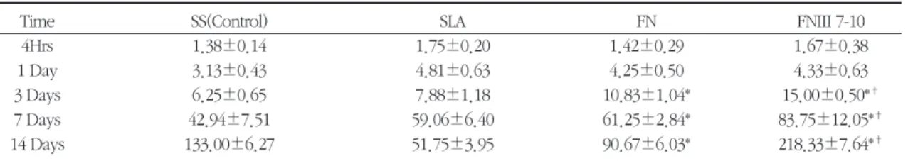
Table 1. Cell Proliferation test by hemocytometry (X 103cell count)
Time SS(Control) SLA FN FNIII 7-10
4Hrs 1.38±0.14 1.75±0.20 1.42±0.29 1.67±0.38
1 Day 3.13±0.43 4.81±0.63 4.25±0.50 4.33±0.63
3 Days 6.25±0.65 7.88±1.18 10.83±1.04* 15.00±0.50*†
7 Days 42.94±7.51 59.06±6.40 61.25±2.84* 83.75±12.05*†
14 Days 133.00±6.27 51.75±3.95 90.67±6.03* 218.33±7.64*†
Data represent mean±S.E. (N=6-7)
SS ; smooth surface, SLA ; sandblasted and acid etched, FN ; native plasma fibronectin, FNIII 7-10 ; fibronectin type III 7-10
* Significantly different from SS group at 3, 7, 14 days (p<0.05)
†Significantly different from SLA group at 3, 7, 14 days (p<0.05)
Table 2. ALPase activity (ρM/μg/30Min)
Days SS(Control) SLA FN FNIII 7-10
3 9.67±5.54 20.08±7.00 27.76±5.33* 33.72±7.73*†
7 19.80±7.29 32.59±8.26 33.9±6.03* 35.42±2.08*†
14 32.94±4.28 32.91±6.78 44.43±3.84* 63.8±10.7*†
Data represent mean±S.E. (N=6-7)
SS ; smooth surface, SLA ; sandblasted and acid etched, FN ; native plasma fibronectin, FNIII 7-10 ; fibronectin type III 7-10
* Significantly different from SS group at 3, 7, 14 days (p<0.05)
†Significantly different from SLA group at 3, 7, 14 days (p<0.05)
Figure 1. Cell Proliferation test by hemocytometry (X 103cell count)
SS ; smooth surface, SLA ; sandblasted and acid etched, FN ; native plasma fibronectin, FNIII 7-10 ; fibronectin type III 7-10
Figure 2. ALPase activity
SS ; smooth surface, SLA ; sandblasted and acid etched, FN ; native plasma fibronectin, FNIII 7-10 ; fibronectin type III 7-10 3
SS(Control) 1000
100
10
1 SLA FN FN III 7-10
7 Days 80
70 60 5040 30 20 10 Pico mole/㎍/30min 0
Cell count(×103)
Hemocytometry
ss(Control) SLA FN FN III 7-10
4 Hrs 1 Day 3 Days 7 Days 14 Days
14
well attached and spread with long cytoplasmic process. Especially cells on SLA spreaded in spike- like fashion compare to other groups(Fig. 3-6).
At 1 day, attached cells on SS, FN and FNIII 7-10 disks were well spread similarly with developed process and cells on on FN and FN III 7-10 main- tained their flattened and elongated pattern. Cells on SLA disks also maintained their spike-like fashion at 1 day (Fig. 7-10). At 3 days, the number of cells on disks was significantly increased in all groups.
Proliferated cells on each disks were grown in a concentric circle pattern due to the turned line on the disk which was occurred during milling(Fig. 11- 14). The confluent cells on SLA had many pieces of cracks and it was assumed that underlying rough surface affected attached cells during sample prepa- ration for SEM(Fig. 12). At 7 and 14 days, all sam- ples had a similar cell proliferation pattern(data are not shown).
3. Cell immunocytochemistry
1) Actin staining
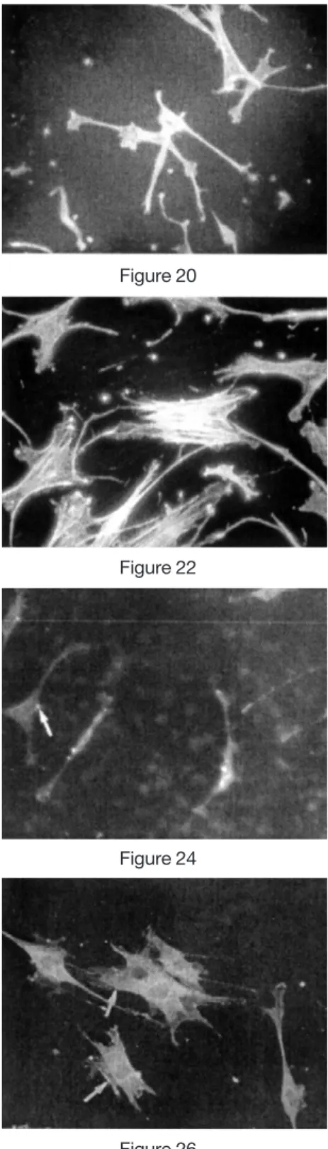
Cytoskeletal organization in adherent osteoblast was observed as an indication of the relative degrees of cell spreading with rhodamine phalloidin. 4 hours after cell plating, cells were already spread on the disks, accordingly bundles of actin fibers forming stress fibers appeared in the cells on all groups (Fig.
15-22). On SLA, however, actin filaments were dis- tributed in elongated spike-like fashion with clumps in proximal ends, which can be predicted as cyto- plasmic process (Fig. 16). At 24 hours of culture, well-defined stress fibers showing regular arrange- ment with particular polarities were found in cells grown on SS, FN and FNIII 7-10 (Fig. 15-22).
2) Immunostaining for FAK
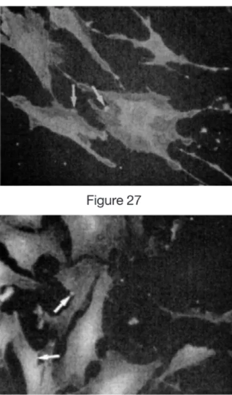
In the cells cultured for 4 hours on Ti disks, many
immunoreactive plaques for FAK were found in the peripheral regions of the cells (Fig. 23, 25, 27, 29).
Cells grown on SS and SLA showed similar immunoreactive in the periphery of the cells, although the immunoreactions for FAK were rela- tively stronger in the cells on FN and FNIII 7-10 disks than in those on SS and SLA.
In the cells cultured for 24 hours, increasing num- bers of plaques with triangular and ovoid shapes were expressed along the periphery of spreaded cells in all samples with, however, comparing the plaque numbers between groups was somewhat difficult due to the limitation of the resolution(Fig.
24, 26, 28, 30).
IV. Discussion
Attachment, spreading, and growth of osteoblast are shown to be modulated by changing surface modification. While previous work came to similar conclusions regarding substrate-dependent cell behavior43), substrate chemistries have rarely been consistent or controlled. These results demonstrate that molecular details in surface chemistry translate to molecular changes in adsorbed ECM, influencing subsequent cell signaling responses. These results indicate that outside-in signaling can be modulated by surface chemistry in anchorage-dependent cells.
Conversely, inhibitory peptides could also be used to block unwanted processes such as tumor cell invasion and metastasis. Conventional thought in this field has focused on the use of fibronectin or its mimetic as adhesive and migration substrates in wounds.
Such small peptide ligand: the sequence, arginine- glycine-aspartic acid or 'RGD' which has high affini- ty RGD peptides increase the overall adhesiveness of the surface for osteoblast44)and enhance the rate of cell spreading compared to YIGSR containing
peptides from laminin45). Integrin adhesion and sig- naling events may contribute to the progressive dif- ferentiation of the osteoblast and to the initiation of a mineralized matrix.
Differential availability and exposure of the sever- al conserved ECM cell binding domains, containing the RGD sequence recognized by cell surface inte- grin receptors, could lead to distinct differences in a material's ability to support cell attachment, spread- ing, and proliferation.
Exposure of this sequence was previously demonstrated to be increased upon adsorption to TCPS surfaces46). The data presented represent the percentage of the total 10th type III sequences found in surface-adsorbed FN exposed and recog- nized by the anti-FN antibody. Although no direct correlation can be made between FN binding domain availability, these results are consistent with reference for the integrin-FN surface and the observed cellular responses on these SAMs. An investigation of these outside-in signaling relation- ships between cell culture surfaces and RhoA activi- ty in attached cells recently demonstrated important correlations between the cell morphological markers described in the study and the molecular signaling pathways responsible for gene regulation47). The clinical success of dental implants is closely associat- ed with the properties of implant surface, which affect cell attachment as well as subsequent cell dif- ferentiation, matrix production, and mineralization.
However, cells in suspension do not possess FAK48). Thus, FAK is a molecule that requires the interaction of integrins with the extracellular matrix for its expression. This suggests that integrins aggre- gate on the cells attached on SLA or pure Ti, bind to the extracellular matrix secreted by these cells, and adsorbed onto the surface of SLA and Ti.
In addition, vitronection or fibronectin derived from serum also play an important role in the attach-
ment between cells and materials. Actin staining by TRITIC-labeled palloidine is normally used to assess cell motility, cell spreading, and cell shape49-52). It is likely that the difference in migration on Ti, SLA, FN and FNIII7-10 disks reflects the difference in organi- zation of actin. Therefore, these observations indi- cate that both surface chemistry and surface topog- raphy are contributing factors in migration of osteoblast. The immobilization of RGD on flat sur- faces and in gels results in increased potency over its parent ligand (e.g. fibronectin) in encouraging cell adhesion 53-57)and supporting cell attachment and growth.58-60)
V. Conclusion
In order to investigate the biologic effect of sur- face modified titanium with fibronectin type III 7-10 to osteoblast MC3T3-E1 cells, cell proliferation, ALPase activity, cell attachment and immunocyto- chemical analysis were done on cells cultured on Ti disks. The results were as follows:
1. Cell proliferation on FN and FNIII 7-10 were significantly higher than that of SS at 3, 7 and 14 days(p<0.05). There were also significant differences between FNIII 7-10 and SLA at 3, 7 and 14days(p<0.05).
2. ALPase activity of cultured cells on FN and FNIII 7-10 were significantly higher than that of SS at 3, 7 and 14 days(p<0.05). There were also significant differences in ALPase activity of cul- tured cells between FNIII 7-10 and SLA(p<0.05).
3. Cells were attached in flat and rhomboidal fash- ion in SS, FN and FNIII 7-10 at early time peri- od and more spreading pattern with elongated cytoplasmic process was seen as time passed.
4. In immunocytochemical analysis for actin fila-
ment and FAK, cells cultured on FN and FNIII 7-10 revealed relatively well-arranged bundles and more plaques, respectively, than those of SS and SLA.
It was concluded that modified titanium surface with FN type III 7-10 fragment had favorable biolog- ic effect on osteoblast cells as comparable as native plasma fibronectin.
VII. References
1. Brighton CT, Albelda SM.: Identification of inte- grin cell-substratum adhesion receptors on cul- tured rat bone cells, J Orthop Res, 10:766-773, 1992
2. Grzesik WJ, robey PG.: Bone matrix RGD glyco- protein: immunolocalization and interaction with human primary osteoblast bone cells in virto. J Bone Miner Res, 9(4):4870-4896, 1994
3. Moursi AM, Globus RK, Damsky CH.:
Interactions between integrin receptors and fibronectin are required for calvarial osteoblast differentiation in vitro. J Cell Sci, 110:2187-2196, 1997
4. Saito T, Albelda SM, Brighton CT.: Identification of integrin receptors on cultured human bone cells. J Orthop Res, 12:384-394, 1994
5. Puleo D, Bizios R.: RGDS tetra peptide binds to osteoblasts and inhibits fibronectin-mediated adhesion. Bone, 12(4):271-276, 1991
6. Romania A, Thomas C, Bringer A, Waters C, Healy K.: The detachment strength and mor- phology of bone cells contacting materials modi- fied with a peptide sequence found within bone sialoprotein. J Boomed Mater Res, 37(1):9-19, 1997
7. Singhvi R, Kumar A, Lopez G.: Engineering cell shape and function. Science, 264(5159):696-698,
1994
8. Brandley B. Schnaar R.: Covalent attachment of an Arg-Gly-Asp sequence peptide to derivatiz- able polyacrylamide surfaces: support of fibrob- last adhesion and long-term growth. Anal Biochem, 172(1):270-278, 1988
9. Rezania A. Healy KE.: Biomimetic peptide sur- faces that regulate adhesion spreading cytoskele- tal organization and mineralization of the matrix deposited by osteoblast-like cells. Biotechnol Prog, 15(1):19-32, 1999
10. Dee KC, Anderson TT, Bizios R.: Osteoblast population migration characteristics on sub- strates modified with immobilized adhesive pep- tides. Biomaterials, 20(3):221-227, 1999
11. Pierschbacher MD, Polarek JW, Craig WS, Tschopp JF, Sipes NJ, Harper JR.: Manipulation of cellular interactions with biomaterials toward a therapeutic outcome: a perspective. J Cell Biochem, 56(2):150-154, 1994
12. Friedman RJ. Bauer TW, Gag K, Kiang M, Yuehuei H, Draughn RA.: Histological and mechanical comparison of hydroxyapatite-coat- ed cobalt-chrome and titanium implants in the rabbit femur. J Appl Biomater, 6:231-235, 1995 13. Nimb L, Jensen JS, Gotfredsen K.: Interface
mechanics and histomorphometric analysis of hydroxyapatite-coated and porous glass-ceramic implants in canine bone. J Biomed Mater Res, 29:1477-1482, 1995
14. Rashmir-Raven AM, Richardson DC, Aberman HM, DeYoung DJ.: The response of cancellous and cortical canine bone to hydroxyapatite-coat- ed and uncoated titanium rods. J Appl Biomater, 6:237-242, 1995
15. Tisdel CL, Goldberg VM, Parr JA, Bensusan JS, Staikoff LS, Stevenson S.: The influence of a hydroxyapatite and tricalcium-phosphate coating on bone growth into titanium fiber-metal
implants. J Bone Jt Surg, 76-A(2):159-171, 1994 16. Wong M, Eulenberger J, Schenk R, Hunziker E.:
Effect of surface topology on the osseointegra- tion of implant materials in trabecular bone. J Biomed Mater Res, 29:1567-1575, 1995
17. Sumner D.: Enhancement of bone ingrowth by transforming growth factor. J Bone Jt Surg, 77- A:1135-1147, 1995
18. Mrksich M, Chen C, Xia Y, Dike L, Ingber D, Whitesides G.: Controlling cell attachment on contoured surfaces with self-assembled mono- layer of alkanethiolates on gold. Proc Natl Acad Sci USA, 93(20): 1075-1078, 1996
19. Mrksich M, Whitesides G.: Using self-assembled monolayers to understand the interactions of man made surfaces with proteins and cells. Ann Rev Biophys Biome Struct, 25:55-78, 1996 20. Prime K, Whitesides G.: Self-assembled organic
monolayers: model systems for studying adsorp- tion of proteins at surfaces. Science, 252(5010):1164-1167, 1991
21. Wink T, van Zuilen S, Bult A, van Bennkom W.:
Self-assembled monolayers for biosensors.
Analyst, 124(4):43-50, 1997
22. Silin V, Weetall H, Vanderah D.: SPR Studies of the nonspecific adsorption kinetics of human IgG and BSA on gold surfaces modified by self- assembled monolayers. J Colloid Interface Sci, 185(1):94-103, 1997
23. Margel S, Vogler E, Firment L, Watt T, Haynie S, Sogah D.: Peptide protein and cellular interac- tions with self-assembled monolayer model sur- faces. J Biomed Mater Res, 27(12):1463-1476, 1993
24. Moodie GD, Sherling MA, Morgan H, Ferris DM, Chen Cy, Valentini RF.: Effects of peptide-immo- bilized gold surfaces on osteoblasts. 24th Annual Meeting of the Society for Biomaterials, 116, 1998
25. Zou L, Moodie GD, Sherling MA, Keeping HS, Ehrlich MG, Valentini RF.: Immobilized RGD peptide modulates integrin and matrix protein gene in primary rat osteoblasts. Biomaterials, 161-164, 1998
26. Fjjisawa R, Mizuno M, Nodasaka Y, Kuboki Y.:
Attachment of osteoblastic cells to hydroxyap- atite crystals by a synthetic peptide (Glu-Pro-Arg- Gly-Asp-Thr) containing two functional sequences of bone sialoprotein. Matrix Biol, 16(1):21-28, 1997
27. Stubbs JT, Mint KP, Eanes ED, Torchia DA, Fisher LW.: Characterization of native and recombinant bone sialoprotein: delineation of the mineral-binding and cell adhesion domains and structural analysis of the RGD domain. J Bone Miner Res, 12(8):1210-1222, 1997
28. Dee KC, Andersen TT, Bizios R.: Design and function of novel osteoblast-adhesive peptides for chemical modification of biomaterials. J Biomed Mater Res, 40(3):371-377, 1998
29. Grant RP, Spitzfaden C, Altroff H, Campbell ID, Mardon HJ.: Structural requirements for biologi- cal activity of the ninth and tenth FIII domains of human fibronectin. J Biol Chem, 272(10):6159- 6166, 1997
30. Miyamoto S, Katz BZ, Lafrenie RM, Yamada KM.: Fibronectin and integrins in cell adhesion signaling and morphogenesis. Ann NY Acad Sci, 857: 119-29, 1998
31. Vuori K, Ruoslahti E.: Association of insulin receptor substrate-1 with integrins. Science, 226:1576-1578, 1994
32. Hunt TR, Schwappach JR, Anderson HC.:
Healing of segmental defect in the rat femur with use of an extract from a cultured human osteosarcoma cell-line (Saos-2). J Bone Jt Surg [Am], 78:41-48, 1996
33. Burridge K, Turner CE, Romer LH.: Tyrosine
phosphorylation of paxillin and pp125FAK accompanies cell adhesion to extracellular matrix: a role in cytoskeletal assembly. J Cell Biol, 119:893-903, 1992
34. Ingber DE, Dike L, Hansen L.: Cellular integrity:
exploring how mechanical changes in the cytoskeleton regulate cell growth, migration, and tissue pattern during morphogenesis. Int Rev Cytol, 150:173-224, 1994
35. Richard P. G, Claus S, Harri Altroff, Iain D. C.
and Helen J. Mardon.: Structural Requirements for Biological Activity of the Ninth and Tenth FIII Domains of Human Fibronectin. 272(10):6159- 6166, 1997
36. Sambra D. Redick, Daniel L. Settles, Gina Briscoe, and Harold P. Erickson.: Defining fibronectin’s cell adhesion synergy site by site- directed mutagenesis. J Cell Biol, 149(2):521-527, 2000
37. Scofield JH.: Hartree-Slater subshell photoioniza- tion cross-sections at 1254 and 1487 eV. J Electron Spectrosc Relat Phenom, 8:129, 1976 38. Vuento M, Vaheri A.: Purification of fibronectin
from human plasma by affinity chromatography under non-denaturing conditions. Biochem J, 183:331-337, 1979
39. Plow E, Ginshberg M. Specific and saturable binding of plasma fibronectin to thrombin-stimu- lated human platelets. J Biol Chem, 256:9477- 9482, 1981
40. Ugarova TP, Zamarron C, Veklich Y, Bowditch RD, Ginsberg MH, Weisel JW, Plow EF.:
Conformational transitions in the cell-binding domain of fibronectin. Biochemistry, 34:4457- 4466, 1995
41. Shuguang Zhang, Lin Yan, Michael Altman, Michael Lassle, Helen Nugent, Felice Frankel, Douglas A. Lauffenburger, George M. Alexander Rich.: Biological surface engineering: a simple
system for cell pattern formation. Biomaterials, 20:1213-1220, 1999
42. Xaoling Sur, Heather Sheardown, John L.
Brash.: Peptide modified gold-coated polyurethanes as thrombin scavenging surfaces.
J Biomed Mater Res, 49:66-78, 2000
43. Nobes CD, Hall A.: Rho, rac, and cdc42 GTPases regulate the assembly of multimolecular focal complexes associated with actin stress fibers, lamellipodia, and filopodia. Cell, 81:53-62, 1995 44. Nobes CD, Hall A.: Rho, rac, and cdc42
GTPases: regulators of actin structures, cell adhe- sion and motility. Biochem Soc Trans, 23:456- 459, 1995
45. Olson MF, Ashworth A, Hall A.: An essential role for Rho, Rac, and CdC42 GTPases in cell cycle progression through G1. Science, 269:1270-1272, 1995
46. McClary KM, Grainger DG.: RhoA-induced changes in fibroblasts cultured on organic monolayers. Biomaterials, 20:2435-2446, 1999 47. Millan Mrksich, Christopher S. Chen, Younan
xia, Laura E. Dike, Donald E. Ingber, and George M. Whitesides.: Controlling cell attach- ment on contoured surfaces with self-assembled monolayers of alkanethiolates on gold Proc Natl Acad Sci, 93:10775-10778, 1976
48. Kevin E. Healya.: Molecular engineering of materials for bioreactivity. Current Opinion in Solid State and Materials Science, 4:381-387, 1999
49. R.L. Edelstein , C.R. Tamanaha , P.E. Sheehan, M.M. Miller , D.R. Baselt, L.J. Whitman , R.J.
Colton.: The BARC biosensor applied to the detection of biological warfare agents.
Biosensors & Bioelectronics, 14:805-813, 2000 50. Richard P.Grant, Claus Spitzfaden, Harri Altroff,
Iain D. Campbell, and Helen J. Mardon.
Structural Requirements for Biological Activity of
the Ninth and Tenth FIII Domains of Human Fibronectin Vol 272, No. 10, Issue of March 7:6159-6166, 1997
51. Mariko Hashimoto-Uoshima, Ying Zhuo Yan, Galen Schneider and Ikramuddin Aukhil.: The alternatively spliced domains EIIIB and EIIIA of human fibronectin affect cell adhesion and spreading. Journal of Cell Science, 110:2271- 2280, 1997
52. Daniel J. Leahy, Ikramuddin Aukhil, and Harold P. Erickson.: 2.0? Crystal Structure of a Four- Domain Segment of Human Fibronectin Encompassing the RGD Loop and Synergy Region. Cell, 84:155-164, 1996
53. Mrksich M, Chen c, Xia Y, Dike L, Ingber D, Whitesides G.: Controlling cell attachment on contoured surfaces with self-as-sembled mono- layers of alkanethiolates on gold. Proc Natl Acad Sci, 93(20):10775-8, 1996
54. MacDonald DE, Markovic B, Allen M, Somasundaran P, Boskey A.: Surface analysis of human plasma fibronectin adsorbed to commer materials. J Biomed Mater Res, 41(1):120-130, 1998
55. Mrksich M, Whitesides G.: Using self-assembled monolayers to understand the interactions of man-made surfaces with proteins and cells. Ann Rev Biophys Biomol Struct, 25:55-78, 1996 56. Laisheng Chou, James D. Firth, Veli-Jukka Uitto,
Donald M. Brunette.: Substratum surface topog- raphy alters cell shape and regulates fibronectin mRNA level, mRNA stability, secretion and assembly in human fibroblasts. Journal of Cell Science, 108:1563-1573, 1995
57. B. Kasemo, J. Gold, C. Minkin, V.C. Marinho.:
Implant surfaces and Interface Processes Role of the Osteoclast at the bone-implant interface Adv
Dent Res, June 13:8-56, 1999
58. M. P. Ferraz, J. C. Knowles, I. Olsen, F. J.
Monteiro, J. D. Santos.: Flow cytometry analysis of effects of glass on response of osteosarcoma cells to plasma-sprayed hydroxyapatite/CaO-P2 O5 coatings. Biomaterial, 20: 2210-2218, 1999 59. A.J. Bergman, K. Zygourakis. Migration of lym-
phocytes on "fibronectin-coated surfaces: tempo- ral evolution of migratory parameters.
Biomaterials 1999;20:2235-2244.
60. Barbara D. Boyan, Victor L. Sylvia, Yuhong Liu, Ruben Sagun, David L. Cochran, Christoph H.
Lohmann, David D. Dean, Zvi Schwartz.:
Surface roughness mediates its elects on osteoblasts via protein kinase A and phospholi- pase. Biomaterials, 20:2305-2310, 1999
61. D. M. Ferris, G. D. Moodie, P. M. Diamond, C.
W. D. Gioranni, M. G. Ehrlich, R. F. Valentini.:
RGD-coated titanium implants stimulate increased bone formation in vivo. Biomaterials, 20:2323-2331, 1999
62. Akira Okumura, Masaaki Goto, Tetsuya Goto, Masao Yoshinari, Sadahiko Masuko, Takeshi Katsuki, Teruo Tanaka.: Substrate affects the ini- tial attachment and subsequent behavior of human osteoblastic cells (Saos-2). Biomaterials, 22:2263-2271, 2001
63. Kristin B. McClary, Tatiana Ugarova, David W.
Grainger.: Modulating fibroblast adhesion, spreading, and proliferation using self-assembled monolayer films of alkylthiolates on gold. J Biomed Mater Res, 50:428-439, 2000
64. Schneider GB, Zaharias R, Stanford C.:
Osteoblast integrin adhesion and signaling regu- late mineralization. J Dent Res, 80(6):1540-1544, 2001
사진부도 설명
Figure 3. Cell morphology, Smooth Surface (SS) 4 hr, Scanning Electron Microscopy (SEM) x3000 Figure 4. Cell morphology, Sandblasted and acid etched surface (SLA) 4 hr, SEM x3000
Figure 5. Cell morphology, Fibronectin (FN) 4 hr, SEM x3000
Figure 6. Cell morphology, Fibronectin type III 7-10 (FNIII 7-10) 4 hr, SEM x3000 Figure 7. Cell morphology, Smooth Surface (SS) 1 day, SEM x3000
Figure 8. Cell morphology, Sandblasted and acid etched surface (SLA) 1 day, SEM x3000 Figure 9. Cell morphology, Fibronectin (FN) 1 day, SEM x3000
Figure 10. Cell morphology, Fibronectin type III 7-10 (FNIII 7-10) 1 day, SEM x3000 Figure 11. Cell morphology, Smooth Surface (SS) 3 day, SEM x200
Figure 12. Cell morphology, Sandblasted and acid etched surface (SLA) 3 day, SEM x200 Figure 13. Cell morphology, Fibronectin (FN) 3 day, SEM x200
Figure 14. Cell morphology, Fibronectin type III 7-10 (FNIII 7-10) 3 day, SEM x200 Figure 15. Actin Filament, Smooth Surface (SS) 4 hr, Actin staining
Figure 16. Actin Filament, Sandblasted and acid etched surface (SLA) 4 hr, Actin staining Figure 17. Actin Filament, Fibronectin (FN) 4 hr, Actin staining
Figure 18. Actin Filament, Fibronectin type III 7-10 (FNIII 7-10) 4 hr, Actin staining Figure 19. Actin Filament, Smooth Surface (SS) 1 day, Actin staining
Figure 20. Actin Filament, Sandblasted and acid etched surface (SLA) 1 day, Actin staining Figure 21. Actin Filament, Fibronectin (FN) 1 day, Actin staining
Figure 22. Actin Filament, Fibronectin type III 7-10 (FNIII 7-10) 1 day, Actin staining
Figure 23. Immunostaining for FA, Smooth Surface (SS) 4 hr, Focal Adhesion Kinase (FAK), arrow indicates FAK positive plaques
Figure 24. Immunostaining for FA, Sandblasted and acid etched surface (SLA) 4 hr, Focal Adhesion Kinase (FAK), arrow indicates FAK positive plaques
Figure 25. Immunostaining for FA, Fibronectin (FN) 4 hr, Focal Adhesion Kinase (FAK), arrow indicates FAK positive plaques
Figure 26. Immunostaining for FA, Fibronectin type III 7-10 (FNIII 7-10) 4 hr, Focal Adhesion Kinase (FAK), arrows indicate FAK positive plaques
Figure 27. Immunostaining for FA, Smooth Surface (SS) 1 day, Focal Adhesion Kinase (FAK), arrowd indi- cate FAK positive plaques
Figure 28. Immunostaining for FA, Sandblasted and acid etched surface (SLA) 1 day, Focal Adhesion Kinase (FAK), arrows indicate FAK positive plaques
Figure 29. Immunostaining for FA, Fibronectin (FN) 1 day, Focal Adhesion Kinase (FAK), arrows indicate FAK positive plaques
Figure 30. Immunostaining for FA, Fibronectin type III 7-10 (FNIII 7-10) 1 day, Focal Adhesion Kinase (FAK), arrows indicate FAK positive plaques
사진부도 ( I )
Figure 3 Figure 4
Figure 5 Figure 6
Figure 7 Figure 8
Figure 9 Figure 10
사진부도 ( II )
Figure 11 Figure 12
Figure 13 Figure 14
Figure 15 Figure 16
Figure 17 Figure 18
사진부도 ( III )
Figure 19 Figure 20
Figure 21 Figure 22
Figure 23 Figure 24
Figure 25 Figure 26
사진부도 ( IV )
Figure 27 Figure 28
Figure 29 Figure 30
-국문초록-
Fibronectin type III 7-10 이 조골세포에 미치는 영향
홍정욱·최상묵·한수부·정종평·류인철,·이용무·구 영 서울대학교 치과대학 치주과학교실
타이태늄은 뛰어난 생체적합성과 적절한 물리적 성질을 바탕으로 치과 및 정형외과 영역의 매식재로 널리 사용되어져 왔으며, 골과 매식재 사이의 골 융합 정도를 증가시킬 목적으로 물리, 화학적인 방법을 이용한 타이 태늄의 표면처리에 관한 많은 연구들이 진행되어 왔다. 최근에는 부착단백질 또는 성장인자를 이용한 생체재 료의 표면개질을 통하여 조직적합성 및 치유 능의 개선을 위한 시도들이 있어왔다. Fibronectin(FN)은 주요 세 포외기질중의 하나로 생체 내 널리 분포하여 세포의 부착, 이동 및 증식에 관여하는 거대 당단백으로, RGD및 PHSRN 펩타이드 서열이 세포의 인테그린과 결합하여 세포의 활성을 조절하는 것으로 알려져 있다. 이 연구에 서는 FN으로 처리된 타이태늄이 조골세포의 부착, 증식 및 분화에 미치는 영향과 이에 따른 석회화 정도에 미 치는 영향을 관찰하여 부착분자를 이용한 타이태늄 표면개질의 효과를 규명하고자 하였다.
상업용 순수 타이태늄을 gold thiol법을 이용하여 표면처리 후, 혈장 FN(plasma FN, pFN)과 유전자재조합법 을 이용하여 얻은 FN조각(FN type III 7-10, FNIII 7-10)을 피복한 시편을 실험군으로, 아무런 처리를 하지 않은 것(smooth surface, SS)과 산 부식(Sandblasted and acid etched, SLA)처리된것을 대조군으로 이용하였다.
배양된 조골세포주(MC3T3-E1)를 사용하여 타이태늄 표면 처리에 따른 세포의 증식, 형태변화, 알칼리성 인 산분해효소(ALPase) 생산 및 세포면역형광법을 이용한 분화정도를 시간 경과에 따라 관찰하였다.
조골세포증식의 경우 FNIII 7-10 처리군에서 pFN 처리군 및 대조군에 비해 시간경과에 따라 유의성있는 세 포수의 증식이 관찰되었으며(p<0.05), ALPase 생성의 경우에도 FNIII 7-10 처리 군에서 아무 처리도 하지 않은 군에 비해 유의성 있게 높은 효소의 생성이 관찰되었다(p<0.05). 주사전자현미경을 이용한 세포의 형태관찰결 과 아무 처리도 하지 않은 군에서는 마름모형태를 나타내었으며, 산 부식 처리된 군에서는 세포가 가시모양의 형태를 보인 반면 FN으로 처리된 두 군에서는 세포의 부착 및 펴짐이 매우 발달되어 있는 모습이 관찰되었다.
세포의 분화정도를 관찰하기 위하여 국소부착키나제(focal adhesion kinase, FAK), 및 actin stress fiber의 분포 양상을 세포면역형광법을 이용하여 관찰한 결과 FN으로 표면처리된 두 군에서 아무런 처리도 하지않은 군 및 산 부식처리 한 군에 비해 프라크의 발현이 높게 나타났으며 잘 발달된 actin stress fiber의 소견을 나타내었다.
이 실험의 결과들은 gold thiol 법을 이용한 표면처리 후 FN부착을 통한 타이태늄의 표면개질이 조골세포의 부착, 증식 및 분화에 중요한 역할을 담당하여 석회화 정도를 촉진시키는 것을 보여주었으며, 이런 결과들은 더 짧은 FN조각을 이용한 다른 생체재료의 표면개질에 폭 넓게 응용할 수 있으리라 생각된다.
주요어 : titanium, fibronectin, osteoblast, cell proliferation, ALP activity, cell differentiation