저작자표시-비영리-변경금지 2.0 대한민국 이용자는 아래의 조건을 따르는 경우에 한하여 자유롭게 l 이 저작물을 복제, 배포, 전송, 전시, 공연 및 방송할 수 있습니다. 다음과 같은 조건을 따라야 합니다: l 귀하는, 이 저작물의 재이용이나 배포의 경우, 이 저작물에 적용된 이용허락조건 을 명확하게 나타내어야 합니다. l 저작권자로부터 별도의 허가를 받으면 이러한 조건들은 적용되지 않습니다. 저작권법에 따른 이용자의 권리는 위의 내용에 의하여 영향을 받지 않습니다. 이것은 이용허락규약(Legal Code)을 이해하기 쉽게 요약한 것입니다. Disclaimer 저작자표시. 귀하는 원저작자를 표시하여야 합니다. 비영리. 귀하는 이 저작물을 영리 목적으로 이용할 수 없습니다. 변경금지. 귀하는 이 저작물을 개작, 변형 또는 가공할 수 없습니다.
Master's Thesis in Molecular Medicine
Effects of dihydrophaseic acid
3
ʹ -
O
-
β
-D-glucopyranoside
isolated from
Lycii radicis
cortex
on
osteoblast differentiation
Ajou University Graduate School
Major in Molecular Medicine Department of
Biomedical Sciences
Effects of dihydrophaseic acid
3
ʹ -
O
-
β
-D-glucopyranoside
isolated from
Lycii radicis
cortex
on
osteoblast differentiation
Hyon J. Kim, Advisor
Seon-Yong Jeong, Advisor
I submit this thesis as the
Master's thesis in Molecular Medicine.
August 2017
Ajou University Graduate School
Major in Molecular Medicine Department of
Biomedical Sciences
i
-
Abstract-
Effects of dihydrophaseic acid 3ʹ-O-β-D-glucopyranoside isolated
from Lycii radicis cortex on osteoblast differentiation
Ethanol extract of Lycii radicis cortex (LRC) prevented the loss of bone mineral density in ovariectomized mice by promoting the differentiation of osteoblast linage cells. In this study, I performed fractionation and isolation of the bioactive compound(s) responsible for the bone formation–enhancing effect of LRC extract. A known sesquiterpene glucoside, (1ʹR,3ʹS,5ʹR,8ʹS,2Z,4E)-dihydrophaseic acid 3ʹ-O-β-D-glucopyranoside (abbreviated as DPA3G), was isolated and identified as a candidate constituent. I investigated the effects of DPA3G on osteoblast and osteoclast differentiation, which play fundamental roles in bone formation and bone resorption, respectively, in bone remodeling. The DPA3G fraction treatment in mesenchymal stem cell line C3H10T1/2 and preosteoblast cell line MC3T3-E1 significantly enhanced cell proliferation and alkaline phosphatase activity in both cell lines compared to the untreated control cells. Furthermore, DPA3G significantly increased mineralized nodule formation and the mRNA expression of osteoblastogenesis markers, Alpl, Runx2, and Bglap, in MC3T3-E1 cells. The DPA3G treatment, however, did not influence osteoclast differentiation in primary-cultured monocytes of mouse bone marrow. Because osteoblastic and osteoclastic precursor cells coexist in
vivo, I tested the DPA3G effects under the co-culture condition of MC3T3-E1
cells and monocytes. Remarkably, DPA3G enhanced not only osteoblast differentiation of MC3T3-El cells but also osteoclast differentiation of
ii
monocytes, indicating that DPA3G plays a role in the maintenance of the normal bone remodeling balance.
Keywords: herbal medicine, bioactive compound, dihydrophaseic acid
iii
TABLE OF CONTENTS
ABSTRACT……….………. i
TABLE OF CONTENTS……….………….…... iii
LIST OF FIGURES……….………….…... v
I. INTORDUCTION……….…….….….……… 1
II. MATERIALS AND METHODS………...……….……....… 6
A. Fractionation, isolation, and structure elucidation of the bioactive component…...……….……...……….6
B. Cell culture………..….………...………...10
C. Water-soluble tetrazolium salt (WST) assay in osteoblast cells...11
D. Alkaline phosphatase (ALP) activity assay in osteoblast cell………...11
E. Mineralized nodule formation in osteoblast cells….………...12
F. Quantitative reverse-transcription PCR (qRT-PCR)….…………..….….12
G. Osteoclastogenesis of primary monocytes and tartrate-resistant acid phosphatase (TRAP) activity assay and staining.…………...……14
H. Co-culture of MC3T3-E1 cells and primary monocytes.…………..……14
I. Statistical analysis...………..…….…….... . ...15
iv
A. DPA3G was isolated and identified from the LRC extract as a bioactive
component for enhancing osteoblast differentiation...16
B. DPA3G increased the cellular proliferation, differentiation, and mineralized nodule formation of osteoblasts………...18
C. DPA3G did not influence differentiation of osteoclasts…………..….… 24
D. DPA3G enhanced both osteoblast and osteoclast differentiation in the MC3T3-E1 and primary monocyte co-culture system……….…. 27
IV. DISCUSSION.……….. 32
V. CONCLUSIONS.………..…. 34
REFERENCES.……….. 35
v
LIST OF FIGURES
Fig. 1. Bone remodeling ……….……… 2 Fig. 2. Fractionation and isolation of the bioactive component enhancing
osteoblast differentiation from 70% ethanol extract of Lycii radices cortex ..… 8 Fig. 3. Analysis of nuclear magnetic resonance ……….………... 9 Fig. 4. Chemical structure of the isolated
(1ʹR,2ʹS,5ʹR,8ʹS,2ʹZ,4ʹE)-dihydrophaseic acid 3ʹ-O-β-D-glucopyranoside (DPA3G).. ………..…17 Fig. 5. Analysis of cell proliferation, ALP activity and alizarin red S staining..20 Fig. 6. Effects of (1ʹR,2ʹS,5ʹR,8ʹS,2ʹZ,4ʹE)-dihydrophaseic acid
3ʹ-O-β-D-glucopyranoside (DPA3G) on the mRNA expression levels of osteoblast
differentiation markers in preosteoblast MC3T3-E1 cells.………...….….23 Fig. 7. Analysis of FACS in monocytes from mouse bone marrow and TRAP assay………....25 Fig. 8. Analysis of Co-culture for monocyte and MC3T3-E1 in Trap activity and ALP activity..………..………28 Fig. 9. Effect of (1ʹR,2ʹS,5ʹR,8ʹS,2ʹZ,4ʹE)-dihydrophaseic acid
3ʹ-O-β-D-glucopyranoside (DPA3G) on osteoblast and osteoclast differentiation in the co-culture of preosteoblasts and primary monocytes…...………29 Fig. 10. Effects of (1ʹR,2ʹS,5ʹR,8ʹS,2ʹZ,4ʹE)-dihydrophaseic acid 3ʹ-O-β-D-glucopyranoside (DPA3G) on Tnfs11 (RANKL) mRNA expression in the co-culture of preosteoblasts and primary monocytes…...………31
-1-
I . INTORDUCTION
Bone is a dynamic tissue that undergoes continuous remodeling with bone formation and resorption to maintain homeostasis in a healthy skeleton (Raggatt & Partridge, 2010a; Feng & McDonald, 2011). Bone remodeling occurs through repeated cycles of reshaping or replacement of bone. Every day through natural processes, aged and damaged bone cells are removed and equal amounts of new mineral deposition are newly formed, resulting in the gradual restructuring of bone.
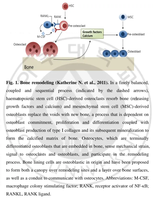
Bone remodeling requires the coordinated action of several types of bone cells, namely bone-lining cells, osteocytes, osteoclasts, and osteoblasts (Katherine N. et al., 2011; Raggatt & Partridge, 2010a; Feng & McDonald, 2011) (Fig. 1). Osteoblasts, the bone-forming cells, are differentiated from mesenchymal stem cells (MSCs); they then differentiate into osteocytes, which play a fundamental role in the initiation of bone remodeling. Osteoclasts, the bone-resorbing cells, are differentiated from mononuclear cells of the monocyte/macrophage lineage (Raggatt & Partridge, 2010a; Feng & McDonald, 2011). Bone formation involves osteoblast proliferation, differentiation with alkaline phosphatase activation, collagen synthesis, and mineralization (Fakhry et al., 2013). Bone resorption involves osteoclast differentiation with tartrate-resistant acid phosphatase activation (Boyce et
al., 2012; Kikuta & Ishii, 2013). Coupling between bone formation and bone
resorption involves the interaction of a wide range of cell types in a basic multicellular unit during bone remodeling (Matsuo & Irie, 2008; Sims & Martin, 2014).
-2-
Fig. 1. Bone remodeling (Katherine N. et al., 2011). In a finely balanced,
coupled and sequential process (indicated by the dashed arrows), haematopoietic stem cell (HSC)-derived osteoclasts resorb bone (releasing growth factors and calcium) and mesenchymal stem cell (MSC)-derived osteoblasts replace the voids with new bone, a process that is dependent on osteoblast commitment, proliferation and differentiation coupled with osteoblast production of type I collagen and its subsequent mineralization to form the calcified matrix of bone. Osteocytes, which are terminally differentiated osteoblasts that are embedded in bone, sense mechanical strain, signal to osteoclasts and osteoblasts, and participate in the remodeling process. Bone lining cells are osteoblastic in origin and have been proposed to form both a canopy over remodeling sites and a layer over bone surfaces, as well as a conduit to communicate with osteocytes. Abbreviations: M-CSF, macrophage colony stimulating factor; RANK, receptor activator of NF-κB; RANKL, RANK ligand.
-3-
Osteoblast and osteoclast lineage precursor cells communicate with each other through cell–cell contact via gap junctions; diffusible paracrine factors, such as growth factors, cytokines, chemokines, and so on; and cell–bone matrix interaction (Matsuo & Irie, 2008). Bone resorption is needed to replace old or damaged bone, and therefore, osteoclast differentiation is the initiation step of bone remodeling. Monocytic precursors’ differentiation into mature osteoclasts on the bone surface depends on diffusible paracrine factors produced by osteoblast-lineage cells, particularly macrophage colony-stimulating factor (M-CSF) and receptor activator of nuclear factor kappa-B ligand (RANKL), indicating that osteoblast differentiation is essential for the initiation of osteoclast differentiation in vivo (Kikuta & Ishii, 2013; Kim & Kim, 2016). In contrast, monocytes contribute to promote the osteoblast formation from MSCs (Matsuo & Irie, 2008; Nicolaidou et al., 2012; Sims & Martin, 2014).
Imbalanced regulation of the bone-remodeling process results in metabolic bone diseases, such as osteoporosis and osteopenia (Feng & McDonald, 2011). Osteoporosis is a common disease characterized by a systemic impairment of bone mass and microarchitecture (Sambrook & Cooper, 2006; Rachner et al., 2011). In osteoporosis patients, particularly postmenopausal women, excessive bone resorption occurs compared to bone rebuilding, which leads to an enhanced risk of bone fragility and susceptibility to fractures (Yoshida et al., 2002). Currently, the mainstay pharmacological treatment for osteoporosis is focused on inhibitors of bone resorption, such as bisphosphonates, or stimulators of bone formation, such as parathyroid hormone analogs. Although a number of effective drugs are currently available for the treatment of osteoporosis, there are still limitations, including side effects and unmet needs (Das & Crockett, 2013; Gennari et al.,
-4- 2016; Harslof & Langdahl, 2016).
Herbal medicine is a term that is widely used for alternative therapy or combination treatment with modern medicine for many diseases (Barnes et
al., 2016; Sammons et al., 2016; Yuan et al., 2016). Discovering the
pharmacologically active compounds from natural products has been a useful strategy for drug discovery and design (Rodrigues et al., 2016; Yuan et al., 2016). For the treatment of bone-related diseases, such as osteoporosis, many Chinese herbal medicines have a long tradition of use, and their bioactive compounds bearing osteoprotective and related properties have been identified (Li et al., 2012; Mukwaya et al., 2014; Che et al., 2016; Zhang et
al., 2016). Because long-term treatment is required for osteoporosis, herbal
medicine has been thought to be a good strategy for alternative treatment of osteoporosis with fewer negative effects.
Our previous study reported that a natural herbal medicine, Lycii radicis cortex (LRC) extract prevented loss of bone mineral density in ovariectomized mice (Park et al., 2014). LRC, which is Lycium chinense root bark, is extensively used in East Asia as a traditional medicine (Potterat, 2010; Zhang et al., 2013). The in vitro study revealed that the LRC extract promotes the differentiation of osteoblast linage cells rather than the inhibition of osteoclastic differentiation (Park et al., 2014). However, another study reported that LRC inhibited RANKL-induced osteoclast differentiation via the suppression of osteoclastogenesis-related markers (Kim et al., 2016). Therefore, to clarify the mechanisms by which the LRC extract affects bone formation and/or resorption, it is necessary to perform experiments under more physiological in vitro conditions, where osteoblastic and osteoclastic precursor cells coexist. The study suggested that a LRC extract may be a
-5-
good candidate as an alternative long-term treatment for osteoporosis without negative effects (Park et al., 2014).
In this study, I aimed to identify the bioactive compound(s) responsible for the bone formation–enhancing effect of LRC extract. I carried out fractionation and isolation of LRC extract and found an uncommon, known single compound with an unknown biological or pharmacological function. Next, I evaluated the effects of the identified compound on osteoblastic and osteoclastic precursor cell differentiation in single-culture and co-culture of preosteoblasts and primary monocytes.
-6-
II. MATERIALS AND METHODS
A. Fractionation, isolation, and structure elucidation of the bioactive component
Seventy percent ethanol extract of LRC (254 g) was evaporated,
suspended in H2O, and then partitioned successively with dichloromethane
(A, 1.6 g), ethyl acetate (B, 3.6 g), n-butanol (C, 120.1 g), and aqueous(D,
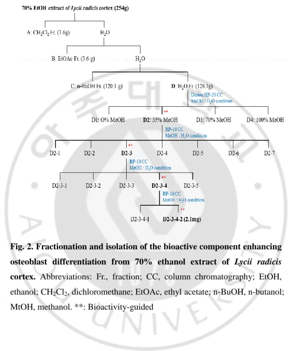
128.1 g) fractions (Fig. 2). The activity of osteoblast differentiation was evaluated in each fraction. Bioactivity-guided fractionations are indicated in Fig. 2. Fraction D was chromatographed using a Diaion HP-20 gel (1,000 g)
column chromatography, eluted with a gradient H2O-methanol (MeOH)
solvent system (O% MeOH, 35% MeOH, 70% MeOH, and 100% MeOH) to give four fractions (D1–D4). Fraction D2 (14.5 g) was subjected to RP-18
gel (200 g) column chromatography eluted with H2O-MeOH (100:0 to 0:100)
to afford seven subfractions (D2-1 to D2-7). Subfraction D2-3 (724.8 mg) was subjected to preparative high-performance liquid chromatography
(HPLC) eluted with H2O-MeOH (100:0 to 0:100) to afford five subfractions
(D2-3-1 to D2-3-5). Subfraction D2-3-4 (63.4 mg) was subjected to
preparative HPLC eluted with MeOH-H2O/0.1% formic acid (10:90) to
afford two subfractions (4-1 and 4-2). Because subfraction D2-3-4-2 significantly enhanced ALP activity in both cell lines, C3H10T1/2 MSCs and MC3T3-E1 preosteoblasts (Fig. 3), this subfraction was deduced to be a “bioactive fraction”.
The structure of the compound in the bioactive fraction was elucidated
-7-
magnetic resonance (13C-NMR), and mass spectrometry analyses (Fig. 3), as
well as by comparison with the previously reported data (Youn et al., 2011). 1
H (700 MHz) and 13C (175 MHz) NMR spectra were recorded on a Bruker
Ascend 700 spectrometer in MeOH-δ4 at 25°C; chemical shifts are given in
values (ppm) based on those of the solvent signals (1H 3.31 and 13C 49.0 ppm). An electrospray ionization (ESI)–tandem mass spectrometry analysis was performed using the Accela liquid chromatographic system (Thermo Fisher Scientific, Waltham, MA, UAS) coupled with the LTQ-Orbitrap XL mass spectrometer (Thermo Fisher Scientific). The data were collected and analyzed using the Thermo Fisher Xcalibur software package (version 2.2). The mass spectrometer equipped with an ESI source was operated in negative ionization mode using the following operating parameters: an electrospray voltage of 4.0 kV, a sheath gas flow rate of 30 arbitrary units, an auxiliary gas flow rate of 8 arbitrary units, a capillary temperature of 275°C, and a capillary voltage of 30 V. Instrument calibration was performed externally prior to each sequence using a calibration solution. Nitrogen (99.95%) was used as a sheath gas and as an auxiliary gas. The nitrogen served as a collision gas in the high-energy collisional dissociation cell and as a bath gas in the C-trap.
-8-
Fig. 2. Fractionation and isolation of the bioactive component enhancing osteoblast differentiation from 70% ethanol extract of Lycii radicis cortex. Abbreviations: Fr., fraction; CC, column chromatography; EtOH,
ethanol; CH2Cl2, dichloromethane; EtOAc, ethyl acetate; n-BuOH, n-butanol;
-9-
Fig. 3. Analysis of nuclear magnetic resonance.Results of proton nuclear magnetic resonance (1H-NMR) (A), carbon-13 nuclear magnetic resonance
(13C-NMR) (B), and mass spectrum (C) analyses of the D2-3-4-2 fraction of
-10-
B. Cell culture
Cells from the murine mesenchymal progenitor cell line C3H10T1/2 were purchased from the Korean Cell Line Bank (Seoul, Korea) and grown in Dulbecco’s Modified Eagle’s (DMEM) medium supplemented with 10% fetal bovine serum (FBS) (Sigma-Aldrich; St. Louis, MO, USA), 100 U/ml of penicillin (Duchefa; RV Haarlem, Netherlands), and 100 μg/ml of streptomycin (Duchefa). Cells from the murine preosteoblast cell line MC3T3-E1 were purchased from the RIKEN Cell Bank (Tsukuba, Japan) and cultured in α-modified minimal essential medium (α-MEM) supplemented with 10% FBS, penicillin (100 U/ml), and streptomycin (100 μg/ml). All cultured cells were incubated in a humidified atmosphere at 37°C
and 5% CO2. The cells were used at passages 5–10 after purchase for all
experiments.
To prepare primary-cultured monocytes, the bone marrow of femoral bones of 6-week-old mice was removed by flushing with a fine-bore syringe into α-MEM medium in the presence of 30 ng/ml of macrophage colony-stimulating factor (M-CSF) (PeproTech; Rocky Hill, NJ, USA) for 3–5 days (Kikuta & Ishii, 2013; Kim & Kim, 2016). The isolated monocytes were validated by immunophenotypic analysis with a CD11b antibody (BioLegend; San Diego, CA, USA) using the FACS Aria III Cell Sorter (BD Biosciences; San Jose, CA, USA) and FACS Diva software (BD Biosciences). The animal research procedures were approved by the Animal Care and Use Committee of the Ajou University School of Medicine (IACUC No. 2014-0066), and all experiments were conducted in accordance with the institutional guidelines established by the committee. All efforts were made to minimize animal suffering and to reduce the number of mice
-11- used.
C. Water-soluble tetrazolium salt (WST) assay in osteoblast cells
The osteoblast-lineage C3H10T1/2 and MC3T3-E1 cells (3×10³
cells/well) were incubated in a 96-well plate overnight and treated with different concentrations of DPA3G fraction (1, 5, and 10 μg/ml) for 3 days. The treatment dose of DPA3G was determined according to a previous study, wherein a stereoisomer of DPA3G isolated from the stem bark of Ginkgo
biloba, (1ʹR,3ʹS,5ʹR,8ʹS,2E,4E)-dihydrophaseic acid
3ʹ-O-β-D-glucopyranoside, was reported to have an anti-inflammatory effect at a concentration of 10.7-11.9 μM (approximately 5 μg/ml) (Ngan et al., 2012). The induction period for the test of cell viability and ALP activity/expression (early osteoblast differentiation marker) of osteoblast-lineage cells was determined according to previous similar studies (Zeng et al., 2015; Guo et
al., 2016). The cell viability was determined with a WST assay. WST
solution (20 μl, 5 mg/ml in phosphate-buffered saline) was added to each well, the cells were incubated for another 4 h, and the media were carefully removed. Formazan crystals were dissolved in acidified isopropyl alcohol (40 mM HCl in isopropanol), and their absorbances were measured at 450 nm and 655 nm using a microplate reader (BIO-RAD; Hercules, CA, USA).
D. Alkaline phosphatase (ALP) activity assay in osteoblast cells
-12-
a 96-well plate overnight. Osteoblast differentiation was induced by adding osteogenic medium containing ascorbic acid (50 μg/ml) and β-glycerophosphate (10 mM). Three days after osteoblast differentiation induction, the cells were treated with different concentrations of DPA3G fraction (1, 5, and 10 μg/ml) for 3 days. ALP activity was measured in total cell lysates after homogenization in buffer containing 1 mmol/l of Tris–HCl
(pH 8.8), 0.5% Triton X-100, 10 mmol/l of Mg2+, and 5 mmol/l of
p-nitrophenylphosphate as the substrate, and the reaction was stopped using 0.5 N NaOH. The absorbance was read at 405 nm with a microplate reader (BIO-RAD).
E. Mineralized nodule formation in osteoblast cells
MC3T3-E1 cells were incubated in a 48-well plate overnight. The cells were treated with 50 μg/ml of ascorbic acid and 10 mM of β-glycerophosphate for the induction of osteoblast differentiation, with or without treatment with the DPA3G fraction (5 μg/ml), for 21 days, and the medium was changed every 2 or 3 days. The colonies were fixed with 70% ethanol for 10 min at room temperature, rinsed with water, and then stained with 40 mM of Alizarin Red S (Sigma-Aldrich). Positive Alizarin Red S staining was determined with a light microscope.
F. Quantitative reverse-transcription PCR (qRT-PCR)
-13-
differentiation was induced by adding osteogenic medium containing ascorbic acid (50 μg/ml) and β-glycerophosphate (10 mM). Three days after osteoblast differentiation induction, the cells were treated with DPA3G fraction (5 μg/ml) for 3 days. Total RNA was extracted from cultured cells using TRIzol reagent (Invitrogen; Carlsbad, CA, USA) following the manufacturer’s instructions, and RNA quality was assessed by the ratio of absorbance at 260 nm and 280 nm and RT-PCR of Gapdh gene. The extracted RNA was subsequently reverse transcribed using a RevertAid™ H Minus First Strand cDNA Synthesis Kit (Fermentas; Hanover, NH, USA),
with oligo(dT)15–18 as a random primer. All real-time reverse transcription
polymerase chain reaction (RT-PCR) measurements were performed using the ABI Prism 7000 Sequence Detection System (Applied Biosystems; Foster City, CA, USA). All PCR amplifications were performed in a total volume of 25 μl containing 150 ng of cDNA using an SYBR Green I qPCR Kit (TaKaRa; Shiga, Japan) according to the manufacturer’s recommendations.
The specific primers for osteoblast markers were as follows: 5′-TCC CAC GTT TTC ACA TTC G3′ and 5′-GGC CAT CCT ATA TGG TAA CGG G-3′ for mouse Alpl (GenBank: NM_007431.3) (117 bp), 5′-TAA AGT GAC AGT GGA CGG TCC C-3′ and 5′-CCT CAG TGA TTT AGG GCG CA-3′ for mouse Runx2 (GenBank: NM_009820.5) (104 bp), 5′-TAG TGA ACA GAC TCC GGC GCT A-3′ and 5′-ATG GCT TGA AGA CCG CCT ACA-3′ for mouse Bglap (GenBank: NM_007541) (135 bp), 5′-CAG CAT CGC TCT GTT CCT GTA-3′ and 5′-CTG CGT TTT CAT GGA GTC TCA-3′ for mouse Tnfsf11 (GenBank: NM_011613.3) (107 bp), and 5′-TGA CCA CAG TCC ATG CCA TC-3′ and 5′-GAC GGA CAC ATT GGG GGT AG-3′ for mouse Gapdh (GenBank: NM_001289726.1) (203 bp). The qRT-PCR
-14-
conditions were as follows: denaturation at 95°C for 5 min; amplification with 40 cycles at 95°C for 5 sec, 60°C for 30 sec, and 72°C for 30 sec; and the terminal step for melting at 72°C to 95°C for 5 sec in each degree. By normalizing to Gapdh, the relative quantification of gene expression was performed using the comparative threshold (Ct) method previously described (Livak & Schmittgen, 2001).
G. Osteoclastogenesis of primary monocytes and tartrate-resistant acid phosphatase (TRAP) activity assay and staining
For osteoclastogenesis of primary-cultured monocytes, the isolated monocytes from the bone marrow of mouse femoral bones were cultured in the presence of 30 ng/ml of M-CSF and 50 ng/ml of RANKL (PeproTech) (Kikuta & Ishii, 2013; Kim & Kim, 2016), with or without DPA3G fraction (5 μg/ml) for 5 days. The cells were fixed in cold 4% paraformaldehyde for 10 min and washed with PBS. The differentiated osteoclast cells from monocytes were measured by a TRAP activity assay and stained using an Acid-Phosphatase Kit (Sigma-Aldrich). TRAP-positive multinucleated cells containing three or more nuclei were counted under a light microscope. The absorbance was measured at 405 nm with a microplate reader (BIO-RAD), and TRAP activity was expressed as the percent of the untreated control.
H. Co-culture of MC3T3-E1 cells and primary monocytes
-15-
overnight. The isolated monocytes (4×10⁴ cells/well) from the bone marrow
of mouse femoral bones were added in the MC3T3-E1 cells and incubated for 1 day. The MC3T3-E1 cells and primary-cultured monocytes were co-cultured in the osteoblast differentiation-induction media containing ascorbic acid (50 µg/ml) and β-glycerophosphate (10 mM) with or without treatment with the DPA3G fraction (5 μg/ml) for 5 days.
I. Statistical analysis
All of the experiments were repeated at least three times with three independent samples, and the results were presented as the means ± standard deviation, as indicated. Statistical analyses were performed using PASW Statistics, version 17.0 (SPSS Inc.; Chicago, IL, USA). Statistical significance between the groups was calculated with a Student’s t-test. A probability value (p) less than 0.05 (p<0.05) was considered statistically significant. Comparisons of multiple groups were done with a one-way analysis of variance (ANOVA), followed by Tukey’s HSD (honest significant difference) post hoc test for correction of multiple comparisons.
-16-
III. RESULTS
A. DPA3G was isolated and identified from the LRC extract as a bioactive component for enhancing osteoblast differentiation
Our previous study demonstrated that ethanol extract of LRC enhanced osteoblast differentiation in MC3T3-E1 preosteoblast cells and prevented the loss of bone mineral density in ovariectomized mice (Park et al., 2014). Several studies have demonstrated that LRC extract contains a variety of physiologically active compounds (Potterat, 2010; Zhang et al., 2013). I also identified the 13 most abundant constituents, including Lyciumoside III, Lyciumin A, and Lyciumin B from the LRC ethanol extract using a high-performance liquid chromatography (HPLC)–electrospray ionization (ESI)– tandem mass spectrometry system (Park et al., 2014).
To identify the bioactive compound(s) responsible for the bone formation–enhancing effect of LRC extract, I conducted fractionation of 70% ethanol extract of LRC. The extract was fractionated into dichloromethane,
ethyl acetate, n-butanol, and aqueousfractions, and the aqueousfraction (D)
was further fractionated (Fig. 2). An alkaline phosphatase (ALP) activity assay of each fraction in preosteoblast MC3T3-E1 cells led to the isolation of bioactive fractions. The constituent of the final active subfraction (D2-3-4-2)
was analyzed by proton nuclear magnetic resonance(1H-NMR), carbon-13
nuclear magnetic resonance (13C-NMR), and mass spectrometry analyses
(Fig. 3). As a result, a known sesquiterpene glucoside, (1ʹR,3ʹS,5ʹR,8ʹS, 2Z,4E)-dihydrophaseic acid 3ʹ-O-β-D-glucopyranoside (abbreviated as DPA3G), was identified (Fig. 4). The molecular formula of DPA3G is
-17- C21H32O10.
Fig. 4. Chemical structure of the isolated (1ʹR,2ʹS,5ʹR,8ʹS,2ʹZ,4ʹE)-dihydrophaseic acid 3ʹ-O-β-D-glucopyranoside (DPA3G).
-18-
B. DPA3G increased the cellular proliferation, differentiation, and mineralized nodule formation of osteoblasts
There are three stages of osteoblast differentiation, as follows: cell proliferation, matrix maturation, and matrix mineralization (Lian & Stein, 1995). The orthodox methods for evaluating osteoblast differentiation include cellular proliferation, ALP activity, mineralization, and mRNA expression of osteoblast differentiation markers, such as Alpl (ALP), Runx2 (runt-related transcription factor 2, Runx2), and Bglap (bone gamma carboxyglutamate protein, Osteocalcin) genes (Balint et al., 2001; Matsubara
et al., 2008; Komori, 2011; Neve et al., 2013).
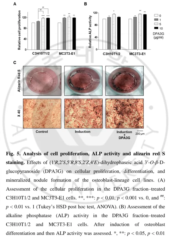
To confirm the bioactivity of the isolated DPA3G in osteoblasts, three different concentrations (1, 5, and 10 μg/ml) of the fraction containing DPA3G were treated in the osteoblast-lineage C3H10T1/2 and MC3T3-E1 cells, and the proliferation and ALP activity of the cells were examined (Fig. 5). After 3 days of incubation, the cell viability and active bone formation were determined by water-soluble tetrazolium salt (WST) and ALP assays, respectively. DPA3G significantly enhanced cellular proliferation in both cell lines (Fig. 5A). The highest ALP activity was observed with the 5 μg/ml DPA3G treatment in both cell lines (Fig. 5B). ALP, a glycoprotein found on the surface of osteoblasts, increases during active bone formation with the induction of osteoblast activity; thus, ALP plays a crucial role in the mineralization of newly formed bone (Watts, 1999; Liu et al., 2014).
Next, I examined whether DPA3G stimulated mineralized nodule formation in MC3T3-E1 cells. Most bone matrix is mineralized by osteoblasts, resulting in the production of calcium and phosphate-based
-19-
minerals; these induce the mineralization of bone and many matrix proteins (Raggatt & Partridge, 2010b). As mineralized matrix and nodule formation are key factors in the development of bone formation (Gough et al., 2004; Raggatt & Partridge, 2010b), Alizarin Red S staining is a common histochemical method for the measurement of calcium deposits in mineralized osteoblast cells (Mori et al., 1997). Positive Alizarin Red S staining signifies the presence of calcium phosphate and osteoblast mineralization, indicating successful in vitro bone formation. After osteoblast induction, the DPA3G fraction (5 μg/ml) was treated in MC3T3-E1 cells for 21 days. Markedly increased mineralized nodule formation was observed in DPA3G-treated cells compared to the untreated cells (Fig. 5C).
-20-
Fig. 5. Analysis of cell proliferation, ALP activity and alizarin red S staining. Effects of (1ʹR,2ʹS,5ʹR,8ʹS,2ʹZ,4ʹE)-dihydrophaseic acid
3ʹ-O-β-D-glucopyranoside (DPA3G) on cellular proliferation, differentiation, and mineralized nodule formation of the osteoblast-lineage cell lines. (A) Assessment of the cellular proliferation in the DPA3G fraction–treated
C3H10T1/2 and MC3T3-E1 cells. **, ***: p < 0.01, p < 0.001 vs. 0, and ##:
p < 0.01 vs. 1 (Tukey’s HSD post hoc test, ANOVA). (B) Assessment of the
alkaline phosphatase (ALP) activity in the DPA3G fraction–treated C3H10T1/2 and MC3T3-E1 cells. After induction of osteoblast differentiation and then ALP activity was assessed. *, **: p < 0.05, p < 0.01
-21-
vs. 0 (Tukey’s HSD post hoc test, ANOVA). (C) Assessment of in vitro bone mineralization in the DPA3G fraction–treated MC3T3-E1 cells. After induction of osteoblast differentiation, and then cells were stained with alizarin red S.
-22-
I examined the effect of DPA3G on the expression of bone remodeling markers Alpl, Runx2, and Bglap (osteocalcin). MC3T3-E1 cells were treated with DPA3G fraction (5 μg/ml) for 3 days. The mRNA expression levels of
Alpl, Runx2, and Bglap were measured by quantitative RT-PCR (qRT-PCR).
The expression levels of Alpl, Runx2, and Bglap were significantly increased in the DPA3G-treated cells compared to the untreated control cells (Fig. 6).
ALP is a central enzyme in the mineralization of newly formed bone (Liu
et al., 2014), and Runx2 is essential for osteoblast differentiation, stimulating
the main bone matrix proteins during the early stages of osteoblast differentiation (Komori, 2006). Differentiated osteoblasts express high levels of Osteocalcin correlated with increases in bone mineral density (Atalay et
-23-
Fig. 6. Effects of (1ʹR,2ʹS,5ʹR,8ʹS,2ʹZ,4ʹE)-dihydrophaseic acid 3ʹ-O-β-D-glucopyranoside (DPA3G) on the mRNA expression levels of osteoblast differentiation markers in preosteoblast MC3T3-E1 cells. After induction
of osteoblast differentiation, cells were treated with 5 µg/ml of DPA3G fraction for 3 days and then total RNA of the cells was extracted. The mRNA expression levels of Alpl (A), Runx2 (B), and Bglap (C) genes were assessed by quantitative reverse-transcription polymerase chain reaction (qRT-PCR) and then normalized to Gapdh mRNA expression. Control: non-DPA3G-treated cells. *: p<0.05 vs. Control (Student’s t-test).
-24-
C. DPA3G did not influence differentiation of osteoclasts
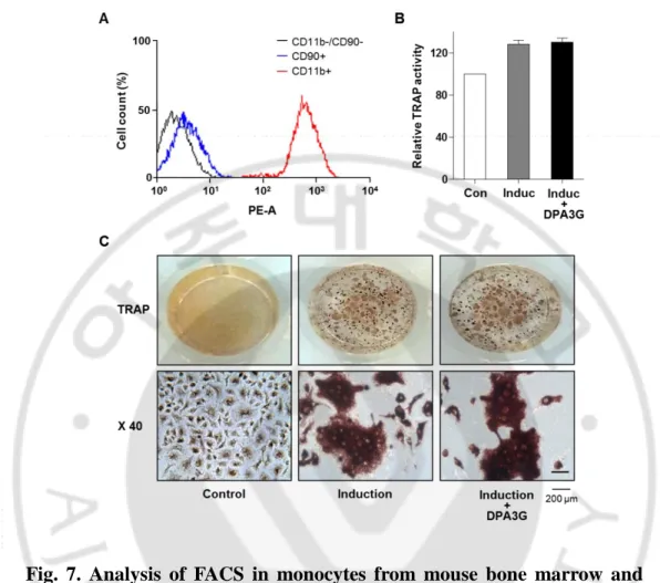
I also investigated the effects of DPA3G on the differentiation of osteoclasts. Monocytic precursors differentiate into mature osteoclasts, and osteoclasts function through the degradation and removal of both the inorganic mineral and organic matrix (Kikuta & Ishii, 2013). Monocytes were isolated from bone marrow of 6-week-old mice, and their successful isolation and culture were confirmed by fluorescence-activated cell sorting (FACS) analysis with monocyte-specific surface markers (anti-CD11b antibody; Fig. 7A). Differentiation of primary-cultured monocytes to osteoclasts was induced by treatment of M-CSF and RANKL (Kikuta & Ishii, 2013; Kim & Kim, 2016). After induction of osteoclast differentiation, DPA3G fraction (5 μg/ml) was treated in the primary monocytes for 5 days. Tartrate-resistant acid phosphatase (TRAP) activity assay and TRAP staining results showed no difference between DPA3G-treated and untreated primary monocytes (Fig. 7B, 7C), indicating that DPA3G plays a role in stimulation of osteoblast differentiation rather than inhibition of osteoclast differentiation.
-25-
Fig. 7. Analysis of FACS in monocytes from mouse bone marrow and TRAP assay. Effects of (1ʹR,2ʹS,5ʹR,8ʹS,2ʹZ,4ʹE)-dihydrophaseic acid
3ʹ-O-β-D-glucopyranoside (DPA3G) on osteoclast differentiation of primary-cultured monocytes. (A) Validation of successful isolation of monocytes from mouse bone marrow. Primary-cultured monocytes were identified by immunophenotypic analysis with a monocyte-specific surface positive marker. The absence of contamination of mesenchymal stem cells (MSCs) was confirmed by an immunophenotypic analysis with an MSC-positive marker using fluorescence-activated cell sorting (FACS) analysis. (B, C) Assessment of tartrate-resistant acid phosphatase (TRAP) activity in the DPA3G fraction-treated monocyte cells. After induction of osteoclast
-26-
differentiation, cells were treated and then TRAP activity was assessed (B). The cells were also stained with a TRAP staining kit (C).
-27-
D. DPA3G enhanced both osteoblast and osteoclast differentiation in the MC3T3-E1 and primary monocyte co-culture system
In the skeletal system, homeostasis of bone remodeling is maintained by the balance of bone resorption and bone formation (Raggatt & Partridge, 2010a; Feng & McDonald, 2011). Finally, I investigated the effects of DPA3G on bone formation and bone resorption under the more physiological conditions in vitro. I established the co-culture system of osteoblast precursor MC3T3-E1 cells and osteoclast precursor monocyte cells based on previous co-culture studies (Bernhardt et al., 2010; Chen et al., 2015; Wu et
al., 2015).
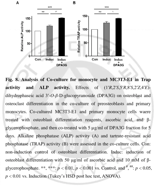
After induction of osteoblast differentiation with ascorbic acid and β-glycerophosphate, the effects of DPA3G on ALP activity for osteoblast differentiation and TRAP activity for osteoclast differentiation were examined. Through osteoblast differentiation induction, both ALP and TRAP activities were increased (Fig. 8), and the ALP and TRAP activities of the tested co-cultured cells were significantly higher than those of the separately cultured MC3T3-E1 and monocyte cells, respectively (Fig. 9), indicating the proper functioning of the co-culture system. Remarkably, treatment of DPA3G fraction (5 μg/ml) significantly enhanced not only ALP activity but also TRAP activity compared with untreated cells (Fig. 8).
-28-
Fig. 8. Analysis of Co-culture for monocyte and MC3T3-E1 in Trap activity and ALP activity. Effects of (1ʹR,2ʹS,5ʹR,8ʹS,2ʹZ,4ʹE)-dihydrophaseic acid 3ʹ-O-β-D-glucopyranoside (DPA3G) on osteoblast and osteoclast differentiation in the co-culture of preosteoblasts and primary monocytes. Co-cultured MC3T3-E1 and primary monocyte cells waere treated with osteoblast differentiation reagents, ascorbic acid, and β-glycerophosphate, and then co-treated with 5 µg/ml of DPA3G fraction for 5 days. Alkaline phosphatase (ALP) activity (A) and tartrate-resistant acid phosphatase (TRAP) activity (B) were assessed in the co-culture cells. Con: non-induction control of osteoblast differentiation. Induc: induction of osteoblast differentiation with 50 μg/ml of ascorbic acid and 10 mM of β-glycerophosphate. **, ***: p < 0.01, p < 0.001 vs. Control, and #, ##: p < 0.05,
-29-
Fig. 9. Effect of (1ʹR,2ʹS,5ʹR,8ʹS,2ʹZ,4ʹE)-dihydrophaseic acid 3ʹ-O-β-D-glucopyranoside (DPA3G) on osteoblast and osteoclast differentiation in the co-culture of preosteoblasts and primary monocytes. MC3T3-E1
preosteoblasts and primary monocytes were cultured separately or co-cultured. In the separate culture, MC3T3-E1 and primary monocyte cells were treated with osteoblast differentiation reagents and osteoclast differentiation reagents, respectively, with DPA3G or without. In co-culture, cells were treated with osteoblast differentiation with DPA3G or without. After 5 day culture, alkaline phosphatase (ALP) activity (A) and tartrate-resistant acid phosphatase (TRAP) activity (B) were assessed in each cell group. Control: non-induction of osteoblast or osteoclast differentiation.
DPA3G: (1ʹR,2ʹS,5ʹR,8ʹS,2ʹZ,4ʹE)-dihydrophaseic acid
-30-
Because DPA3G did not affect osteoclast differentiation in single-culture of monocytes (Fig. 5), the enhancement of osteoclast differentiation by DPA3G treatment in co-culture may be due to the increased M-CSF and RANKL in the co-culture media, which resulted from the enhanced osteoblast differentiation by DPA3G treatment. To confirm this, mRNA expression levels of Tnfs11 (tumor necrosis factor superfamily member 11, RANKL) gene were compared between DPA3G-treated and untreated co-culture cells. As expected, the DPA3G treatment significantly increased expression of Tnfs11 (Fig. 10).
All the results of co-culture experiments indicate that DPA3G may contribute to enhanced coupling between osteoblasts and osteoclasts in a paracrine fashion.
-31-
Fig. 10. Effects of (1ʹR,2ʹS,5ʹR,8ʹS,2ʹZ,4ʹE)-dihydrophaseic acid 3ʹ-O-β-D-glucopyranoside (DPA3G) on Tnfs11 (RANKL) mRNA expression in the co-culture of preosteoblasts and primary monocytes. MC3T3-E1
preosteoblast and primary monocyte cells were co-cultured for 1 day and then added with 50 μg/ml of ascorbic acid and 10 mM of β-glycerophosphate for induction of osteoblast differentiation. Cells were treated with 5 µg/ml of DPA3G fraction for 5 days and then total RNA of the cells was extracted. The mRNA expression level of Tnfs11 gene was assessed by quantitative reverse-transcription PCR and then normalized to Gapdh mRNA expression. Control: non-DPA3G-treated cells. *: p < 0.05 vs. Control.
-32-
IV. DISCUSSION
Our previous study found that ethanol extract of Lycii radicis cortex prevented the loss of bone mineral density in ovariectomized mice. In this study, I performed fractionation and isolation of the bioactive compound(s) responsible for the bone formation–enhancing effect of LRC extract. A known sesquiterpene glucoside, (1ʹR,3ʹS,5ʹR,8ʹS,2Z,4E)-dihydrophaseic acid 3ʹ-O-β-D-glucopyranoside (abbreviated as DPA3G), was isolated from LRC extract and identified as a candidate constituent. I demonstrated that DPA3G increased the proliferation, differentiation, and mineralization of preosteoblast cells. Co-culture of osteoblast precursor cells and osteoclast precursor cells revealed that DPA3G may contribute to enhancing coupling between osteoblasts and osteoclasts in a paracrine fashion, thereby playing a role in maintenance of normal bone turn over balance.
DPA3G was previously isolated from Nelumbo nucifera Gaertner (Nymphaeaceae, lotus) and Zizyphus jujube var. spinose (jujube) (Youn et al., 2011; Lee et al., 2013). The biological function of DPA3G has not yet been determined, but its stereoisomer isolated from the stem bark of Ginkgo
biloba, (1ʹR,3ʹS,5ʹR,8ʹS,2E,4E)-dihydrophaseic acid 3ʹ-O-β-D-gluco- pyranoside, was reported to have an anti-inflammatory effect via the inhibition of tumor necrosis factor-alpha (TNFα)-induced nuclear factor kappa-B (NF-κB) transcriptional activity and a transactivational effect of peroxisome proliferator-activated receptor-β/δ (PPARβ/δ) (Ngan et al., 2012). A previous study reported that NF-κB reduced Runx2 and β-catenin binding to osteocalcin and bone sialoprotein promoters and that NF-κB inhibition in
-33-
osteoblasts increased osteocalcin expression in mice with periodontal disease (Tarapore et al., 2016). Further, inhibition of NF-κB by orthosilicic acid treatment resulted in activation of Runx2, the master transcription factor for osteoblast precursor differentiation, and suppression of NFATc1 expression, the key transcription gene for osteoclast precursor differentiation (Zhou et al., 2016). PPARβ/δ is known to govern Wnt signaling and bone turnover (Scholtysek et al., 2013). Activation of PPARβ/δ by agonist treatment in ovariectomized osteoporotic mice led to rebalancing of bone turnover and restoration of normal bone density (Scholtysek et al., 2013). These results suggest that DPA3G is one of the candidate constituents responsible for the anti-osteoporotic effect of LRC extract and may function via NF-κB inhibition and/or PPARβ/δ activation. Our results indicate that DPA3G enhances the proliferation, differentiation, and mineralized nodule formation of bone–forming osteoblasts. Although the exact molecular mechanisms of pharmacological action of DPA3G on these effects remain unclear, based on the stereoisomer’s function (Ngan et al., 2012), DPA3G seems to play a role in the stimulation of osteoblastogenesis via NF-κB inhibition and/or PPARβ/δ activation
Further research using ovariectomized mice treated with DPA3G will help elucidate how DPA3G is responsible for the bone formation–enhancing effect of the LRC extract in vivo. However, because a large amount of DPA3G is required for an in vivo experiment and DPA3G is not commercially available, we could not conduct an in vivo experiment using DPA3G. Establishment of a mass production system of DPA3G from the LRC extract is needed to investigate the function of DPA3G in vivo.
-34-
V. CONCLUSIONS
DPA3G, (1ʹR,3ʹS,5ʹR,8ʹS,2Z,4E)-dihydrophaseic acid
3ʹ-O-β-D-glucopyranoside, was isolated and identified as one of the candidate bioactive compounds responsible for the bone formation–enhancing effect of
Lycii radicis cortex (LRC) extract.
DPA3G increased the proliferation, differentiation, and mineralized nodule formation of preosteoblast cells. In co-culture of osteoblast precursor MC3T3-E1 cells and osteoclast precursor monocytes, DPA3G enhanced both osteoblast and osteoclast differentiation, indicating that DPA3G may contribute to enhanced coupling between osteoblasts and osteoclasts in a paracrine fashion, thereby playing a role in the maintenance of normal bone turnover balance.
In conclusion, this study demonstrated the role of DPA3G isolated from LRC on enhancing osteoblast differentiation.
-35-
REFERENCES
1. Ata Atalay, S., Elci, A., Kayadibi, H., Onder, C.B. & Aka, N. (2012) Diagnostic utility of osteocalcin, undercarboxylated osteocalcin, and
alkaline phosphatase for osteoporosis in premenopausal and
postmenopausal women. Ann Lab Med, 32, 23-30.
2. Balint, E., Szabo, P., Marshall, C.F. & Sprague, S.M. (2001) Glucose-induced inhibition of in vitro bone mineralization. Bone, 28, 21-28. 3. Barnes, J., McLachlan, A.J., Sherwin, C.M. & Enioutina, E.Y. (2016)
Herbal medicines: challenges in the modern world. Part 1. Australia and New Zealand. Expert Rev Clin Pharmacol, 1-11.
4. Bernhardt, A., Thieme, S., Domaschke, H., Springer, A., Rosen-Wolff, A. & Gelinsky, M. (2010) Crosstalk of osteoblast and osteoclast precursors on mineralized collagen--towards an in vitro model for bone remodeling.
Journal of biomedical materials research. 2010, 95, 848-856.
5. Boyce, B.F., Rosenberg, E., de Papp, A.E. & Duong, L.T. (2012) The osteoclast, bone remodelling and treatment of metabolic bone disease.
Eur J Clin Invest, 42, 1332-1341.
6. Che, C.T., Wong, M.S. & Lam, C.W. (2016) Natural Products from Chinese Medicines with Potential Benefits to Bone Health. Molecules, 21, 239.
7. Chen, S., Ye, X., Yu, X., Xu, Q., Pan, K., Lu, S. & Yang, P. (2015) Co-culture with periodontal ligament stem cells enhanced osteoblastic
-36-
differentiation of MC3T3-E1 cells and osteoclastic differentiation of RAW264.7 cells. Int J Clin Exp Pathol, 8, 14596-14607.
8. Das, S. & Crockett, J.C. (2013) Osteoporosis - a current view of pharmacological prevention and treatment. Drug design, development
and therapy, 7, 435-448.
9. Fakhry, M., Hamade, E., Badran, B., Buchet, R. & Magne, D. (2013) Molecular mechanisms of mesenchymal stem cell differentiation towards osteoblasts. World J Stem Cells, 5, 136-148.
10. Feng, X. & McDonald, J.M. (2011) Disorders of bone remodeling. Annu.
Rev. Pathol., 6, 121-145.
11. Gennari, L., Rotatori, S., Bianciardi, S., Nuti, R. & Merlotti, D. (2016) Treatment needs and current options for postmenopausal osteoporosis.
Expert Opin Pharmacother, 17, 1141-1152.
12. Gough, J.E., Jones, J.R. & Hench, L.L. (2004) Nodule formation and mineralisation of human primary osteoblasts cultured on a porous bioactive glass scaffold. Biomaterials, 25, 2039-2046.
13. Guo, C., Yang, X.G., Wang, F. & Ma, X.Y. (2016) IL-1alpha induces apoptosis and inhibits the osteoblast differentiation of MC3T3-E1 cells through the JNK and p38 MAPK pathways. Int J Mol Med, 38, 319-327. 14. Harslof, T. & Langdahl, B.L. (2016) New horizons in osteoporosis
therapies. Curr Opin Pharmacol, 28, 38-42.
-37-
Cancer to bone: a fatal attraction. Nat Rev Cancer 2011, 11(6), 411–425. 16. Kikuta, J. & Ishii, M. (2013) Osteoclast migration, differentiation and
function: novel therapeutic targets for rheumatic diseases. Rheumatology
(Oxford), 52, 226-234.
17. Kim, J.H., Kim, E.Y., Lee, B., Min, J.H., Song, D.U., Lim, J.M., Eom, J.W., Yeom, M., Jung, H.S. & Sohn, Y. (2016) The effects of Lycii Radicis Cortex on RANKL-induced osteoclast differentiation and activation in RAW 264.7 cells. Int J Mol Med, 37, 649-658.
18. Kim, J.H. & Kim, N. (2016) Signaling Pathways in Osteoclast Differentiation. Chonnam Med J, 52, 12-17.
19. Komori, T. (2006) Regulation of osteoblast differentiation by transcription factors. J Cell Biochem, 99, 1233-1239.
20. Komori, T. (2011) Signaling networks in RUNX2-dependent bone development. J. Cell. Biochem., 112, 750-755.
21. Lee, S.Y., Kim, J.S., Lee, J.H., Kim, Y.S. & Kang, S.S. (2013) A New Saponin from the Seeds of Zizyphus jujuba var. spinosa. Bull Korean
Chem Soc 34, 657-660.
22. Li, T.M., Huang, H.C., Su, C.M., Ho, T.Y., Wu, C.M., Chen, W.C., Fong, Y.C. & Tang, C.H. (2012) Cistanche deserticola extract increases bone formation in osteoblasts. J Pharm Pharmacol, 64, 897-907.
23. Lian, J.B. & Stein, G.S. (1995) Development of the osteoblast phenotype: molecular mechanisms mediating osteoblast growth and differentiation.
-38-
Iowa Orthop J, 15, 118-140.
24. Liu, J., Nam, H.K., Campbell, C., Gasque, K.C., Millan, J.L. & Hatch, N.E. (2014) Tissue-nonspecific alkaline phosphatase deficiency causes abnormal craniofacial bone development in the Alpl(-/-) mouse model of infantile hypophosphatasia. Bone, 67, 81-94.
25. Livak, K.J. & Schmittgen, T.D. (2001) Analysis of relative gene expression data using real-time quantitative PCR and the 2(-Delta Delta C(T)) Method. Methods, 25, 402-408.
26. Matsubara, T., Kida, K., Yamaguchi, A., Hata, K., Ichida, F., Meguro, H., Aburatani, H., Nishimura, R. & Yoneda, T. (2008) BMP2 regulates Osterix through Msx2 and Runx2 during osteoblast differentiation. J.
Biol. Chem., 283, 29119-29125.
27. Matsuo, K. & Irie, N. (2008) Osteoclast-osteoblast communication. Arch
Biochem Biophys, 473, 201-209.
28. Mori, S., Harruff, R., Ambrosius, W. & Burr, D.B. (1997) Trabecular bone volume and microdamage accumulation in the femoral heads of women with and without femoral neck fractures. Bone, 21, 521-526. 29. Mukwaya, E., Xu, F., Wong, M.S. & Zhang, Y. (2014) Chinese herbal
medicine for bone health. Pharm Biol, 52, 1223-1228.
30. Neve, A., Corrado, A. & Cantatore, F.P. (2013) Osteocalcin: skeletal and extra-skeletal effects. J. Cell. Physiol., 228, 1149-1153.
-39-
(2012) Anti-inflammatory and PPAR transactivational effects of components from the stem bark of Ginkgo biloba. J Agric Food Chem, 60, 2815-2824.
32. Nicolaidou, V., Wong, M.M., Redpath, A.N., Ersek, A., Baban, D.F., Williams, L.M., Cope, A.P. & Horwood, N.J. (2012) Monocytes induce STAT3 activation in human mesenchymal stem cells to promote osteoblast formation. PLoS One, 7, e39871.
33. Park, E., Jin, H.S., Cho, D.Y., Kim, J., Kim, M.C., Chio, C.W., Lee, J.W., Park, J.H., Chung, Y.S., Huh, D. & Jeong, S.Y. (2014) The effect of Lycii Radicis Cortex extract on bone formation in vitro and in vivo Molecules, 19, 19594-19609.
34. Potterat, O. (2010) Goji (Lycium barbarum and L. chinense): Phytochemistry, pharmacology and safety in the perspective of traditional uses and recent popularity. Planta Med, 76, 7-19.
35. Rachner, T.D., Khosla, S. & Hofbauer, L.C. (2011) Osteoporosis: now and the future. Lancet, 377, 1276-1287.
36. Raggatt, L.J. & Partridge, N.C. (2010a) Cellular and molecular mechanisms of bone remodeling J Biol Chem, pp. 25103-25108.
37. Raggatt, L.J. & Partridge, N.C. (2010b) Cellular and molecular mechanisms of bone remodeling. The Journal of biological chemistry, 285, 25103-25108.
-40-
on natural products for drug design. Nat Chem, 8, 531-541.
39. Sambrook, P. & Cooper, C. (2006) Osteoporosis. Lancet, 367, 2010-2018. 40. Sammons, H.M., Gubarev, M.I., Krepkova, L.V., Bortnikova, V.V.,
Corrick, F., Job, K.M., Sherwin, C.M. & Enioutina, E.Y. (2016) Herbal medicines: challenges in the modern world. Part 2. European Union and Russia. Expert Rev Clin Pharmacol, 1-11.
41. Scholtysek, C., Katzenbeisser, J., Fu, H., Uderhardt, S., Ipseiz, N., Stoll, C., Zaiss, M.M., Stock, M., Donhauser, L., Bohm, C., Kleyer, A., Hess, A., Engelke, K., David, J.P., Djouad, F., Tuckermann, J.P., Desvergne, B., Schett, G. & Kronke, G. (2013) PPARbeta/delta governs Wnt signaling and bone turnover. Nat Med, 19, 608-613.
42. Shieh, A., Han, W., Ishii, S., Greendale, G.A., Crandall, C.J. & Karlamangla, A.S. (2016) Quantifying the Balance Between Total Bone Formation and Total Bone Resorption: An Index of Net Bone Formation.
J Clin Endocrinol Metab, jc20154262.
43. Sims, N.A. & Martin, T.J. (2014) Coupling the activities of bone formation and resorption: a multitude of signals within the basic multicellular unit. Bonekey Rep, 3, 481.
44. Tarapore, R.S., Lim, J., Tian, C., Pacios, S., Xiao, W., Reid, D., Guan, H., Mattos, M., Yu, B., Wang, C.Y. & Graves, D.T. (2016) NF-kappaB Has a Direct Role in Inhibiting Bmp- and Wnt-Induced Matrix Protein Expression. Journal of bone and mineral research : the official journal of
-41-
45. Watts, N.B. (1999) Clinical utility of biochemical markers of bone remodeling. Clin Chem, 45, 1359-1368.
46. Wu, L., Feyerabend, F., Schilling, A.F., Willumeit-Romer, R. & Luthringer, B.J. (2015) Effects of extracellular magnesium extract on the proliferation and differentiation of human osteoblasts and osteoclasts in coculture. Acta Biomater, 27, 294-304.
47. Yoshida, K., Oida, H., Kobayashi, T., Maruyama, T., Tanaka, M., Katayama, T., Yamaguchi, K., Segi, E., Tsuboyama, T., Matsushita, M., Ito, K., Ito, Y., Sugimoto, Y., Ushikubi, F., Ohuchida, S., Kondo, K., Nakamura, T. & Narumiya, S. (2002) Stimulation of bone formation and prevention of bone loss by prostaglandin E EP4 receptor activation. Proc
Natl Acad Sci U S A, 99, 4580-4585.
48. Youn, U.J., Lee, J., Nam, J.W., Lee, Y.J. & Seo, E.K. (2011) Identification of a New Isomer of Dihydrophaseic Acid 3'-O-β-D-Glucopyranoside from Nelumbo nucifera. Bull Korean Chem Soc, 32, 4083-4085.
49. Yuan, H., Ma, Q., Ye, L. & Piao, G. (2016) The Traditional Medicine and Modern Medicine from Natural Products. Molecules, 21.
50. Zeng, Q., Guo, Y., Liu, Y., Li, R., Zhang, X., Liu, L., Wang, Y., Zhang, X. & Zou, X. (2015) Integrin-beta1, not integrin-beta5, mediates osteoblastic differentiation and ECM formation promoted by mechanical tensile strain. Biol Res, 48, 25.
-42-
Chen, X.H., Bi, K.S. & Guo, D.A. (2013) Neolignanamides, lignanamides, and other phenolic compounds from the root bark of Lycium chinense. J Nat Prod, 76, 51-58.
52. Zhang, N.D., Han, T., Huang, B.K., Rahman, K., Jiang, Y.P., Xu, H.T., Qin, L.P., Xin, H.L. & Zhang, Q.Y. (2016) Traditional Chinese Medicine formulas for the treatment of osteoporosis: Implication for antiosteoporotic drug discovery. Journal of ethnopharmacology.
53. Zhou, X., Moussa, F.M., Mankoci, S., Ustriyana, P., Zhang, N., Abdelmagid, S., Molenda, J., Murphy, W.L., Safadi, F.F. & Sahai, N. (2016) Orthosilicic acid, Si(OH)4, stimulates osteoblast differentiation in vitro by upregulating miR-146a to antagonize NF-kappaB activation. Acta Biomater, 39, 192-202.lay, S., Elci, A., Kayadibi, H., Onder, C.B. & Aka, N. (2012) Diagnostic utility of osteocalcin, undercarboxylated osteocalcin, and alkaline phosphatase for osteoporosis in premenopausal and postmenopausal women. Ann Lab Med, 32, 23-30.
-43- - 국문요약 –
지골피추출물에서 분리한 dihydrophaseic acid
3'-O-β-D-glucopyranoside 의 조골세포 분화 촉진 효과
이 륜 진
아주대학교 대학원 의생명과학과 (지도교수 김 현 주, 정 선 용)
선행 연구에서 지골피 추출물(Lycii radicis cortex)이 조골세포 의 분화를 촉진하고 난소를 절제한 골다공증 모델마우스의 골밀도 감소를 억제한다는 것을 발견하였다. 본 연구는 지골피 추출물에서 조골세포의 분화를 촉진하는 효능성분(bioactive compound)을 분 리·동정하고 그 효과를 조사하는 연구를 수행하였다. 70% 에탄올 지골피 추출물에서 다양한 용매를 사용한 분획과 양성자핵자기공명 (1H-NMR), 탄소핵자기공명 (13C-NMR), 질 량스펙트럼 방법을 통한 분획물의 분석을 통해 조골세포 분화 촉 진 효능성분으로 (1ʹR,3ʹS,5ʹR,8ʹS,2Z,4E)-dihydrophaseic acid 3ʹ-O-β-D-glucopyranoside(DPA3G)를 동정하였다.
-44-
DPA3G가 골 형성(bone formation)에 관여하는 조골세포 (ososteobla)의 분화와 골 흡수(bone resorption)에 관여하는 파 골세포(osteoclast) 분화에 미치는 영향을 각각 연구하였다. 조골 세포의 전구세포인 중간엽 줄기세포주 C3H10T1 / 2 와 조골모세 포주 MC3T3-E1에서 DPA3G 분획을 처리하여 세포의 증식 및 알칼리성 포스파타아제 활성(alkaline phosphatase)을 분석한 결 과, DPA3G가 조골세포의 세포증식과 분화 증가에 유의한 효과가 있었으며, MC3T3-E1 세포의 석회화 결절형성(mineralized
nodule formation)과 조골세포 분화의 유전적 마커인 Alpl, Runx2
및 Bglap의 mRNA 발현을 크게 증가시켰다. 한편, DPA3G는 마우
스 골수에서 분리·배양한 초대배양 단핵세포(primary monocyte) 의 파골세포 분화에는 영향을 미치지 않았다.
생체 내에서는 조골세포와 파골세포가 공존하여 뼈 리모델링을 조절하기 때문에, 조골세포 전구체인 MC3T3-E1 세포와 파골세 포 전구체인 초대배양 단핵세포의 공배양(co-culture) 조건하에서 DPA3G 효과를 실험 하였다. DPA3G 처리에 의해 MC3T3-E1 세포의 조골세포로의 분화뿐만 아니라 단핵세포의 파골세포로의 분화도 함께 증가되었다. 특히, 공배양에서 파골세포의 분화촉진에 필요한 RANKL의 발현 증가가 확인 되었다. 이는 DPA3G에 의한 조골세포의 분화 촉진으로 RANKL의 분비가 증가되고, RANKL에 의해 파골세포의 분화가 촉진 됨으로써 전체적인 뼈 리모델링이 촉진되었을 가능성을 시사한다.
-45-
이러한 결과로부터, DPA3G가 정상적인 뼈 리모델링의 균형 유 지에 중요한 역할을 하는 것으로 사료된다.
핵심어: 천연물 의약품, 효능성분, dihydrophaseic acid 3ʹ-O-β-D-glucopyranoside (DPA3G), 조골세포, 파골세포, 뼈 리모델링