• 검색 결과가 없습니다.

Comparison of surface roughness effects upon the attachment of osteoblastic progenitor MC3T3-E1 cells and inflammatory RAW 264.7 cells to a titanium disc

N/A
N/A
Protected

Academic year: 2021

Share "Comparison of surface roughness effects upon the attachment of osteoblastic progenitor MC3T3-E1 cells and inflammatory RAW 264.7 cells to a titanium disc"

Copied!
6
0
0

로드 중.... (전체 텍스트 보기)

전체 글

(1)

Copyright ⓒ 2009, The Korean Academy of Oral Biology

37

Journal of Oral Biology

Comparison of surface roughness effects upon the attachment of osteoblastic progenitor MC3T3-E1 cells and inflammatory RAW 264.7 cells to a titanium disc

Se Ra Noh, Tae Yoon Im, Eun Young Lee, Ha Na Jang, Tran. D. Dung, Myung Soo Kim, and Hoon Yoo*

Department of Pharmacology and Dental Therapeutics, School of Dentistry, Chosun University, Gwangju 501-759, Korea (received November 28, 2008 ; revised March 12, 2009 ; accepted March 20, 2009)

The attachment and adhesion of RAW 264.7 and MC3T3- E1 cells to titanium (Ti) discs with various degrees of roughness was investigated. The attachment, adhesion, and proliferation of these cells were evaluated after 4 hr, 24 hr and 7 day incubations. Both RAW 264.7 and MC3T3-E1 cells showed a time-dependant correlation between attachment and adhesion on the surface of the titanium discs. Both types of cells tended to have higher survival rate on these discs as the surface roughness increased. The percentage of adherent inflammatory RAW 264.7 cells was greater than MC3T3-E1 cells at 24 hr, but this was reversed at 7 days in culture. The morphology of osteoblastic MC3T3-E1 cells at 24 hr, determined using a surface emission microscope (SEM), appeared flattened and spread out while inflammatory RAW 264.7 cells were predominantly spherical in shape. The adhesion of both cell types on the titanium discs was dependant on the levels of fibronectin adsorbed on the disc surface, indicating that serum constituents modulate the efficient adhesion of these cells. Our data indicate that the cellular response to the titanium surface is dependent on the types of cells, surface roughness and serum constituents.

Key words: Titanium, Attachment; Adhesion; Roughness, Fibronectin

Introduction

Titanium, the representative biomaterial used for the dental implants, is well-known for its high biocompatibility and effective osteointegration (Petrie et al., 2008; Jimbo et al., 2007). Surface characteristics of titanium are one of the critical factors for the biological response of cells such as cell attachment, cell growth, proliferation, differentiation and other cellular signaling (Keselowsky et al., 2007;

Duewelhenke et al., 2007; Degasne et al., 1999; Martin et al., 1995). Several reports showed that cellular response for the adhesion to the biomaterial, like titanium, was enhanced by increasing the roughness on its surfaces (Lincks et al., 1998; Anselme et al., 2000). A variety of cell membrane adhesion molecules, including subtypes of integrins, collagen type I, vitronectin, fibronectin and laminin, were involved in the extracellular signaling (Ku et al., 2005;

Sousa et al., 2008; Park et al., 2005; Park et al., 2006). The initial contact of cells with titanium surface, via integrin receptors, was critical for the process of osteointegration on the surface of tissue-titanium implant (Petrie et al., 2008;

Galli et al., 2005). However, it was not clear whether the effect of surface roughness on cells was mainly due to the surface roughness itself or other factors such as cell type and functionality of serum proteins. In this report, we questioned how the foreign surface of titanium with different degrees of roughness might response to two independent cell types, osteoblastic progenitor MC3T3-E1 cells and inflammatory RAW 264.7 cells (Jayaraman et al., 2004; ). In addition, we attempted to determine the contribution of plasma proteins in the adhesion of living cells on titanium discs, by choosing a constituent of plasma proteins, fibronectin, as an external addition (Jimbo et al., 2007; MacDonald et al., 1998). Here, we provide the evidence that cellular response to the

*Corresponding author: Hoon Yoo, Department of Pharmacology and Dental Therapeutics, School of Dentistry, Chosun University, 375 Seosuk-dong, Dong-gu, Gwangju 501-759, Korea.

Phone and Fax: 82-62-230-6894, E-mail: [email protected].

(2)

38 Se Ra Noh et al.

titanium surface was dependant on the surface roughness, cell types and serum constituents.

Materials and Methods

Materials

Commercial titanium bars (ASTM B348) with diameter of 100 mm were purchased from Daido Co. (Nagoya, Japan).

Oregon green 488 were purchased from Molecular Probes (Eugene, OR, USA) and the stock solution was prepared in DMSO (1 mg/ml). DMEM and α-MEM were from Welgene (Daegue, Korea). All other chemicals including fibronectin were from Sigma Chemical (St. Louis, MO, USA).

Preparation of titanium discs

Commercial bar was cut into discs with height of 1 mm.

All discs were polished with silicon carbide papers in the sequence of 240, 600 and 2000 grit. Discs were subsequently subjected to the following treatments: Ti-smooth, polished with Al

2

O

3

to a final grain of 0.05 µm; Ti-25, blasted with 25 µm particles of Al

2

O

3

(average roughness 1.343 µm); Ti- 125, blasted with 75-µm particles of Al

2

O

3

(average roughness 2.135 µm); Ti-250, blasted with 250-µm particles of Al

2

O

3

(average roughness 3.209 µm). All discs were cleaned in an ultrasonic bath and autoclaved before use in the cell culture experiments. The titanium surfaces were evaluated by scanning electron microscopy (SEM) and six discs of each group were used for Ra measurement using a profilometer. Discs were preserved under 20 % nitric acid for 1 hr and followed with sonication for 10 minutes. Finally discs were washed with 70 % ethanol ( × 2) and autoclaved before taking SEM and using for cell culture.

Cell attachment and proliferation

For cell attachment evaluation, 1 × 10

4

cells were cultured for various periods on titanium discs (MC3T3-E1 preosteoblast cells in α-MEM with 10 % FBS, and RAW 264.7 macrophage cells in DMEM with 10 % FBS). The culture medium was removed at appropriate time and the wells were washed with PBS ( × 3) at 37

o

C to eliminate unattached cells. The adherent cells were enzymatically released from the titanium discs and counted by using a hemacytometer. For proliferation evaluation cells were cultured for 7 days on titanium discs, substituting the medium in every two days.

Preparation of Oregon green 488 conjugated fibronectin Appropriate amount of Oregon green 488 carboxylic acid succinimidyl ester in DMSO (1 mg/ml) were added to the fibronectin solution in 1 ml of 50 mM sodium borate buffer (pH 8.3). The reaction was continued for 4 hr at 40

o

C. The formation of conjugated product was monitored by thin layer chromatography (TLC), using EtOH as mobile phase.

The Oregon green-tagged fibronectin was purified by PD- 10 column sephadex 25, using mobile phase as 20 % MeOH in DDW (1 % TFA), pH 2.0. The collected fraction was vaporized under vacuum at room temperature.

Assessment of Oregon green-tagged fibronectin deposition on the titanium disc

The titanium surfaces were rinsed by ultrasonic cleansing with distilled water and ethanol. A 150 µl of the prepared Oregon-green tagged fibronectin solution (0.2 mg/m l) was place on the titanium disc surface in 48-well plate for 24 hr at 36

o

C. After deposition, the extra solution were removed from the disc surface, and then the discs were completely dried prior to the measurement of FL intensity at 580 nm (exciting wavelength: 485 nm), using fluorescence microplate reader (TECAN). Percent cell viability was calculated using the equation:

% Cell Viability =

(number of living cells on disc × 100 %)/(total cells on disc) Fibronectin dependence of cell attachment on titanium disc surface

1 × 10

5

cells in DMEM (with 10 % FBS and fibronectin (0.2 mg/ml)) ware seeded into the 24-well plate containing the titanium disc. After 24 hr incubation, the cells were carefully rinsed with PBS and harvested with 0.5 % trypsin- EDTA solution. Harvested cells were neutralized in PBS or medium and then centrifuged for 1 min at room temperature. The resuspended cells in PBS were treated with lysis buffer, stabilizing buffer, and then finally transferred into cassette for the count of cells by using NucleoCounter (Chemometec).

Results

Morphology of titanium discs on the degree of roughness Discs of titanium with different surface roughness were prepared by blasting with Al

2

O

3

particles. Blasting with large particles of Al

2

O

3

produced discs with high surfaces roughness, as described in Materals and Methods. The surface images of titanium discs by scanning electron micrographs (SEM) showed different surface characteristics as the degree of roughness increases (Fig. 1).

Comparison of cell attachment of MC3T3-E1 and RAW 264.7 cells on titanium discs

For the evaluation of cell attachment, cells were seeded on

the titanium discs in culture media and the cells attached on

the titanium discs were counted at various periods of time by

using either a hemacytometer or a cell counter. As shown in

Fig. 2, the degree of surface roughness affected cell

attachment and cell adhesion. Both MC3T3-E1 and RAW

264.7 cells showed increase in the percent of cell attached

on the surface of discs at 4 hr and this trend continued at 24

(3)

hr, 48 hr (data not shown) and even after 7 days of incubation. At early stage of adhesion (24 hr), inflammatory RAW 264.7 cells outnumbered MC3T3-E1 cells in the percent of cell attachment. However, at 7 days in culture, the

attached percent cell population of MC3T3-E1 cells slightly surpassed the RAW 264.7 cells regardless the surface roughness. The percent cell attachment on the disc with high degree of roughness was always higher than the one with Fig. 1. Scanning Electron Microscope (SEM) micrographs of titanium discs with various degree of roughness: (a) Ti-smooth; (b) Ti-25; (c) Ti-125; (d) Ti-250.

Fig. 2. Comparison of percent cell adhesion of MC3T3-E1 cells and RAW 264.7 cells on titanium discs with different roughness at various

periods. (a) 4 hr; (b) 24 hr; (c) 7 days. Black bar, RAW 264.7 cells; white bars, MC3T3-E1 cells. *p < 0.05 and **p < 0.1 versus Raw 264.7,

(n > 3).

(4)

40 Se Ra Noh et al.

smooth surface with the maximum survival rate of about 70 percent (Fig. 2).

SEM view of MC3T3-E1 cells and RAW 264.7 cells on the surface of titanium discs

Morphological images of MC3T3-E1 cells and RAW 264.7 cells attached on the surface of titanium disc were taken by SEM, after incubating cells for 24 hr in culture media. As shown in Fig. 3, osteoblastic MC3T3-E1 cells at 24 hr were well spread and flattened on the disc surface while majority of inflammatory RAW 264.7 cells were in round and spherical shapes.

Assessment of Oregon green-tagged fibronectin deposition on the titanium discs

In order to assess the deposition of plasma proteins on the surface of titanium disc, fibronectin, one of the adhesive plasma proteins, was chosen as an external marker protein.

The degree of fibronectin deposition on the disc surface was investigated by preparing Oregon green 488 conjugated fluorescent fibronectin as described in Materials and Methods. The ability of Oregon green 488 conjugated fibronectin to deposit on the surface of titanium disc was evaluated by measuring fluorescence intensity, after incubating discs with media containing fluorescent fibronectin for 24 hr. The disc with high-roughness surface showed strong fluorescence intensity in progenitor MC3T3- E1 cells, while the smooth-surfaced disc showed less intensity (Fig. 4), indicating that the deposition of

fibronectin on rough surface was preferred to the smooth- surfaced one.

Fig. 3. Scanning electron micrographs view of MC3T3-E1 cells and RAW 264.7 cells on the surface of titanium discs at 24 hr: (a) MC3T3- E1, Ti-smooth; (b) RAW 264.7, Ti-smooth; (c) MC3T3-E1, Ti-250; (d) RAW 264.7, Ti-250.

Fig. 4. Detection of adsorbed fibronectins on surface of Ti-disc.

Oregon green conjugated fibronectin (0.2 mg/ml) was added into the 10 % serum containing DMEM and then 150 µl of medium was loaded on the surface of titanium disc. After air-drying, FL intensity was measured at 580 nm (exciting wavelength: 485 nm) by using fluorescence microplate reader. #0 indicates Ti-smooth.

*p < 0.05 versus Ti-smooth (n > 3).

(5)

Fibronectin dependence of cell adhesion on rough surface discs

Fibronectin was further used for the quantitative measurement of cell attachment on the surface of titanium discs. The discs were treated with the media containing with 10 % serum plus externally added fluorescent fibronectin for 24 hr before seeding with cells. Cells on the disc were incubated for 24 hr at 36

o

C, and then the harvested cells (as described in Materials and Methods) were used for the evaluation of the cell viability by using NucleoCounter (Chemometec). Pre-deposition of fibronectin on the surface of discs clearly promoted the cell adhesion of both MC3T3- E1 and RAW 264.7 on the disc surface with high-roughness (Fig. 5).

Discussion

In this study, we investigated cell response of two independent types of cells, MC3T3-E1 and RAW 264.7, to the foreign surface of titanium with different degree of roughness, and addressed the potential contribution of plasma proteins in the adhesion of living cells on titanium discs. The relative percent of cell attachment and adhesion were compared by exposing cells on the surface of titanium disc at various periods of time. Both RAW 264.7 and MC3T3-E1 cells showed roughness dependency in the initial attachment and adhesion on the titanium disc surface.

Cells on disc with high degree of roughness showed higher survival rate than the smooth-surfaced one with the maximum survival rate of about 70 percent. The percent adhesion rate of inflammatory RAW 264.7 cells at 24 hr

surpassed the one of MC3T3-E1 cells, suggesting that inflammatory cells have strong response to the foreign environment of titanium disc surface in the early stage of the culture. Both types of cells were surface roughness dependant on the proliferation. Cell proliferation analysis at 7 days showed slightly higher percent cell population of MC3T3-E1 cells than RAW 264.7 cells. The reversed order in the cellular response of MC3T3-E1 cells and RAW 264.7 between 24 hr and 7 days is an interesting result in this study. Certainly, surface microtopography of titanium disc influenced the function of adhesion molecules (e.g., integrins). However, what particular adhesion molecules are involved for the mediation of different cellular response remains to be answered in future study. Cell morphology of osteoblastic MC3T3-E1 cells at adhesion stage (24 hr) were flattened and started to spread while inflammatory RAW 264.7 cells were predominantly spherical shape. In addition, cell adhesion was dependent on the deposition of fibronectin on the surface of titanium disc (Jimbo et al., 2007; MacDonald et al., 1998). The fibronectin dependency on the adhesion of cells implicates that other adhesive plasma proteins such as fibrinogen and albumin probably are also involved for the cell attachment and adhesion to the titanium disc surface (Sousa et al., 2004; Sousa et al., 2005;

Yang et al., 2003). A variety of integrin receptors are known to be expressed on the cell surface of MC3T3-E1 preosteoblast cells and inflammatory RAW 264.7 cells.

RAW 264.7 cells expressed α

1

- α

5

β

1

integrins as well as β

2

integrins (Brian et al., 2000) and MC3T3-E1 preosteoblast

cells express multiple integrins including α

5

β

1

, which is

dependent of RGD sequence of fibronectin (Lüthen et al.,

2005; Petrie et al., 2008, Baek et al., 2002). The high

Fig. 5. Fibronectin dependence of cell attachment on rough surface disc. (a) MC3T3-E1 cells; (b) RAW 264.7 cells. *p < 0.05 versus Ti-

smooth (n > 3).

(6)

42 Se Ra Noh et al.

expression of these integrin receptors is linked to cytoskeletal rearrangement to form focal adhesion. Thus the strong adhesion property of both types of cells and the difference on the cellular response to the surface of titanium disc may reflect that the expression level of integrin receptors on the cell membrane are associated with adhesive plasma proteins adsorbed on the surface cell-type in a cell- type dependent manner.

In conclusion, our study showed that the cellular response of cells to the titanium surface was dependant on not only the surface roughness itself, but also cell types and serum constituents.

Acknowledgements

This study was supported by research funds from Chosun University, 2008.

References

Anselme K, Bigerelle M, Noel B, Dufresne E, Judas D, Iost A, Hardouin P. Qualitative and quantitative study of human osteoblast adhesion on materials with various surface roughnesses. J Biomed Mater Res. 2000;49:155-66.

Baek JH, Cho MG, Kim YH, Kim GS. Interactions between fibronectin and osteoblast are important in osteoblast differentiation and mineralized nodule formation. Int J Oral Biol. 2002;27:61-7.

Degasne I, Baslé MF, Demais V, Huré G, Lesourd M, Grolleau B, Mercier L. Chappard D. Effects of roughness, fibronectin and vitronectin on attachment, spreading, and proliferation of human osteoblast-like cells (Saos-2) on titanium surfaces.

Calcif Tissue Int. 1999;64:499-507.

Duewelhenke N, Eysel P. Serum-free cultivation of osteoprogenitor cells and osteoblasts to test biomaterials.

Orthopade. 2007;36:220-26.

Galli C, Guizzardi S, Passeri G, Martini D, Tinti A, Mauro G, Macaluso GM. Comparison of human mandibular osteoblasts grown on two commercially available titanium implant surfaces. J Periodontol. 2005;76(3):364-72.

Gowen BB, Borg TK, Ghaffar A, Mayer EB. Selective adhesion of macrophages to denatured forms of type I collagen is mediated by scavenger receptors. Matrix Biol.

2000;19:61-71.

Jayaraman M, Meyer U, Bühner M, Joos U, Wiesmann HP.

Influence of titanium surfaces on attachment of osteoblast- like cells in vitro. Biomaterials. 2004;25:625-31.

Jimbo R, Sawase T, Shibata Y, Hirata K, Hishikawa Y, Tanaka Y, Bessho K, Ikeda T, Atsuta M. Enhanced osseointegration by the chemotactic activity of plasma fibronectin for cellular fibronectin positive cells. Biomaterials. 2007;28:3469-77.

Keselowsky BG, Wang L, Schwartz Z, Garcia AJ, Boyan BD.

Integrin alpha(5) controls osteoblastic proliferation and

differentiation responses to titanium substrates presenting different roughness characteristics in a roughness indepen- dent manner. J Biomed Mater Res A. 2007;80:700-10.

Ku Y, Chung CP, Jang JH. The effect of the surface modification of titanium using a recombinant fragment of fibronectin and vitronectin on cell behavior. Biomaterials.

2005;26:5153-57.

Lincks J, Boyan BD, Blanchard CR, Lohmann CH, Liu Y, Cochran DL, Dean DD, Schwartz Z. Response of MG63 osteoblast-like cells to titanium and titanium alloy is dependent on surface roughness and composition. Biomaterials.

1998;19:2219-32.

Lüthen F, Lange R, Becker P, Rychly J, Beck U, Nebe JG. The influence of surface roughness of titanium on beta1- and beta3-integrin adhesion and the organization of fibronectin in human osteoblastic cells. Biomaterials. 2005;26:2423-40.

MacDonald DE, Markovic B, Allen M, Somasundaran P, Boskey AL. Surface analysis of human plasma fibronectin adsorbed to commercially pure titanium materials. J Biomed Mater Res. 1998;41:120-30.

Martin JY, Schwartz Z, Hummert TW, Schraub DM, Simpson J, Lankford J Jr, Dean DD, Cochran DL, Boyan BD. Effect of titanium surface roughness on proliferation, differentiation, and protein synthesis of human osteoblast-like cells (MG63). J Biomed Mater Res. 1995;29:389-401.

Park BS, Heo SJ, Kim CS, Oh JE, Kim JM, Lee G, Park WH, Chung CP, Min BM. Effects of adhesion molecules on the behavior of osteoblast-like cells and normal human fibroblasts on different titanium surfaces. J Biomed Mater Res A. 2005;74:640-51.

Park JM, Koak JY, Jang JH, Han CH, Kim SK, Heo SJ.

Osseointegration of anodized titanium implants coated with fibroblast growth factor-fibronectin (FGF-FN) fusion protein. Int J Oral Maxillofac Implants. 2006;21:859-66.

Petrie TA, Reyes CD, Burns KL, García AJ. Simple application of fibronectin-mimetic coating enhances osseointegration of titanium implants. J Cell Mol Med.

2008;Epub ahead of print.

Petrie TA, Raynor JE, Reyes CD, Burns KL, Collard DM, García AJ. The effect of integrin-specific bioactive coatings on tissue healing and implant osseointegration. Biomaterials.

2008;29:2849-57.

Sousa SR, Moradas-Ferreira P, Saramago B, Melo LV, Barbosa MA. Human serum albumin adsorption on TiO2 from single protein solutions and from plasma. Langmuir.

2004;20:9745-54.

Sousa SR, Moradas-Ferreira P, Barbosa MA. TiO2 type influences fibronectin adsorption. J Mater Sci Mater Med.

2005;16:1173-78.

Sousa SR, Lamghari M, Sampaio P, Moradas-Ferreira P, Barbosa MA. Osteoblast adhesion and morphology on TiO2 depends on the competitive preadsorption of albumin and fibronectin. J Biomed Mater Res A. 2008;84:281-90.

Yang Y, Cavin R, Ong JL. Protein adsorption on titanium

surfaces and their effect on osteoblast attachment. J Biomed

Mater Res A. 2003;67:344-49.

수치

Fig. 2. Comparison of percent cell adhesion of MC3T3-E1 cells and RAW 264.7 cells on titanium discs with different roughness at various periods
Fig. 4. Detection of adsorbed fibronectins on surface of Ti-disc.

참조

관련 문서

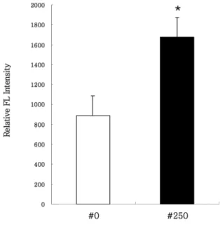
mRNA expression of osteonectin, Runx2 and BSP(bone sialoprotein) in MC3T3-E1 cells cultured for 24 hours on 4 different titanium surfaces.. The mRNA were analyzed

MC3T3-E1 cell morphorologies on the (a) CP-Ti surface and (b) nano-mesh surface for 20 min culturing time.. MC3T3-E1 cell morphorologies on the (a) CP-Ti surface and

The untreated surfaces and AA plasma treated with 3D-PCL surfaces were also examined for their in vitro pre-osteoblast (MC3T3-E1) cells proliferation and

The effects of the acceleration voltage, the probe current and the travel speed on the formation, the ingredient variation and the surface roughness of the deposited

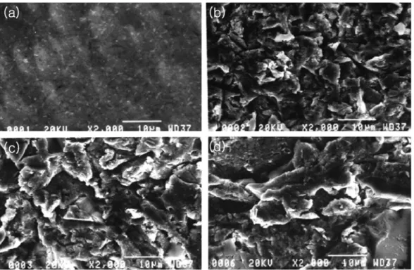
Determining optimal surface roughness of TiO₂blasted titanium implant material for attachment, proliferation and differentiation of cells derived from

 Often found connected to other molecules on the outsides of cells --- cellular recognition, cell signaling, cell

Effect of cell migration of JMJD6 transcript variants over- expressed MCF-7 cells using Transwell.. Effect of cell migration of JMJD6 transcript variants

: Bone response to unloaded titanium implants with a sandblasted and acid-etched surface : A histomorphometric study in the canine mandible.. : Early