대한소화기학회지 2009;53:1-4 □ IMAGE OF THE MONTH □
연락처: 김동준, 200-704, 강원도 춘천시 교동 153 한림대학교 의과대학 내과학교실 Tel: (033) 252-9970, Fax: (033) 256-4291 E-mail: [email protected]
Correspondence to: Dong Joon Kim, M.D.
Department of Internal Medicine, Hallym University College of Medicine, 153, Gyo-dong, Chuncheon 200-704, Korea Tel: +82-33-252-9970, Fax: +82-33-256-4291
E-mail: [email protected]
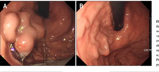
Fig. 1. (A) Initial endoscopic finding. Huge gastric varices which were continuous with esophageal varices and extending along less- er curvature are seen. There is a shallow ulcer on the variceal surface. (B) Follow-up endo- scopic finding after 2 months.
Previous gastric varices disap- peared.
역행 경정맥 폐색술로 치료된 위정맥류
한림대학교 의과대학 내과학교실
이주용ㆍ김동준
Gastric Varices Treated with Balloon-occluded Retrograde Transvenous Obliteration (BRTO)
Jue Yong Lee, M.D. and Dong Joon Kim, M.D.
Department of Internal Medicine, Hallym University College of Medicine, Chuncheon, Korea
증례: 61세 여자가 내원 20일 전 발생한 우측 옆구리 통 증과 호흡곤란으로 입원하였다. 내원 7년 전 만성B형간염을 진단받았으며 이후 간경변으로 진행한 과거력이 있었다. 신 체 검사에서 우측 폐야의 호흡음이 감소되어 있었으며 경골 앞 함요부종이 있었다. 복부는 전반적으로 부드러웠으나 팽 만되어 있었으며 이동탁음은 없었다. 활력징후는 혈압 120/80 mmHg, 맥박수 88회/분, 호흡수 20회/분, 체온 36.7oC 였고 의식은 명료했다. 말초 혈액검사에서 혈색소 13.0 g/dL, 헤마토크리트 40%였고 혈소판수는 116,000/μL로 감 소되어 있었다. 생화학 검사에서 총 빌리루빈 1.82 mg/dL, ALP 387 IU/L로 상승되었고 총 단백 6.3 g/dL, 알부민 2.8 g/dL로 감소되어 있었으며 AST/ALT는 정상 범위였다. 혈액
응고 검사에서 INR은 1.34로 증가되어 있었다. HBsAg, Anti-HBe가 각각 양성이었으며 Anti-HBs, HBeAg은 모두 음 성이었다. 상부위장관내시경에서 식도하부에 위치한 경도 의 정맥류와 함께 위분문부와 소만곡부에 걸쳐 존재하는 위 정맥류를 관찰할 수 있었고 뚜렷한 출혈 소견은 없었으나 정맥류 표면에 얕은 궤양이 있었다(Fig. 1A). 복부전산화단 층촬영에서 불규칙한 표면의 수축된 간과 비장 종대가 관찰 되었으며 비장문과 췌장, 신장주위에 사행 정맥이 있었다 (Fig. 2). 대퇴정맥을 천자하여 폐쇄풍선도관을 위신정맥단 락 기시부에 위치시키고 좌측 신정맥조영술을 시행하였으 며 조영제가 위정맥류 내에 4분 이상 저류하는 것을 관찰할 수 있었고(Fig. 3A, B) 풍선 팽창 후에 시행한 조영술에서는
2 대한소화기학회지: 제53권 제1호, 2009
Fig. 2. Abdominal CT scan shows (A) gastric varices with dilated tortuous left gastric vein and (B) gastrorenal shunt on kidney level.
Fig. 3. Angiographic findings.
Left adrenal venography is per- formed. (A) Initial finding. (B) Contrast medium remained in gastric varices after 4 minutes.
(C) Balloon-occluded retrograde venography shows the gastric varices and their small feeding and draining vessels. (D) A mix- ture of sclerosant with contrast medium is injected. Gastric vari- ces are completely filled with the mixture.
위정맥류와 함께 작은 크기의 측부 정맥이 발견되었다(Fig 3C). 투시 유도하에 풍선을 팽창시켜 위신정맥단락을 폐쇄 하고 50% glucose 20 mL를 주입 후 조영제(Pamiray 300)와 경화제(5% Ethanolamine oleate)를 1:1 비율로 섞은 용액 10
mL를 위정맥류에 가득 찰 때까지 주입하였다(Fig. 3D). 시 술 2달 후 시행한 상부위장관내시경에서 위정맥류는 거의 소실되었다(Fig. 1B). 진단 및 치료방법은?
이주용 외 1인. 역행 경정맥 폐색술로 치료된 위정맥류 3
진단: 위신정맥단락을 동반한 위정맥류
치료방법: 역행 경정맥 폐색술(balloon-occluded re- trograde transvenous obliteration, BTRO)
문맥고혈압 환자에게 위정맥류는 식도정맥류에 비해 덜 발생하고 출혈률 또한 식도정맥류에 비해 낮은 것으로 알려 져 있다. 그러나 풍부한 측부 정맥에 의해 혈류량이 많고 재 출혈률이 높기 때문에 위저부에 발생한 위정맥류의 출혈에 의한 사망은 45%에 이른다.1 위정맥류 출혈 환자의 치료에 는 간내문맥정맥단락술, 수술, 내시경 경화요법 등이 있다.
간내문맥정맥단락술의 경우 식도정맥류의 치료 효과에 비 해 위정맥류에서는 효과가 떨어진다는 보고가 있으며2 내시 경 경화요법은 위신정맥단락에 의한 빠른 혈류로 인해 경화 제 소실과 주입 부위 출혈이라는 문제가 있다.
위정맥류는 식도정맥류와는 다른 혈류 공급을 받게 되는 데 원칙적으로 위정맥류는 어떤 위정맥에 의해서도 발생할 수 있으나 대부분의 위정맥류는 좌측 위정맥(left gastric vein)과 후위정맥(posterior gastric vein)의 혈액 공급을 받게 된다. 좌측 위정맥에 의한 위정맥류는 주로 위분문부(cardia) 에 발생하며 후위정맥 및 단위정맥(short gastric vein)에 의한 위정맥류는 주로 위기저부(fundus)에 발생하게 된다.3 대부 분의 기저부에 발생한 위정맥류의 경우는 하위 횡경정맥 (inferior phrenic vein)으로 유출되어 좌측 신정맥(left renal vein)과 만나 위신정맥단락(80-85%)을 형성하거나 횡경막 직하부에서 하대정맥과 만나 위대정맥단락(gastrocaval shunt) (10-15%)을 형성한다.4,5
BRTO은 Olson과 Klatte6가 ethanol을 이용하여 1984년 처 음 보고한 이후 Kanagawa 등7이 최초로 경화제를 이용한 방 법을 소개하였다. 이후 BRTO의 임상 효용성에 관한 국내의 여러 보고가 있었다.8-11
BRTO의 표준 방법은 먼저 시술 전 복부전산화단층촬영 을 통해 위신정맥단락의 존재와 측부 혈류를 확인한다. 우 측 내경정맥(internal carotid vein) 혹은 대퇴정맥을 이용하여 폐쇄풍선도관을 좌측 부신정맥에 삽입한 후 풍선을 확장시 켜 정맥을 막은 다음 좌측 부신정맥조영술을 시행하고 위정 맥류와 측부 정맥을 확인한다. 좌측 부신정맥조영술에서 위 정맥류 정도와 측부 정맥에 따라 Hirota 등12의 분류를 기준 으로 5단계로 구분할 수 있다. Grade 1은 측부 정맥이 없는 위정맥류인 경우, Grade 2는 작은 크기의 측부 정맥이 있으 면서 조영제가 위정맥류에 3분 이상 저류하는 경우, Grade 3는 중등도의 크기를 가진 측부 정맥이 있으면서 조영제가 위정맥류에 3분 이내에 소실되는 경우, Grade 4는 크고 많 은 측부 정맥이 있어 위정맥류가 보이지 않는 경우, Grade 5 는 좌측 부신정맥이 풍선으로 막히지 않을 만큼 직경이 큰 경우로 구분하였다. Grade 1, 2의 경우 바로 경화제를 주입 하고 Grade 3, 4의 경우 측부 정맥을 코일로 색전한 후 경화
제를 주입한다. 경화제는 5% ethanolamine oleate를 주로 사 용하며 조영제와 1:1로 혼합하여 보통 20-30 mL를 주입하게 된다.
BRTO의 치료 효과에 대해 70-90%에서 위정맥류 소실 혹 은 상당한 정도의 크기 감소를 이루었고9-11,13 이와 더불어 문맥과 체정맥 간의 단락을 감소시켜 간성뇌증 치료에 도움 이 되며7 문맥을 통한 혈류 증대로 간 기능이 호전된다는 부가적인 장점을 얻을 수 있다는 보고들이 있다.14 그러나 문맥압의 증가로 인한 복수의 악화, 식도정맥류를 악화시킬 수 있는 단점도 발생한다.15,16
참고문헌
1. Sarin SK, Lahoti D, Saxena SP, Murthy NS, Makwana UK.
Prevalence, classification and natural history of gastric vari- ces: a long-term follow-up study in 568 portal hypertension patients. Hepatology 1992;16:1343-1349.
2. Sanyal AJ, Freedman AM, Luketic VA, et al. The natural history of portal hypertension after transjugular intrahepatic portosystemic shunts. Gastroenterology 1997;112:889-898.
3. Kiyosue H, Mori H, Matsumoto S, Yamada Y, Hori Y, Okino Y. Transcatheter obliteration of gastric varices. Part 1.
Anatomic classification. Radiographics 2003;23:911-920.
4. Chikamori F, Kuniyoshi N, Shibuya S, Takase Y. Correlation between endoscopic and angiographic findings in patients with esophageal and isolated gastric varices. Dig Surg 2001;
18:176-181.
5. Koito K, Namieno T, Nagakawa T, Morita K. Balloon-oc- cluded retrograde transvenous obliteration for gastric varices with gastrorenal or gastrocaval collaterals. AJR Am J Roentgenol 1996;167:1317-1320.
6. Olson E YH, Klatte EC. Transrenal-vein reflux ethanol scle- rosis of gastroesophageal varices. AJR Am J Roentgenol 1984;143:627-628.
7. Kanagawa H, Mima S, Kouyama H, Gotoh K, Uchida T, Okuda K. Treatment of gastric fundal varices by balloon-oc- cluded retrograde transvenous obliteration. J Gastroenterol Hepatol 1996;11:51-58.
8. Choi YS, Lee JH, Sinn DH, et al. Effect of balloon-occluded retrograde transvenous obliteration on the natural history of coexisting esophageal varices. J Clin Gastroenterol 2008;42:
974-979.
9. Park KS, Kim YH, Choi JS, et al. Therapeutic efficacy of balloon-occluded retrograde transvenous obliteration in pa- tients with gastric variceal bleeding. Korean J Gastroenterol 2006;47:370-378.
10. Kim YH, Seong CK, Kim YJ, Shin TB, Park NH, Choi JS.
4 The Korean Journal of Gastroenterology: Vol. 53, No. 1, 2009
Balloon-occluded retrograde transvenous obliteration for gas- tric variceal bleeding patient. J Korean Radiol Soc 2003;
48:225-233.
11. Kim ES, Park SY, Kwon KT, et al. The clinical usefulness of balloon occluded retrograde transvenous obliteration in gastric variceal bleeding. Korean J Hepatol 2003;9:315-323.
12. Hirota S, Matsumoto S, Tomita M, Sako M, Kono M. Retro- grade transvenous obliteration of gastric varices. Radiology 1999;211:349-356.
13. Baik GH, Kim DJ, Lee HG, et al. Therapeutic efficacy of balloon-occluded retrograde transvenous obliteration in the treatment of gastric varices in cirrhotic patients with gastrore- nal shunt. Korean J Gastroenterol 2004;43:196-203.
14. Akahane T, Iwasaki T, Kobayashi N, et al. Changes in liver function parameters after occlusion of gastrorenal shunts with balloon-occluded retrograde transvenous obliteration. Am J Gastroenterol 1997;92:1026-1030.
15. Ninoi T, Nishida N, Kaminou T, et al. Balloon-occluded ret- rograde transvenous obliteration of gastric varices with gastro- renal shunt: long-term follow-up in 78 patients. AJR Am J Roentgenol 2005;184:1340-1346.
16. Fukuda T, Hirota S, Matsumoto S, et al. Application of bal- loon-occluded retrograde transvenous obliteration to gastric varices complicating refractory ascites. Cardiovasc Intervent Radiol 2004;27:64-67.