Copyrights © 2015 The Korean Society of Radiology
344
Case Report
pISSN 1738-2637 / eISSN 2288-2928 J Korean Soc Radiol 2015;72(5):344-347 http://dx.doi.org/10.3348/jksr.2015.72.5.344
INTRODUCTION
Stomal varices can develop secondary to portal hypertension in patients with a stoma. The bleeding is often recurrent and may be fatal with mortality rates of 3–4% (1). Treatment options in- clude 1) direct pressure or ligation (1), 2) direct percutaneous em- bolization of the feeding portal branch using a coil or of the var- ix using glue (2, 3), 3) systemic therapy with vasoactive drugs (4), 4) surgical management (1, 5), 5) transjugular intrahepatic por- tosystemic shunt (TIPS) or balloon occluded retrograde trans- venous obliteration (BRTO) (6, 7).
We present a patient with recurrent bleeding from a stomal var- ix that was controlled with BRTO using sodium tetradecyl sulfate (STS) (Omega Laboratories Ltd., Quebec, ONT, Canada) foam.
CASE REPORT
The patient was a 52-year-old man who underwent colostomy 27 years ago due to Crohn’s disease with an extensive anal fistula.
He later developed signs of portal hypertension, such as spleno- megaly and stomal varix, on computed tomography (CT) imag- es. The etiology of the portal hypertension was unclear but was presumed to be due to drug taken to treat Crohn’s disease. Hep- atitis B virus and hepatitis C virus serological markers were neg- ative, and serum liver enzyme levels were normal. The varix bleed- ing from the stoma had started 1 year ago. Several bleeding episodes were managed conservatively with manual compression.
Finally, the patient was referred to our department to emboli- ze the stomal varix. An abdominal CT scan showed multiple peristomal varices with a large feeding vein arising from the in- ferior mesenteric vein (IMV) and drainage through superficial epigastric veins into the left common femoral vein (Fig. 1).
The left common femoral vein was accessed using ultrasound guidance and a 7 Fr introducer catheter was placed in the drain- ing epigastric vein. Then, a 6 Fr occlusion balloon catheter (Bos- ton Scientific, Cork, Ireland) was inserted and inflated. The varix and feeding veins were not visualized due to several competing collateral veins on the initial retrograde balloon occluded venog-
Balloon Occluded Retrograde Transvenous Obliteration of Bleeding Stomal Varices Using Sodium Tetradecyl Sulfate Foam: A Case Report
장루 환자의 구멍 정맥류에 Sodium Tetradecyl Sulfate 거품을 사용하여 풍선 폐색 역행성 경정맥 경화요법 치료: 증례 보고Jichang Kim, MD, Posong Yang, MD, Yeonsoo Lee, MD, Hyunjeong Kim, MD, Gun Park, MD
Department of Radiology, The Catholic University of Korea College of Medicine, Daejeon St. Mary’s Hospital, Daejeon, Korea
A stomal varix is an uncommon complication with a high mortality rate occurring secondary to portal hypertension in patients with a stoma. We describe a case of re- current stomal varix bleeding successfully managed by balloon occluded retrograde transvenous obliteration using sodium tetradecyl sulfate foam.
Index terms Stomal Varix
Balloon Occluded Retrograde Transvenous Obliteration
Received September 24, 2014 Accepted March 12, 2015
Corresponding author: Jichang Kim, MD
Department of Radiology, The Catholic University of Korea College of Medicine, Daejeon St. Mary’s Hospital, 64 Daeheung-ro, Jung-gu, Daejeon 301-723, Korea.
Tel. 82-42-220-9645 Fax. 82-42-220-9087 E-mail: [email protected]
This is an Open Access article distributed under the terms of the Creative Commons Attribution Non-Commercial License (http://creativecommons.org/licenses/by-nc/3.0) which permits unrestricted non-commercial use, distri- bution, and reproduction in any medium, provided the original work is properly cited.
Jichang Kim, et al
345
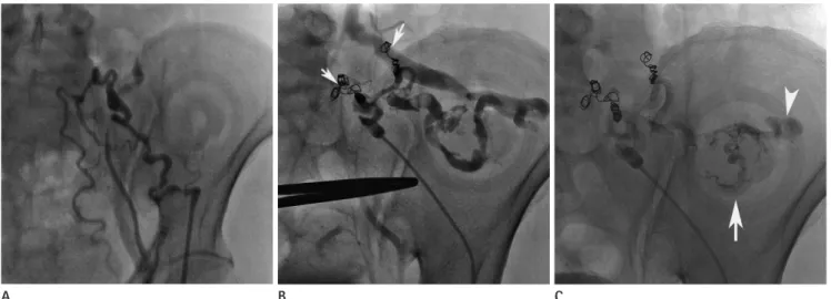
jksronline.org J Korean Soc Radiol 2015;72(5):344-347 raphy (Fig. 2A). Two main competing abdominal wall collaterals were selectively catheterized using a microcatheter (Renegade;
Boston Scientific), and embolization was performed using sev- eral microcoils (Tornado embolization microcoil; Cook Medi- cal, Bloomington, IN, USA). After successfully embolizing two collateral veins, a repeat venography with manual compression of a persistent small collateral superficial vein showed opacifica- tion of the stomal varices and the feeding branch from the IMV (Fig. 2B). STS foam (5 mL 1% STS, lipiodol, and air were mixed 1:2:3) was infused into the stomal varices under fluoroscopic guidance until the IMV was visualized. The occlusion balloon was deflated and removed after 1 hr when the contrast material stagnated (Fig. 2C).
A noncontrast-enhanced CT scan acquired just after remov-
ing the catheter showed complete filling of the target varices and collateral veins with sclerosant (data not shown). A follow-up contrast-enhanced CT scan obtained 1.5 years after the proce- dure showed complete obliteration of the stomal varices and a shrunken inflow vein (Fig. 3). The patient has remained well for 1.5 years without further stomal bleeding episodes.
DISCUSSION
The risk of stomal bleeding in patients with cirrhosis under- going colectomy with a stoma is approximately 27%. Various treatment strategies to manage bleeding stomal varices have been reported. Although local treatments, such as direct embo- lization, are effective to control the bleeding initially, they may
Fig. 1. Computed tomography of abdomen (A, B) shows stomal varix at left lower quadrant abdominal wall and inflow vein (arrow in B) arising from inferior mesenteric vein (IMV). Reconstructed CT angiogram (C) shows varix (arrowheads), inflow vein from IMV (white arrow) and out flow vein drain to left femoral vein (black arrow).
A B C
Fig. 2. Varix and inflow veins are not visualized due to collateral competing veins at the initial venogram (A). After coil embolization of competing collateral veins (arrows) and manual compression of small remaining superficial collateral veins, retrograde venogram clearly demonstrates varices and inflow vein (B). Fluoroscopy image obtained immediately after STS foam infusion (C) sclerosant filled varices (arrow) and partially visualized inflow vein (arrowhead).
STS = sodium tetradecyl sulfate
A B C
Balloon Occluded Retrograde Transvenous Obliteration of Bleeding Stomal Varices Using Sodium Tetradecyl Sulfate Foam
346
J Korean Soc Radiol 2015;72(5):344-347 jksronline.orgnot prevent a recurrent hemorrhage. Collaterals reform because of the underlying portal hypertension, re-bleeding occurs later with a high risk of porto-mesenteric bleeding (2, 3, 5). TIPS is an effective treatment option for stomal varix bleeding. Howev- er, TIPS may result in secondary hepatic encephalopathy or liver failure in the presence of decompenstated liver function (5, 7).
BRTO is an endovascular technique that was developed in Ja- pan as a therapeutic adjunct or alternative to TIPS to manage gastric varices. The BRTO technique involves occluding the out- flow veins of the portosystemic shunt using an occlusion balloon followed by endovascular injection of a sclerosing agent directly into the varix. Stagnation of the sclerosant within the varix or shunt without reflux into either the portal or systemic vasculature is crit- ical because this can result in serious complications. Thus, occlu- sion balloons are strategically placed to modulate the flow with- in the varix and/or shunt. Additionally, microcatheters and em- bolization coils are used to administer a high concentration of sclerosant within the varix and prevent reflux to nontarget sites (6).
The BRTO technique can be applied to a stomal varix as with a gastric varix. A draining epigastric vein approach through the left femoral vein and venous occlusion is easier than that of the left renal vein. In this case, two main competing abdominal col- laterals were embolized using microcoils after selecting the mi- crocatheter. Several persistent small collaterals were compressed manually using a Kelly clamp because they were located superfi- cially. The manual compression was removed rather quickly but the occlusion balloon was deflated after 1 hr when the sclerosant stagnated within the varices. The development of portal hyper- tension in this patient appeared to be related to azathioprine treat-
ment. Nodular regenerative hyperplasia leading to portal hyper- tension has been described previously in patients taking azathio- prine (8).
Possible complications from the BRTO procedure include re- nal failure, hypertension, hypotension, nausea, pulmonary em- bolism, chills, and fever. We detected no definite complications in the present case.
The BRTO procedure is a treatment option for bleeding sto- mal varices but additional study is required to evaluate its safety and effectiveness.
REFERENCES
1. Norton ID, Andrews JC, Kamath PS. Management of ecto- pic varices. Hepatology 1998;28:1154-1158
2. Naidu SG, Castle EP, Kriegshauser JS, Huettl EA. Direct per- cutaneous embolization of bleeding stomal varices. Cardio- vasc Intervent Radiol 2010;33:201-204
3. Thouveny F, Aubé C, Konaté A, Lebigot J, Bouvier A, Oberti F.
Direct percutaneous approach for endoluminal glue embo- lization of stomal varices. J Vasc Interv Radiol 2008;19:774- 777
4. Noubibou M, Douala HC, Druez PM, Kartheuzer AH, Detry RJ, Geubel AP. Chronic stomal variceal bleeding after colonic surgery in patients with portal hypertension: efficacy of beta-blocking agents? Eur J Gastroenterol Hepatol 2006;18:
807-808
5. Spier BJ, Fayyad AA, Lucey MR, Johnson EA, Wojtowycz M, Rikkers L, et al. Bleeding stomal varices: case series and sys- Fig. 3. Follow-up CT obtained after 1.5 years after sclerotherapy shows near completely obliterated stomal varices (arrow in A) and inflow vein (ar- row in B).
A B
Jichang Kim, et al
347
jksronline.org J Korean Soc Radiol 2015;72(5):344-347 tematic review of the literature. Clin Gastroenterol Hepatol 2008;6:346-352
6. Minami S, Okada K, Matsuo M, Kamohara Y, Sakamoto I, Kane- matsu T. Treatment of bleeding stomal varices by balloon- occluded retrograde transvenous obliteration. J Gastroen- terol 2007;42:91-95
7. Vangeli M, Patch D, Terreni N, Tibballs J, Watkinson A, Davies
장루 환자의 구멍 정맥류에 Sodium Tetradecyl Sulfate 거품을 사용하여 풍선 폐색 역행성 경정맥 경화요법 치료: 증례 보고
김지창 · 양보성 · 이연수 · 김현정 · 박 건
구멍 정맥류는 장루가 있는 문맥 고혈압 환자의 구멍이나 구멍 주변에서 발생하는 드문 합병증으로 출혈에 의한 사망률이 높다. 저자들은 반복적인 구멍 정맥류 출혈이 있는 환자를 sodium tetradecyl sulfate 거품을 이용하여 풍선 폐색 역행성 경 정맥 경화요법으로 치료한 증례를 보고하고자 한다.
가톨릭대학교 의과대학 대전성모병원 영상의학과
N, et al. Bleeding ectopic varices--treatment with transjug- ular intrahepatic porto-systemic shunt (TIPS) and embolisa- tion. J Hepatol 2004;41:560-566
8. Calabrese E, Hanauer SB. Assessment of non-cirrhotic por- tal hypertension associated with thiopurine therapy in in- flammatory bowel disease. J Crohns Colitis 2011;5:48-53