507 서 론
요석은 비뇨기과 분야에서 가장 흔한 질환 중의 하나로 빈도는 0.1-0.3%이고, 유병률은 5-10%로 평가되며,1 대부분
의 보고에 의하면 여자보다 남자에서 요석 발생률이 약 2-3 배 더 높다고 알려져 있다.2,3 최근 내비뇨기과학의 발달 및 체외충격파쇄석술의 출현으로 요석의 외과적 치료는 커다 란 발전을 이룩한 반면 요석의 생성기전 및 예방법에 대해 서는 아직 명확한 규명이 되어 있지 않아, 요석대사검사를
위험인자의 분석
Metabolic Stone Risk Factors Associated with Papillary Calcification on Unenhanced Spiral Computed
Tomography
Jai Seong Cha, Sang Bong Jeon, Myung Ki Kim, Young Beom Jeong, Young Gon Kim
From the Department of Urology, Chonbuk National University Medical School, Jeonju, Korea
Purpose: Increasing evidence suggests that Randalls plaque contributes to the pathogenesis of urinary stone formation. The purpose of our study is to compare the incidence of the abnormal metabolic stone risk factors between the calcium stone former with papillary calcification and the calcium stone former without papillary calcification on unenhanced spiral computed tomography (CT).
Materials and Methods: A series of patients with calcium stones (n=49) underwent unenhanced spiral CT and complete metabolic evaluation after they consumed a random diet for 1 month after stone removal. Of the 49 patients, 38 patients showed papillary calcification on unenhanced spiral CT and 11 patients did not. Their blood was evaluated by using a multichannel analysis sequential multichannel autoanalyzer (SMA)-20 and PTH tests. The 24-hour urinary constituents were assayed for calcium, oxalate, citrate, total volume, phosphorus and sodium. We compared the incidence of abnormal metabolic risk factors between the two groups.
Statistical analysis was performed by chi-square tests.
Results: The incidences of hyperuricemia were 27.3% in the patients without papillary calcification and 31.6% in the patients with papillary calcification. The incidences of hypernatriuria were 18.2% versus 42.1%, the incidences of hypercalciuria were 36.4% versus 26.3%, the incidences of hyperuricosuria were 18.2% versus 18.4%, the incidences of hyper- oxaluria were zero versus 28.9%, and the incidences of hypocitraturia were 45.5% versus 44.7%, respectively. The difference between the two groups was statistically significant only for hyperoxaluria (p=0.043).
Conclusions: Hyperoxaluria is the only metabolic risk factor that more frequently occurred in the patients with papillary calcification. Our date suggest that hyperoxaluria may be an important factor in the pathogenesis of papillary calcification. (Korean J Urol 2006;47:507-511)
ꠏꠏꠏꠏꠏꠏꠏꠏꠏꠏꠏꠏꠏꠏꠏꠏꠏꠏꠏꠏ
Key Words: Urinary stone, Risk factor, Hyperoxaluria
대한비뇨기과학회지 제 47 권 제 5 호 2006
전북대학교 의과대학 비뇨기과학교실
차재성․전상봉․김명기 정영범․김영곤
접수일자:2005년 12월 2일 채택일자:2005년 12월 31일
교신저자: 김영곤
전북대학교병원 비뇨기과 전라북도 전주시 덕진구 금암동 634-18
ꂕ 561-712 TEL: 063-250-1567 FAX: 063-250-1564 E-mail: ygkim@chonbuk.
ac.kr
통해 생리적인 요인들을 분석한 후 내과적 치료를 실시하 는 것이 현재까지 재발을 방지하는 중요한 방법으로 여겨 지고 있다.
최근 들어 활발히 연구되고 있는 요중 대사적 검사에 따 르면 약 90%의 요석 환자에서 대사 이상이 발견되고 있으 며, 특히 고칼슘뇨증, 고수산뇨증, 고요산뇨증, 저구연산요 증 등이 주요한 원인으로 주목되고 있다.4,5 요로결석 중 가 장 많은 성분은 수산칼슘석으로 한국에서 전체 요석 중 81.6%를 차지하고 있으며,6 신장의 수산칼슘석은 1년에 약 10%, 5년에 35%, 10년이 지나면 50% 이상의 높은 재발률을 보이고 있다.7 서구에서는 요석의 병태생리에 대한 연구가 1980년 이후로 매우 활발하게 이루어졌는데,8,9 특히 요석 형성의 병리 기전으로서 Randall's papillary plaque10의 중요 성은 점차 커지고 있다. Randall's papillary plaque는 신 유두 끝 또는 그 근처의 간질 내에 결정이 침착된 부위로 정의되 는데, 수산칼슘석을 가진 환자의 신장에서는 거의 100%에 서 발견되고 있으며, 다른 성분의 요석을 가지고 있는 환자 의 신장에서도 43% 정도에서 보고되고 있다.11
본 연구에서는 비조영증강 전산화단층촬영에서 관찰되 는 신 유두 석회화가 Randall's plaque와 강한 연관성이 있으 며 요석 형성에 있어서 중요한 역할을 할 것으로 추측하고, 요석 위험인자와의 관련성을 알아보고자 하였다.
대상 및 방법
2003년 10월부터 2004년 7월까지 본원에서 요석의 진단 을 위해 시행했던 비조영증강 전산화단층촬영에서 요석과 함께 신 유두 석회화를 보인 환자 중 혈중 검사와 24시간 요 대사검사를 시행한 38명과 신 유두 석회화가 없이 요석 이 있으면서 혈중 검사와 24시간 요 대사검사를 시행한 11
명의 환자를 대상으로 비교분석하였다.
모든 검사대상자는 일반 식이를 하였고 요배양검사 결과 음성이었으며, 요석의 재발 예방에 관여하는 어떤 약물도 투여받지 않았고, 요석 치료 후 실시한 요석 분석결과 칼슘 석으로 확인된 경우만을 포함시켰다. 요 채취는 요석이 완 전히 해결된 후 3-4주 지나서, 오전 7시에 배뇨해서 버리고 다음날 오전 7시까지의 마지막 소변을 포함하여 24시간 요 를 수집하도록 하였으며 가능한 즉시 병원에 가져오게 하 였다.
요석대사를 위한 혈중 검사로는 나트륨, 칼륨, 염소, 요소 질소, 크레아티닌, 요산, 칼슘, 인산, 중성지방과 부갑상선 호르몬의 수치를 측정하였고, 24시간 요 대사검사로는 나 트륨, 칼륨, 염소, 크레아티닌, 칼슘, 인산, 요산, 구연산, 수 산을 측정하였다.
혈중에서 각 물질의 정상 참고치는 요산은 남자의 경우 3.2-7.7mg/dl, 여자의 경우 2.4-5.3mg/dl, 칼슘은 8.4-10.2mg/dl, 인산은 2.5-4.5mg/dl, 중성지방은 200mg/dl 이하, 부갑상선호 르몬은 11-62pg/ml를 적용하였다. 또한 요 중 대사 이상의 조사를 위한 각 물질의 정상 참고치로는 24시간 요에서 나 트륨은 220mEq/day 이하, 칼슘은 4mg/kg 이하, 요산은 남자 의 경우 800mg/day 이하, 여자의 경우 750mg/day 이하, 인산 은 1,300mg/day 이하, 수산은 45mg/day 이하, 구연산은 320mg/day 이상을 적용하였다.
각 인자들의 값은 평균±표준편차로 표시하였으며 혈중 요산, 칼슘, 인산, 중성지방, 부갑상선호르몬 및 요 중 나트 륨, 칼슘, 요산, 인산, 수산, 구연산은 대사이상 수 및 빈도를 구하였다.
통계학적 분석으로 수치의 분석은 Independent samples t-test를, 빈도 비교는 chi-square test를 이용하여 p값이 0.05 미만인 경우에 의미가 있는 것으로 간주하였다.
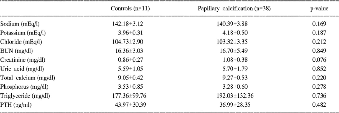
Table 1. Results of serum analysis for the patients with and without papillary calcification (mean±SD)
ꠚꠚꠚꠚꠚꠚꠚꠚꠚꠚꠚꠚꠚꠚꠚꠚꠚꠚꠚꠚꠚꠚꠚꠚꠚꠚꠚꠚꠚꠚꠚꠚꠚꠚꠚꠚꠚꠚꠚꠚꠚꠚꠚꠚꠚꠚꠚꠚꠚꠚꠚꠚꠚꠚꠚꠚꠚꠚꠚꠚꠚꠚꠚꠚꠚꠚꠚꠚꠚꠚꠚꠚꠚꠚꠚꠚꠚꠚꠚꠚꠚꠚꠚꠚꠚꠚꠚꠚꠚꠚꠚꠚꠚꠚꠚꠚꠚꠚꠚꠚꠚꠚꠚꠚꠚꠚꠚꠚꠚꠚꠚꠚꠚꠚꠚ
Controls (n=11) Papillary calcification (n=38) p-value
ꠏꠏꠏꠏꠏꠏꠏꠏꠏꠏꠏꠏꠏꠏꠏꠏꠏꠏꠏꠏꠏꠏꠏꠏꠏꠏꠏꠏꠏꠏꠏꠏꠏꠏꠏꠏꠏꠏꠏꠏꠏꠏꠏꠏꠏꠏꠏꠏꠏꠏꠏꠏꠏꠏꠏꠏꠏꠏꠏꠏꠏꠏꠏꠏꠏꠏꠏꠏꠏꠏꠏꠏꠏꠏꠏꠏꠏꠏꠏꠏꠏꠏꠏꠏꠏꠏꠏꠏꠏꠏꠏꠏꠏꠏꠏꠏꠏꠏꠏꠏꠏꠏꠏꠏꠏꠏꠏꠏꠏꠏꠏꠏꠏꠏꠏ
Sodium (mEq/l) 142.18±3.12 140.39±3.88 0.169
Potassium (mEq/l) 3.96±0.31 4.18±0.50 0.187
Chloride (mEq/l) 104.73±2.90 103.32±3.35 0.212
BUN (mg/dl) 16.36±3.03 16.70±5.49 0.849
Creatinine (mg/dl) 0.86±0.27 1.08±0.38 0.076
Uric acid (mg/dl) 5.59±1.05 5.70±1.79 0.852
Total calcium (mg/dl) 9.05±0.42 9.27±0.53 0.220
Phosphorus (mg/dl) 3.53±0.85 3.28±0.60 0.278
Triglyceride (mg/dl) 177.36±99.76 192.03±132.36 0.736
PTH (pg/ml) 43.97±30.39 36.99±28.35 0.482
ꠏꠏꠏꠏꠏꠏꠏꠏꠏꠏꠏꠏꠏꠏꠏꠏꠏꠏꠏꠏꠏꠏꠏꠏꠏꠏꠏꠏꠏꠏꠏꠏꠏꠏꠏꠏꠏꠏꠏꠏꠏꠏꠏꠏꠏꠏꠏꠏꠏꠏꠏꠏꠏꠏꠏꠏꠏꠏꠏꠏꠏꠏꠏꠏꠏꠏꠏꠏꠏꠏꠏꠏꠏꠏꠏꠏꠏꠏꠏꠏꠏꠏꠏꠏꠏꠏꠏꠏꠏꠏꠏꠏꠏꠏꠏꠏꠏꠏꠏꠏꠏꠏꠏꠏꠏꠏꠏꠏꠏꠏꠏꠏꠏꠏꠏ BUN: blood urea nitrogen, PTH: parathyroid hormone
결 과
전체 환자의 평균 연령은 46.3±13.9세였고, 비조영증강 전산화 단층 촬영에서 확인된 신 유두 석회화 환자의 평균 연령은 48.1±12.9세였으며 대조군의 평균 연령은 39.8±
15.9세였다. 신 유두 석회화 환자와 대조군에서 혈중 검사 및 24시간 뇨 대사검사의 결과는 다음과 같았다 (Table 1, 2). 혈중 및 24시간 요 대사 검사의 모든 요석 위험 인자에 서 두 군 간의 평균값은 거의 비슷하였으며 통계적 의의도 없었다. 두 군 간의 요석 위험 인자의 빈도 비교에서 혈중 검사에서는 수치적 또는 통계적인 의의를 찾을 수 없었으 나, 24시간 요 대사 검사에서는 신 유두 석회화 환자에서 대조군에 비하여 과나트륨뇨증, 과수산뇨증이 높은 비율로 발생하였으며, 특히 과수산뇨증의 경우 통계적으로 의의가 있었다 (p=0.043) (Table 3, 4).
고 찰
요석은 다양한 원인에 의하여 발생하며 여러 가지 위험 인자들이 알려져 있다. 역학 조사에 의하면 요석의 형성은 지역이나 종족 등에 따라 빈도가 매우 다양하며 또한 식이 습관, 기후, 가족력, 직업 등이 관련되어 있다.12,13 특히 성별 이나 연령에 따라 분포가 뚜렷하여 여자보다 남자에서 호 발하고 20-50대의 나이에서 주로 발병하게 된다.14 최근 내 비뇨기과학의 발달 및 체외충격파쇄석술의 출현으로 치료 에 괄목할 만한 발전을 이룬 반면, 많은 임상 관찰이나 실험 에도 불구하고 요석의 정확한 발생기전 및 예방법이 아직 까지 명확히 알려져 있지 않으며 22.6-51%의 재발률을 보
인다.15,16 현재까지는 혈중 및 24시간 요를 이용한 요석대사
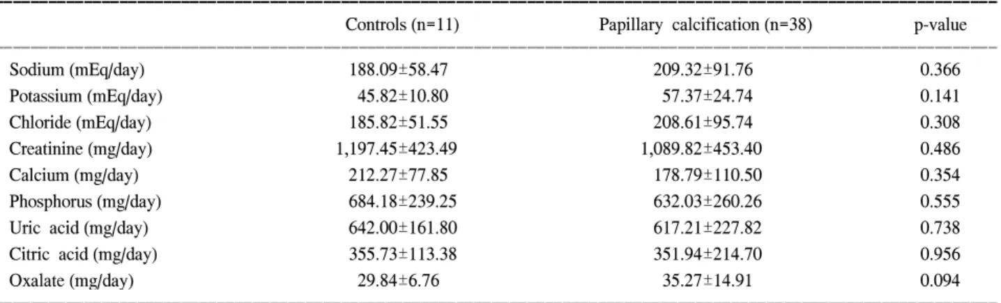
Table 2. Results of 24-hour urine analysis for the patients with and without papillary calcification (mean±SD)
ꠚꠚꠚꠚꠚꠚꠚꠚꠚꠚꠚꠚꠚꠚꠚꠚꠚꠚꠚꠚꠚꠚꠚꠚꠚꠚꠚꠚꠚꠚꠚꠚꠚꠚꠚꠚꠚꠚꠚꠚꠚꠚꠚꠚꠚꠚꠚꠚꠚꠚꠚꠚꠚꠚꠚꠚꠚꠚꠚꠚꠚꠚꠚꠚꠚꠚꠚꠚꠚꠚꠚꠚꠚꠚꠚꠚꠚꠚꠚꠚꠚꠚꠚꠚꠚꠚꠚꠚꠚꠚꠚꠚꠚꠚꠚꠚꠚꠚꠚꠚꠚꠚꠚꠚꠚꠚꠚꠚꠚꠚꠚꠚꠚꠚꠚ Controls (n=11) Papillary calcification (n=38) p-value ꠏꠏꠏꠏꠏꠏꠏꠏꠏꠏꠏꠏꠏꠏꠏꠏꠏꠏꠏꠏꠏꠏꠏꠏꠏꠏꠏꠏꠏꠏꠏꠏꠏꠏꠏꠏꠏꠏꠏꠏꠏꠏꠏꠏꠏꠏꠏꠏꠏꠏꠏꠏꠏꠏꠏꠏꠏꠏꠏꠏꠏꠏꠏꠏꠏꠏꠏꠏꠏꠏꠏꠏꠏꠏꠏꠏꠏꠏꠏꠏꠏꠏꠏꠏꠏꠏꠏꠏꠏꠏꠏꠏꠏꠏꠏꠏꠏꠏꠏꠏꠏꠏꠏꠏꠏꠏꠏꠏꠏꠏꠏꠏꠏꠏꠏ
Sodium (mEq/day) 188.09±58.47 209.32±91.76 0.366
Potassium (mEq/day) 45.82±10.80 57.37±24.74 0.141
Chloride (mEq/day) 185.82±51.55 208.61±95.74 0.308
Creatinine (mg/day) 1,197.45±423.49 1,089.82±453.40 0.486
Calcium (mg/day) 212.27±77.85 178.79±110.50 0.354
Phosphorus (mg/day) 684.18±239.25 632.03±260.26 0.555
Uric acid (mg/day) 642.00±161.80 617.21±227.82 0.738
Citric acid (mg/day) 355.73±113.38 351.94±214.70 0.956
Oxalate (mg/day) 29.84±6.76 35.27±14.91 0.094
ꠏꠏꠏꠏꠏꠏꠏꠏꠏꠏꠏꠏꠏꠏꠏꠏꠏꠏꠏꠏꠏꠏꠏꠏꠏꠏꠏꠏꠏꠏꠏꠏꠏꠏꠏꠏꠏꠏꠏꠏꠏꠏꠏꠏꠏꠏꠏꠏꠏꠏꠏꠏꠏꠏꠏꠏꠏꠏꠏꠏꠏꠏꠏꠏꠏꠏꠏꠏꠏꠏꠏꠏꠏꠏꠏꠏꠏꠏꠏꠏꠏꠏꠏꠏꠏꠏꠏꠏꠏꠏꠏꠏꠏꠏꠏꠏꠏꠏꠏꠏꠏꠏꠏꠏꠏꠏꠏꠏꠏꠏꠏꠏꠏꠏꠏ
Table 3. Frequency of serum metabolic abnormalities for the patients with and without papillary calcification
ꠚꠚꠚꠚꠚꠚꠚꠚꠚꠚꠚꠚꠚꠚꠚꠚꠚꠚꠚꠚꠚꠚꠚꠚꠚꠚꠚꠚꠚꠚꠚꠚꠚꠚꠚꠚꠚꠚꠚꠚꠚꠚꠚꠚꠚꠚꠚꠚꠚꠚꠚꠚꠚꠚꠚ Papillary
Controls
calcification p-value (%) (n=11)
(%) (n=38)
ꠏꠏꠏꠏꠏꠏꠏꠏꠏꠏꠏꠏꠏꠏꠏꠏꠏꠏꠏꠏꠏꠏꠏꠏꠏꠏꠏꠏꠏꠏꠏꠏꠏꠏꠏꠏꠏꠏꠏꠏꠏꠏꠏꠏꠏꠏꠏꠏꠏꠏꠏꠏꠏꠏꠏ Hyperuricemia
3 (27.3) 12 (31.6) 0.785 (>6.3mg/dl)
Hypercalcemia
0 (0.0) 2 (5.3) 1.000
(>10.2mg/dl) Hyperphosphatemia
1 (9.1) 0 (0.0) 0.224
(>4.5mg/dl) Hypertriglyceridemia
4 (36.4) 14 (36.8) 0.977 (>200mg/dl)
Hyperparathyroidemia
1 (9.1) 5 (13.2) 1.000 (>62pg/ml)
ꠏꠏꠏꠏꠏꠏꠏꠏꠏꠏꠏꠏꠏꠏꠏꠏꠏꠏꠏꠏꠏꠏꠏꠏꠏꠏꠏꠏꠏꠏꠏꠏꠏꠏꠏꠏꠏꠏꠏꠏꠏꠏꠏꠏꠏꠏꠏꠏꠏꠏꠏꠏꠏꠏꠏ
Table 4. Frequency of 24-hour urine metabolic abnormalities for the patients with and without papillary calcification
ꠚꠚꠚꠚꠚꠚꠚꠚꠚꠚꠚꠚꠚꠚꠚꠚꠚꠚꠚꠚꠚꠚꠚꠚꠚꠚꠚꠚꠚꠚꠚꠚꠚꠚꠚꠚꠚꠚꠚꠚꠚꠚꠚꠚꠚꠚꠚꠚꠚꠚꠚꠚꠚꠚꠚ Papillary
Controls
calcification p-value (%) (n=11)
(%) (n=38)
ꠏꠏꠏꠏꠏꠏꠏꠏꠏꠏꠏꠏꠏꠏꠏꠏꠏꠏꠏꠏꠏꠏꠏꠏꠏꠏꠏꠏꠏꠏꠏꠏꠏꠏꠏꠏꠏꠏꠏꠏꠏꠏꠏꠏꠏꠏꠏꠏꠏꠏꠏꠏꠏꠏꠏ Hypernatriuria
2 (18.2) 16 (42.1) 0.147 (>220mEq/day)
Hypercalciuria
4 (36.4) 10 (26.3) 0.516 (>250mg/day)
Hyperuricosuria
2 (18.2) 7 (18.4) 0.986 (>750mg/day)
Hyperphosphaturia
0 (0.0) 1 (2.6) 1.000 (>1,300mg/day)
Hyperoxaluria
0 (0.0) 11 (28.9) 0.043 (>45mg/day)
Hypocitraturia
5 (45.5) 17 (44.7) 0.966 (<320mg/day)
ꠏꠏꠏꠏꠏꠏꠏꠏꠏꠏꠏꠏꠏꠏꠏꠏꠏꠏꠏꠏꠏꠏꠏꠏꠏꠏꠏꠏꠏꠏꠏꠏꠏꠏꠏꠏꠏꠏꠏꠏꠏꠏꠏꠏꠏꠏꠏꠏꠏꠏꠏꠏꠏꠏꠏ
위험인자의 정확한 분석이 요석의 치료와 재발방지 및 예 방에 중요한 것으로 인정받고 있으며,17 이런 대사적 검사가 활발히 진행되면서 많은 위험인자들이 알려지고 있다. 요 석의 생성기전은 요중에 요석 구성성분이 과포화상태로 용 해된 후 침전되면서 결정체가 형성되며, 이로 인해 요석이 형성되는 것으로 생각되고 있고, 요중 요석 형성인자로는 칼슘, 수산, 인산, 요산, 암모늄, 시스틴 등이 있다. 한편 과 포화상태에서 요석 형성을 억제하는 물질도 알려져 있는데 결정화 억제물질로는 구연산, 마그네슘, pyrophosphate, glycosaminoglycan (GAG) 등이 있고,18 그중에서도 구연산이 가장 중요한 것으로 알려져 있다.
요석의 대부분을 차지하는 칼슘결석은 수산칼슘 또는 인 산칼슘으로 구성되어 있고, 특히 수산칼슘석은 전체 요석 의 80%를 차지하는데, 이것 또한 다른 요석들처럼 발생원 인 및 생성기전이 완전히 규명되지 않아서 결석의 치료 및 재발을 예방하는데 장애가 되고 있다. 하지만 Cochran 등19 이 수산칼슘석 형성에 고수산뇨증의 역할을 강조한 이래 요 중 수산 농도는 수산칼슘석 형성의 중요한 인자로 생각 되고 있다.
요석 형성의 병리기전으로 그 중요성이 커지고 있는 가설 인 Randall's papillary plaque는 1937년 Randall이 부검신의 연구를 통해 처음 발표한10,20 이래 서구의 많은 연구자들에 의해서 그 가설을 증명하려는 노력이 계속되어 왔다. Evan 등21은 수산칼슘석 환자에서 경피적 신절석술 시에 신 유두 의 결정 침착 부위에서 조직 검사를 시행하여 헨레의 고리 (Henle's loop)의 thin-limb의 기저막에서부터 요석형성이 시 작되며, 요로상피의 하방을 따라서 간질내에 요석이 자라 남을 보고하였다.또한 Kuo 등22은 Randall's plaque를 증명 하기 위한 방법으로 경피적 신절석술 시에 신 유두 부위의 조직 검사를 시행하는 것이 합병증이 없는 안전한 방법이 며, 이를 통하여 요석 형성 과정의 이해에 도움을 줄 수 있
다고 보고하였다. Low 등23은 전체 143명의 환자를 대상으 로 결석 제거 또는 다른 목적으로 내비뇨기과적 시술을 하 는 동안 신 유두 부위의 조직 검사를 시행하여, Randall's plaque의 발생률과 양상이 요석의 성분 및 요중 요석 위험 인자와 연관성이 있는지를 알아보는 연구 결과를 보고하였 다. 이 연구에서 Randall's plaque는 수산칼슘석 환자에서 다 른 성분의 요석을 가지고 있는 환자에서보다 더 많이 발견 되었으며, Randall's plaque와 연관된 통계적으로 의미 있는 요석 위험인자는 없었지만 모든 위험 인자들이 Randall's plaque를 가진 환자에서 높은 빈도로 나타났으며, 특히 과 칼슘요증이 높은 빈도로 나타났음을 보고하였다.
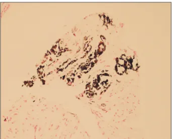
저자들의 연구에서는 신 유두 석회화를 가진 환자를 2mm 간격의 비조영증강 전산화단층촬영을 통하여 선별하였고
(Fig. 1), 이 중 8명에서는 경피적 신절석술 시에 신 유두 결정 침착 부위에서 조직 검사를 시행하여 von Kossa stain 으로 석회화를 확인하였다 (Fig. 2). 앞서 언급한 Low 등23의 연구에서와 마찬가지로 저자들의 연구에서도 혈중 및 24시 간 뇨 대사검사에서 모든 위험인자들의 평균값은 신 유두 석회화 환자군과 대조군 간의 의미 있는 차이는 보이지 않 았다. 또한 고요산혈증, 고칼슘혈증, 고인산혈증, 고중성지 방혈증, 고부갑상선혈증에 대한 두 군 간의 차이는 보이지 않았다. 하지만 과나트륨뇨증, 과칼슘뇨증, 과요산뇨증, 과 인산뇨증, 과수산뇨증, 저구연산뇨증의 비교에서는 과나트 륨뇨증, 과수산뇨증이 높은 비율로 발생하였으며, 특히 과 수산뇨증의 경우 통계적인 차이가 발견되었다 (p=0.043). 과 수산뇨증은 요중에서 수산칼슘석이 결정화하는 데 있어서
Fig. 1. Papillary calcification in the left kidney on unenhanced spiral computed tomography in our study (white arrows).
Fig. 2. von Kossa stain. This is a low magnification light micro- scopic image of a papillary biopsy specimen from a calcium oxa- late stone patient with papillary calcification on unenhanced spiral computed tomography. The site of calcium deposits is seen in the interstitial tissue and it stained black by the von Kossa stain for calcium histochemistry (Magnification, x100).
과칼슘뇨증보다 더 중요한 인자라고 알려져 있는데, 그 이 유는 칼슘보다 상대적으로 적은 배설량으로 인하여 배설의 조그만 차이에도 쉽게 요중 용해도의 변화를 일으킬 수 있 기 때문인 것으로 설명되고 있다.
비록 본 연구의 대상자 수가 적고 모든 환자에서 Ran- dall's plaque를 확인하기 위한 조직 검사를 시행하지 못한 면이 있으나, 앞으로 더 많은 환자를 대상으로 연구가 이루 어진다면, 요석 형성의 중요한 기전인 Randall's plaque와 요 석 위험 인자와의 관계를 좀 더 명확하게 밝히고 요석을 치료하고 예방하는데 있어 중요하게 사용될 것이다.
결 론
신 유두 석회화 환자에 있어서 혈중 검사를 통해 요석의 위험인자와의 관련성을 찾아보기 위한 대조군과의 비교에 서 통계적으로 유의한 차이는 없었다. 그러나 신 유두 석회 화 환자의 24시간 요 대사검사에서 과나트륨뇨증, 과수산 뇨증이 대조군에 비해 높은 비율로 발생하였으며, 특히 과 수산뇨증의 경우 통계적으로 의미 있게 높았다. 따라서 지 속적인 연구를 통하여 신 유두 석회화 환자의 내과적 원인 을 찾아내어 이를 교정한다면 요석의 재발을 줄이는 데 도 움을 줄 수 있을 것으로 생각한다.
REFERENCES
1. Leusmann DB, Blaschke R, Schmandt W. Results of 5,035 stone analyses: a contribution to epidemiology of urinary stone disease. Scand J Urol Nephrol 1990;24:205-10
2. Westbury EJ. Some observations on the quantitative analysis of over 1000 urinary calculi. Br J Urol 1974;46:215-27 3. Yoshida O, Okada Y. Epidemiology of urolithiasis in Japan:
a chronological and geotraphical study. Urol Int 1990;45:104- 11
4. Borghi L, Meschi T, Amato F, Briganti A, Novarini A, Giannini A. Urinary volume, water and recurrences in idiopa- thic calcium nephrolithiasis: a 5-year randomized prospective study. J Urol 1996;155:839-43
5. Cupisti A, Morelli E, Lupetti S, Meola M, Barsotti G. Low urine citrate excretion as main risk factor for recurrent calcium oxalate nephrolithiasis in males. Nephron 1992;61:73-6 6. Byeon SS, Kim HH, Kim SH. Analysis of the urinary stone
components using chemical analysis method. Korean J Urol 1996;37:179-86
7. Uribarri J, Oh MS, Carroll HJ. The first kidney stone. Ann Intern Med 1989;111:1006-9
8. Coe FL, Parks JH Asplin JR. The pathogenesis and treatment of kidney stones. N Engl J Med 1992;327:1141-52
9. Smith LH. The pathophysiology and medical treatment of urolithiasis. Semin Nephrol 1990;10:31-52
10. Randall A. The origin and growth of renal calculi. Ann Surg 1937;105:1009-27
11. Low RK, Stoller ML. Endoscopic mapping of renal papillae for Randall's plaques in patients with urinary stone disease.
J Urol 1997;158:2062-4
12. Whalley NA, Moraes MF, Shar TG, Pretorius SS, Meyers AM.
Lithogenic risk factors in the urine of black and white subjects.
Br J Urol 1998;82:785-90
13. Seftel A, resnick MI. Metabolic evaluation of urolithiasis. Urol Clin North Am 1990;17:159-69
14. Johnson CM, Wilson DM, O'Fallon WM, Malek RS, Kurland LT. Renal stone epidemiology: a 25-years study in Rochester, Minnesota. Kidney Int 1979;16:624-31
15. Carr LK, D'A Honey J, Jewett MA, Ibanez D, Ryan M, Bombardier C. New stone formation: a comparison of extra- corporeal shock wave lithotripsy and percutaneous nephroli- thotomy. J Urol 1996;155:1565-7
16. Sun BY, Lee YH, Jiaan BP, Chen KK, Chang LS, Chen KT.
Recurrence rate and risk factors for urinary calculi after extracorporeal shock wave lithotripsy. J Urol 1996;156:903-5 17. Hering LC. Observation on the analysis of ten thousand
urinary calculi. J Urol 1962;88:545-62
18. Francois B, Cahen R, Pascal B. Inhibitors of urinary stone formation in 40 recurrent stone formers. Br J Urol 1986;58:
479-83
19. Cochran M, Hofhkinson A, Zarembski PM, Anderson CK.
Hyperoxaluria in adults. Br J Surg 1968;55:121-8
20. Randall A. The etiology of primary renal calculus. Int Abstr Surg 1940;71:209-40
21. Evan AP, Lingeman JE, Coe FL, Parks JH, Bledsoe SB, Shao Y, et al. Randall's plaque of patients with nephrolithiasis begins in basement membranes of thin loops of Henle. J Clin Invest 2003;111:607-16
22. Kuo RL, Lingeman JE, Evan AP, Paterson RF, Bledsoe SB, Kim SC, et al. Endoscopic renal papillary biopsies: a tissue retrieval technique for histological studies in patients with nephrolithiasis. J Urol 2003;170:2186-9
23. Low RK, Stoller ML, Schreiber CK. Metabolic and urinary risk factors associatied with Randall's papillary plaques. J Endourol 2000;14:507-10