Blood Res2017;52:62-76. bloodresearch.or.kr
74 Letters to the Editor
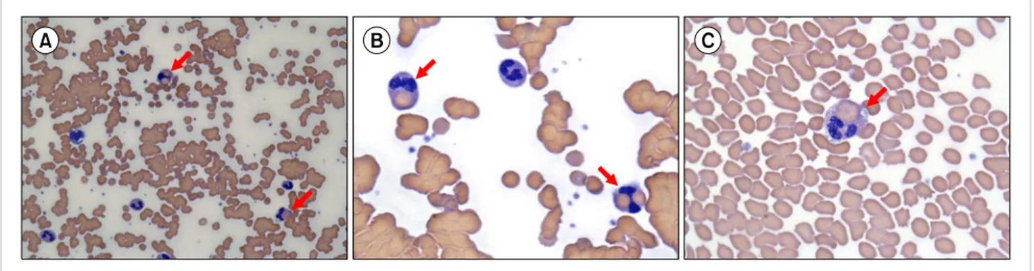
Fig. 1. The peripheral blood smear showed red blood cell agglutination (A, B) and erythrophagocytosis by neutrophils (A, B, C; red arrows).
A rare case of erythrophagocytosis by neutrophils on the peripheral blood smear
TO THE EDITOR: Erythrophagocytosis by monocytes or macrophages is occasionally seen in hematologic malig- nancy, autoimmune hemolytic anemia, and hemophagocytic lymphohistiocytosis [1, 2]. However, erythrophagocytosis by neutrophils on the peripheral blood smear is an unusual morphological phenomenon. Erythrophagocytosis by neu- trophils has been reported in patients with some hemolytic anemias, particularly paroxysmal cold hemoglobinuria (PCH), and other conditions including poisoning with potas- sium chlorate, sickle cell disease, and incompatible trans- fusion [3-6]. Cases of erythrophagocytosis by neutrophils have rarely been observed in cold agglutinin disease (CAD) [7-9]. We report a female patient with CAD and the presence of erythrophagocytosis by neutrophils on the peripheral blood smear.
An 80-year-old woman was referred to our hospital be- cause of dizziness. A few days prior to referral, she had cold symptoms, with a cough and rhinorrhea. She had a history of hypertension. Physical examination revealed ic- teric sclera and jaundice of her torso. Her white blood cell count was 15.88×10⁹/L (83% segmented neutrophils, 8%
band forms, 1% metamyelocytes, 2% myelocytes, 3% lym- phocytes, and 3% monocytes), hemoglobin concentration was 9 g/dL, and platelet count was 369×109/L. Other labo- ratory tests showed the following: increased lactate de- hydrogenase (2,530 IU/L), total bilirubin (5.14 mg/dL), and direct bilirubin (2.05 mg/dL), and decreased haptoglobin (2 mg/dL). The peripheral blood smear showed red blood cell (RBC) agglutination with a few nucleated RBCs and erythrophagocytosis by approximately 10% of the neu- trophils (Fig. 1). Cold agglutinin titer was 1:256. The direct Coombs’ test returned positive results (3+) for C3d and weak- ly positive results for IgG. The indirect Coombs’ test returned negative results. Sepsis was suspected and empirical was administered. However, the patient’s condition deteriorated rapidly and she died two days after admission. Although
the Donath-Landsteiner test was not examined and PCH could not completely be excluded, a diagnosis of CAD was made based on the available laboratory results.
CAD is generally classified as primary (idiopathic) or secondary. The latter is associated with underlying con- ditions such as malignancy, infection, or immune disorders [10]. Therefore, after diagnosis of CAD, patients should be evaluated for underlying conditions. Two sets of blood cul- tures returned negative results. Mycoplasma pneumoniae and Epstein-Barr virus were not detected. Evaluation for underlying malignancy or other disease was not performed because of the short clinical course. Therefore, we could not determine whether underlying disease was associated with the CAD.
The CR1 receptor of neutrophils can react with RBC- bound C3b [11]. However, the mechanism underlying eryth- rophagocytosis by neutrophils is unclear. To the best of our knowledge, this is the first reported case in Korea of CAD with erythrophagocytosis by neutrophils on a periph- eral blood smear.
Jong Ho Lee Department of Laboratory Medicine, Yeungnam University
College of Medicine, Daegu, Korea Correspondence to: Jong Ho Lee Department of Laboratory Medicine, Yeungnam University College of Medicine, Hyunchoongro 170, Nam-gu, Daegu 42415, Korea
E-mail: ae4207@naver.com
Received on Apr. 1, 2016; Revised on Apr. 11, 2016; Accepted on May 31, 2016 https://doi.org/10.5045/br.2017.52.1.74
AuthorsÊ Disclosures of Potential Conflicts of Interest No potential conflicts of interest relevant to this article were reported.
REFERENCES
1. Salama R, Girgis G, Zhou J. Erythrophagocytosis by neutrophils associated with Clostridium perfringens-induced hemolytic anemia. Ann Hematol 2015;94:521-2.
bloodresearch.or.kr Blood Res 2017;52:62-76.
Letters to the Editor 75
2. Lewandowski K, Homenda W, Mital A, Complak A, Hellmann A. Erythrophagocytosis by neutrophils-a rare morphological phenomenon resulting in acquired haemolytic anaemia? Int J Lab Hematol 2011;33:447-50.
3. Santos F, Costa E, Pinto RM, Barbot J, Freitas I. Erythrophagocy- tosis by neutrophils in paroxysmal cold haemoglobinuria. Eur J Haematol 2012;89:371.
4. Mamtani M, Sharma M, Amin M, Amin A, Jawahirani A, Kulkarni H. Erythrophagocytosis in sickle cell anemia: statistical evidence for a biological phenomenon. Med Hypotheses 2007;68:1065-70.
5. Mukhopadhyay S, Keating L, Souid AK. Erythrophagocytosis in paroxysmal cold hemoglobinuria. Am J Hematol 2003;74:196-7.
6. Hernandez JA, Steane SM. Erythrophagocytosis by segmented neutrophils in paroxysmal cold hemoglobinuria. Am J Clin Pathol 1984;81:787-9.
7. Walia H, Jain R, Bansal RK, Gupta GN. Cold agglutinin disease with erythrophagocytosis by neutrophils occurring during re- covery phase of chickenpox. J Lab Physicians 2013;5:146-7.
8. Beyá MD, Pereira A, Merino A. Transfusion medicine illustrated.
Erythrophagocytosis in cold agglutinin disease. Transfusion 2010;50:2310.
9. Keramati MR, Sadeghian MH, Ayatollahi H, Zarmehri AM. Case of erythrophagocytosis in a patient with mantle cell lymphoma and cold agglutinin disease. Leuk Res 2010;34:e167-8.
10. Swiecicki PL, Hegerova LT, Gertz MA. Cold agglutinin disease.
Blood 2013;122:1114-21.
11. Garratty G. Erythrophagocytosis on the peripheral blood smear and paroxysmal cold hemoglobinuria. Transfusion 2001;41:
1073-4.
The first case report of a patient with coexisting hemophilia B and Down syndrome
TO THE EDITOR: Hemophilia B, also known as Christmas disease, is an X-linked disorder caused by either the absence or reduced biosynthesis of clotting factor IX. This disorder affects approximately 1 in 30,000 male individuals world- wide [1]. It is five times less common than hemophilia A. On the other hand, Down syndrome (DS), the most common human chromosomal anomaly, results from tris- omy of chromosome 21 and leads to mental retardation.
Besides, it represents many consistent phenotypes including characteristic facies, intellectual disability, congenital heart diseases, and gastrointestinal abnormalities. In particular, hematological abnormalities include transient abnormal myelopoiesis, acute megakaryoblastic leukemia, and tran- sient thrombocytopenia/polycythemia/neutrophilia [2]. We present a case with a rare phenotype, i.e., the coexistence of hemophilia B and DS: one X-linked disorder and the other a chromosomal disorder. Here, we also describe the
management of this rare coexistence.
A 2-year-old male child born of a non-consanguineous marriage with a mixed ethnic background (father is a Punjabi and mother is from Orissa), visited to the pediatric emer- gency department with a history of spontaneous gum bleed- ing over the previous 4 days which was not resolved by general remedies. In addition, the patient suffered from episodic ecchymotic patches over the anterior abdominal wall in the previous month. There was no history suggestive of any bleeding disorders in close relatives (maternal/pater- nal sides). He was the second born child with an asympto- matic elder sister. On physical examination, the child had delayed developmental milestones, mongoloid slant, flat oc- ciput, depressed nasal bridge, short hands, and simian crease, all suggestive of the DS phenotype. However, no abnormal- ity was found in review of systems.
Karyotype analysis confirmed DS (47, XY, +21). Imaging studies confirmed the absence of any renal or cardiac malformations. Thyroid profile showed normal T3, T4, and thyroid stimulating hormone levels of 1.72, 10.57, and 3.5 units, respectively. Complete blood cell count (CBC) re- vealed hemoglobin level of 13.0 g/dL, white blood cell (WBC) count of 5.6×10⁹/L, and platelet count of 292×109/L.
Coagulation test showed normal prothrombin time (PT), 14 sec (reference range, 12–16 sec); prolonged activated partial thromboplastin time (aPTT), 85 sec (reference range, 26–32 sec); and normal fibrinogen level, 1.75 g/L (reference range, 2–4 g/L). Mixing study using normal pooled plasma and patient’s plasma was suggestive of factor IX deficiency.
Factor IX quantitative assay revealed a concentration of
<1%, indicative of severe deficiency (Hemophilia B).
Sequence analysis of peripheral blood for the F9 gene (exon 7) revealed c.760G>A (p.Gly254Ser). This mutation has been predicted as pathogenic variant by in silico program, Polyphen-2 (http://genetics.bwh.harvard.edu/pph2), and multiple sequence alignment has shown the glycine to be conserved across multiple species. The mother and sister of the patient were also found to be the carriers of the same mutation. The child, given two units of fresh frozen plasma along with vitamin K supplements, became stable and was discharged after a hospital stay of 7 days. Currently, he grows up 5 years old with regularly attending speech as well as physiotherapy clinics, and shows normal growth parameters except for small head, occasional skin bleedings, and joint bleedings. His parents have been counseled regard- ing the carrier state of his sister and further prenatal diagnosis.
The hematological abnormalities in DS have been studied in order to understand their pathophysiology. The spectrum of these abnormalities includes benign conditions (neutro- philia, thrombocytopenia, and polycythemia) which usually resolve by 3 weeks of age, as well as malignancies like acute megakaryoblastic leukemia [3]. The likely explanation for all these manifestations may be secondary to the extra copy of chromosome 21 or because of mutations involving the GATA1 gene [2]. The exact mechanism of how trisomy