3-dimensional reconstruction of mandibular canal at the interforaminal region using micro- computed tomography in Korean
Yong Hyun Jeon1, Chul Kwon Lee2, Hee-Jung Kim3, Jae-Heon Chung3, Heung-Joong Kim2, Sun-Kyoung Yu2*
1Department of Anatomy, College of Medicine, Chosun University, Gwangju, Republic of Korea
2Department of Oral Anatomy, College of Dentistry, Chosun University, Gwangju, Republic of Korea
3Department of Dental Prosthetics, College of Dentistry, Chosun University, Gwangju, Republic of Korea
PURPOSE. The purpose of this study was to identify the complex course of the mandibular canal using 3D reconstruction of microCT images and to provide the diagram for clinicians to help them understand at the interforaminal region in Korean. MATERIALS AND METHODS. Twenty-six hemimandibles obtained from cadavers were examined using microCT, and the images were reconstructed. At both the midpoint of mental foramen and the tip of anterior loop, the bucco-lingual position, the height from the mandibular inferior border, the horizontal distance between two points, and position relative to tooth site on the mandibular canal were measured. The angle that the mental canal diverges from the mandibular canal was measured in posterior- superior and lateral-superior direction. RESULTS. The buccal distance from the mandibular canal was significantly much shorter than lingual distance at both the mental foramen and the tip of anterior loop. The mandibular canal at the tip of anterior loop was significantly located closer to buccal side and higher than at the mental foramen. And the mental canal most commonly diverged from the mandibular canal below the first premolar by approximately 50º posterior-superior and 41º lateral-superior direction, which had with a mean length of 5.19 mm in front of the mental foramen, and exited to the mental foramen below the second premolar.
CONCLUSION. These results suggest that it could form a hazardous tetrahedron space at the interforaminal region, thus, the clinician need to pay attention to the width of a premolar tooth from the mental foramen during dental implant placement. [J Adv Prosthodont 2017;9:470-5]
KEYWORDS: Mandibular canal; Anterior loop; Interforaminal region; 3D-reconstruction; MicroCT
INTRODUCTION
Since July 1, 2014, treatment with two dental implants as well as complete removable denture has been covered by Korea national health insurance in old people over 75 years old. As a result, the number of patients treated with such prostheses increases gradually, and therefore, detailed knowledge on anatomical structure is required. In particular, the mandible has a much faster absorption rate than the maxilla, and the residual bone height in posterior mandible is insufficient for implant placement compared with that of anterior mandi- ble.1 Therefore, in a severely atrophied mandible, it is recom- mended to be treated with complete removable prostheses with a two-implant-supported overdenture which is placed between the mental foramen.2
Around the mental foramen, the mandibular canal divides into the mental and incisive canals and continues on
Corresponding author:
Sun-Kyoung Yu
Department of Oral Anatomy, College of Dentistry, Chosun University 309 Pilmun-daero, Dong-gu, Gwangju 61452, Republic of Korea Tel. +82622306357: e-mail, [email protected]
Received November 24, 2016 / Last Revision July 10, 2017 / Accepted July 11, 2017
© 2017 The Korean Academy of Prosthodontics
This is an Open Access article distributed under the terms of the Creative Commons Attribution Non-Commercial License (http://creativecommons.
org/licenses/by-nc/3.0) which permits unrestricted non-commercial use, distribution, and reproduction in any medium, provided the original work is properly cited.
This research was supported by Basic Science Research Program through the National Research Foundation of Korea (NRF) funded by the Ministry of Science and ICT (No. 2017R1A1A3A04069265).
to the region of the anterior teeth.3 The mental canal curves upward, backward, and lateral to reach the mental foramen, which is located below the second premolar,4,5 and the inci- sive canal continues to run toward the anterior teeth in a slightly downward direction, eventually reaching the chin.6 This means that the mandibular canal runs along the lingual cortical plate at the mandibular ramus and body, and then continues toward the mental canal with three dimensional (3D) complex course to exit the buccal side toward the mental foramen.7,8
At this ramification point, the canal forms an anterior loop and transitions back posteriorly to the mental canal;
the mandibular and mental canals exist as two canals simul- taneously.9 Therefore, even though the mental foramen is well known to clinician and the bone thickness on anterior mandible is thickest in canine distal region,10 at the interfo- raminal region between the mental foramen and the point at which the anterior loop develops, special care should be required during surgical procedures such as dental implant placement and genioplasty to avoid damaging the neurovas- cular bundle.5,7,9
In addition, the mandibular canal still includes the men- tal and dental nerves simultaneously at the interforaminal region across the mental foramen.11 The incisive nerve is totally separated from the surrounding epineurium of the mental nerve in the premolar region, being located lingually and inferiorly thereto, and continued into the incisive canal, which is incompletely formed and has a smaller diameter than the mandibular canal.3,12-14 Thus, on panoramic imag- ing, which is used widely for preoperative evaluation of the jaw, the mandibular canal and mental foramen are reported- ly readily visible in 49% of images, while the anterior loop is readily visible in only 3%.12 And, although cone beam com- puted tomography imaging was recently found to enhance the visualization of the mandibular anatomical structures,5,15 detailed evaluation remains difficult due to low cortical bone density and an incomplete bony canal at the interfo- raminal region.
Micro-computed tomography (microCT) can produce high-resolution images and is an effective method for detailed evaluation of the internal structure of bones.16,17 Furthermore, since 3D reconstructions are possible with these acquired high-resolution images, it is a very effective tool for examining the small facial canals.18 Therefore, the purposes of this study were to identify using 3D recon- struction of microCT images and to provide the diagram for clinicians to help them understand the complex course of the mandibular canal at the interforaminal region.
MATERIALS AND METHODS
Twenty-six hemimandibles from 19 cadavers that had been donated to the Department of Anatomy, School of Medicine, Chosun University for educational purposes were examined in this study. They were comprised of 16 males and 3 females, with a mean age at death of 54.4 years (range, 29 - 75 years), harvested all dentulous specimens which were
from the first molar to the lateral incisor, and were subject- ed to microCT scanning. This study followed the Declaration of Helsinki with respect to the medical proto- col and ethics.
The specimens were placed onto the holder so that the inferior border of the mandible was touching and perpendicu- lar to the floor, and were scanned using microCT (TVX- IMT225CT Dual type Micro CT, Techvalley, Seongnam, Korea) with a focus size of 1 μm. The obtained serial images were three-dimensionally reconstructed using 3D Doctor Software (3D-Doctor V 3.5 demo version, Able Software Corporation, MA, USA). Every fifth image was used for recon- struction because the three-dimensionally reconstructed results did not affect the original morphology of the mandible.
On the three-dimensionally reconstructed images, the mandibular and mental canals, mental foramen, and tip of the anterior loop, which coincides with the starting point of the mental canal, were identified. According to the result of Yu et al.19 that the mean diameters of the mandibular and mental canals are 2.80 and 2.63 mm, respectively, the tip of anterior loop was set a cutoff point of 2 mm for its mini- mum diameter. After that, at both the midpoint of mental foramen and the tip of anterior loop, the bucco-lingual dis- tances from the mandibular canal to external cortex and the height from the inferior border of the mandibular canal to the inferior border of the mandible were measured. At the midpoint of mental foramen, the vertical distance from the inferior border of the mandibular canal to the superior bor- der of the mental foramen was also measured. And, the horizontal distance between the midpoint of mental fora- men and the tip of anterior loop, like as the anterior loop length of previous researches, was investigated. The posi- tions of the midpoint of mental foramen and the tip of anterior loop were also examined relative to tooth site.
After the mandibular canal was set as the horizontal line, the specimens were classified into three types according to the divergent shape of the mental canal from the mandibu- lar canal in the posterior-superior direction. And the angle that the mental canal diverges from the mandibular canal was also measured in lateral-superior direction.
After microCT scanning, the specimens were decalcified for 3 days in 10% nitric acid and then neutralized in distilled water for 12 hours. While conserving the outer shape of the mental foramen, the buccal cortical and cancellous bone were carefully removed with the aid of a surgical micro- scope (OPMI-FC, Carl Zeiss, Oberkochen, Germany) to prevent damage to the inferior alveolar neurovascular bun- dle. The shape of the divergence of the mental canal from the mandibular canal and the positions of the mental fora- men and the anterior loop of the mental canal relative to tooth site were re-examined on the dissected specimens.
One-way ANOVA was used to analyze differences between observers, bucco-lingual distances at each mea- sured locations, and buccal distance and height between two locations using IBM SPSS Statistics (version 23.0, IBM Corporation, Somers, NY, USA). The significance level was set at P < .05.
RESULTS
The mandibular and mental canals were observed running anteriorly past the mental foramen on the serial, coronal- plane microCT images (Fig. 1). While the cortical wall that surrounds the mandibular canal was well formed, the corti- cal wall of the mental and incisive canals was partially bro- ken down, rendering it difficult to identify the canal borders.
The buccal distance from the mandibular canal was sig- nificantly shorter than lingual distance at both the mental foramen and the tip of anterior loop (P < .05). The man- dibular canal at the tip of anterior loop was significantly
located closer to buccal side (P < .05) and higher (P = .101) than at the mental foramen (Table 1). And the mean vertical distance from the mandibular canal to the mental foramen was 8.38 ± 2.16 mm.
The mean horizontal distance between the mental fora- men and the tip of anterior loop was 5.19 ± 1.48 mm, rang- ing from 2.98 to 9.45 mm. The mental foramen was observed most commonly below the second premolar (54.6%, n = 12), and the tip of the anterior loop was observed most commonly below the first premolar (45.5%, n = 10; Table 2).
As a result of 3D reconstruction, the divergent shape of
Table 1. Topography of the mandibular canal at coronal section of the midpoint of the mental foramen and the tip of the anterior loop
Midpoint of mental foramen Tip of anterior loop
Minimum Maximum Mean ± SD Minimum Maximum Mean ± SD
Buccal distance 2.52 6.59 4.24 ± 1.10 1.76 4.92 3.36 ± 0.94
Lingual distance 4.22 9.98 6.09 ± 1.66 3.73 9.69 6.56 ± 1.55
Height 11.11 16.78 13.11 ± 1.54 11.06 18.25 13.93 ± 1.61
Data are mean ± SD values.
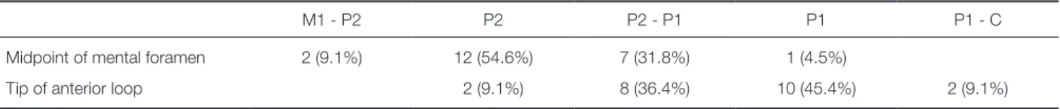
Table 2. Positions of the midpoint of the mental foramen and the tip of the anterior loop relative to tooth site
M1 - P2 P2 P2 - P1 P1 P1 - C
Midpoint of mental foramen 2 (9.1%) 12 (54.6%) 7 (31.8%) 1 (4.5%)
Tip of anterior loop 2 (9.1%) 8 (36.4%) 10 (45.4%) 2 (9.1%)
Abbreviations: C, canine; P1, first premolar; P2, second premolar; M1, first molar.
Data are n (%) values for each tooth site.
Fig. 1. Serial images (coronal views) of the mandibular canal obtained using microCT scanning. The dashed circle indicates the course of the mandibular canal at each tooth site. The arrow indicates the opening (mental foramen) of mental canal. (A) M1, first molar; (B) P2, second premolar; (C) P1, first premolar; (D) C, canine. Bu, buccal side; Li, lingual side.
A B C D
the mental canal, which runs a curved course posteriosupe- riorly from the mandibular canal, was classified into three types according to the angle of that curvature. Type 1 (0 - 30º) was observed in 18% (n = 4) of the specimens, with the mental canal lying almost parallel to the mandibular canal. Type 2 (30 - 60º) was the most common type, occur- ring in 59% (n = 13) of the specimens; the mode was 50º.
Type 3 (60 - 90º) appeared in 23% (n = 5) of the specimens, and since the mental canal in these cases was almost per- pendicular to the mandibular canal, the anterior loop of the mental canal was very short or absent (Fig. 2). And the mean angle that the mental canal diverges from the mandib- ular canal was 41.6 ± 9.22º in lateral-superior direction.
DISCUSSION
The mandibular canal crosses the mental foramen, then forms the anterior loop at the interforaminal region, and separates into the mental canal and the incisive canal.20 When the clinicians give a surgical treatment in the interfo- raminal region including the parasymphyseal area, the men- tal canal with its complex course, the anterior overextension
of the anterior loop beyond the mental foramen, and the large size of the incisive canal should be carefully taken into account.6,7,21 Repetitive and empirical surgeries performed without an accurate preoperative understanding of these anatomical structures may result in discomfort and postop- erative pain for patients.
As the residual bone height is not sufficient for implant placement, the bucco-lingual distance from the mandibular canal is more important.8 In previous research, the mandibu- lar canal courses close to the lingual side in molar region leans to the buccal side toward the premolar region,7,8,11,22 and then runs close to the middle portion in incisor region.11 In the present study, at both the mental foramen and the tip of anterior loop, it was also located close to the buccal side.
Moreover, at the tip of anterior loop more anteriorly, it was significantly closer to buccal side than the mental foramen.
In particular, at the mental foramen, the height from the inferior border of the mandibular canal to the inferior bor- der of the mandible was 13.11 mm and the vertical distance to mental foramen was 8.38 mm. The combined value was approximately 21 mm similar to the value of Liang et al.’s study.1 The distance from the superior border of the man- Fig. 2. Three-dimensional reconstructed images of the mandibular canal. (A - C, capital letter of top line) Lateral views showing the 3 types of the mandibular canal according to the divergent in the posterior-superior direction: type 1 (A), type 2 (B), and type 3 (C). (a - c, small letter of bottom line) Anteroposterior views of the mandibular canal
corresponding to the images in A - C, respectively.
A B C
a b c
dibular canal to the cementoenamel junction was approxi- mately 19 mm.22 Therefore, it was thought that the height of alveolar bone above the mental foramen was only about 10 mm in normal patient without periodontal diseases.
As the anterior loop length in previous researches, the horizontal distance from the mental foramen to the tip of the anterior loop varies in the range 0.2 - 7.6 mm,19 and its prevalence has been reported an wide range of 0 - 88%.5 In the present study, the midpoint of the mental foramen was most commonly found below the second premolar and the tip of the anterior loop was most commonly found below the first premolar, with a mean length of 5.19 mm ranged from 2.98 to 9.45 mm. Therefore, careful attention needs to be paid to the width of a premolar tooth, which is approxi- mately 7 mm,23 anteriorly from the mental foramen in older edentulous patients.
The mandibular canal has direction with 67.2º superior, 39.4º lateral, and 80.2º posterior based on the mental fora- men, forms the anterior loop in 61.5%, and transitions into the mental canal.7,13 In the present study, the mental canal from the mandibular canal diverged into an angle of approximately 50º posterior-superior and 41º lateral-superi- or similar to previous research. Therefore, de Freitas et al.24 recommended that the needle be inclined to around 55º from back to front and around 40º from outward to inward when blocking the mental nerve. During implant placement, Krekmanov et al.25 described that if the fixture is tilted 25 - 35º from the anterior loop, an average distance of 6.5 mm can be earned for prosthetic support.
The mental canal was almost parallel (0 - 30º) with the mandibular canal in type 1 cases, meaning that there may be sufficient alveolar bone height superior to the alveolar crest to enable a relatively stable implant placement. However, in type 3 the mental canal was almost perpendicular to the mandibular canal (60 - 90º), and so no anterior loop was formed; this could have a negative effect on implant place- ment around the mental foramen, but since the anterior mandible at the interforaminal region is relatively extended horizontally, it could have a positive effect on genioplasty.
Hu et al.26 noted a vertically formed anterior loop of the mental nerve in 15.4% of cases, and a straight form toward the anterior teeth in 23.1% of the cases. Hence, additional research is needed to evaluate the correlation between the divergent angle of the mental canal relative to the mandibu- lar canal and the length from the mental foramen to the anterior loop, and to identify cases in which the mental canal continues toward the anterior teeth with an angle of more than 90º.
CONCLUSION
Since the mandibular canal divides into two terminal canals, the mental canal including the anterior loop presents a Y-shaped or delta-shaped divergence.20,27 In the present study, the mental canal diverged from the mandibular canal below the first premolar by approximately 50º posterior- superior and 41º lateral-superior direction, in which was
located about 5 mm in front of the mental foramen, and exited to the mental foramen below the second premolar.
Therefore, it could form a hazardous tetrahedron space at the interforaminal region as diagrammed in Figure 3 in which there is the potential for simultaneous damage to the mental and dental branches. The clinician need to pay atten- tion to the width of a premolar tooth from the mental fora- men anteriorly during dental implant placement and genio- plasty at the interforaminal region.
ORCID
Hee-Jung Kim https://orcid.org/0000-0002-2015-1530 Sun-Kyoung Yu https://orcid.org/0000-0003-0801-1663 REFERENCES
1. Liang XH, Kim YM, Cho IH. Residual bone height measured by panoramic radiography in older edentulous Korean pa- tients. J Adv Prosthodont 2014;6:53-9.
2. Feine JS, Carlsson GE, Awad MA, Chehade A, Duncan WJ, Gizani S, Head T, Lund JP, MacEntee M, Mericske-Stern R, Mojon P, Morais J, Naert I, Payne AG, Penrod J, Stoker GT Jr, Tawse-Smith A, Taylor TD, Thomason JM, Thomson WM, Wismeijer D. The McGill Consensus Statement on Overdentures. Montreal, Quebec, Canada. May 24-25, 2002.
Int J Prosthodont 2002;15:413-4.
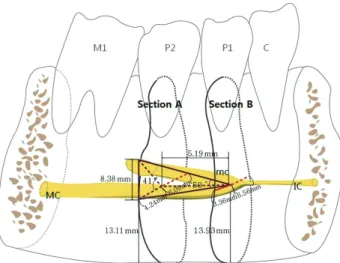
Fig. 3. Schematic drawing shows the major course of the mandibular canal which forms a hazardous tetrahedron space at the interforaminal region. The measurement was performed in the reference position where is at both the midpoint of mental foramen (Section A) and the tip of anterior loop (Section B). The data represent mean values of each measurement items. MC, mandibular canal; mc, mental canal; IC, incisive canal.
3. Watanabe H, Mohammad Abdul M, Kurabayashi T, Aoki H.
Mandible size and morphology determined with CT on a premise of dental implant operation. Surg Radiol Anat 2010;
32:343-9.
4. Kim MK. Head and neck anatomy. 5th ed. Seoul; Dental &
Medical Publishing.: 2011. p. 86.
5. Li X, Jin ZK, Zhao H, Yang K, Duan JM, Wang WJ. The prevalence, length and position of the anterior loop of the inferior alveolar nerve in Chinese, assessed by spiral comput- ed tomography. Surg Radiol Anat 2013;35:823-30.
6. Juodzbalys G, Wang HL, Sabalys G. Anatomy of mandibular vital structures. part II: Mandibular incisive canal, mental fo- ramen and associated neurovascular bundles in relation with dental implantology. J Oral Maxillofac Res 2010;1:e3.
7. Hwang K, Lee WJ, Song YB, Chung IH. Vulnerability of the inferior alveolar nerve and mental nerve during genioplasty:
an anatomic study. J Craniofac Surg 2005;16:10-4.
8. Kim ST, Hu KS, Song WC, Kang MK, Park HD, Kim HJ.
Location of the mandibular canal and the topography of its neurovascular structures. J Craniofac Surg 2009;20:936-9.
9. Apostolakis D, Brown JE. The anterior loop of the inferior alveolar nerve: prevalence, measurement of its length and a recommendation for interforaminal implant installation based on cone beam CT imaging. Clin Oral Implants Res 2012;23:
1022-30.
10. Kim HJ, Yu SK, Lee MH, Lee HJ, Kim HJ, Chung CH.
Cortical and cancellous bone thickness on the anterior region of alveolar bone in Korean: a study of dentate human cadav- ers. J Adv Prosthodont 2012;4:146-52.
11. Yu SK, Lee MH, Jeon YH, Chung YY, Kim HJ. Anatomical configuration of the inferior alveolar neurovascular bundle: a histomorphometric analysis. Surg Radiol Anat 2016;38:195- 201.
12. Jacobs R, Mraiwa N, Van Steenberghe D, Sanderink G, Quirynen M. Appearance of the mandibular incisive canal on panoramic radiographs. Surg Radiol Anat 2004;26:329-33.
13. Mardinger O, Chaushu G, Arensburg B, Taicher S, Kaffe I.
Anterior loop of the mental canal: an anatomical-radiologic study. Implant Dent 2000;9:120-5.
14. Lee MH, Kim HJ, Kim DK, Yu SK. Histologic features and fascicular arrangement of the inferior alveolar nerve. Arch Oral Biol 2015;60:1736-41.
15. Uchida Y, Noguchi N, Goto M, Yamashita Y, Hanihara T, Takamori H, Sato I, Kawai T, Yosue T. Measurement of ante- rior loop length for the mandibular canal and diameter of the mandibular incisive canal to avoid nerve damage when install- ing endosseous implants in the interforaminal region: a sec- ond attempt introducing cone beam computed tomography. J Oral Maxillofac Surg 2009;67:744-50.
16. Choi DY, Sun KH, Won SY, Lee JG, Hu KS, Kim KD, Kim HJ. Trabecular bone ratio of the mandibular condyle accord- ing to the presence of teeth: a micro-CT study. Surg Radiol Anat 2012;34:519-26.
17. Engelke K, Song SM, Glüer CC, Genant HK. A digital model of trabecular bone. J Bone Miner Res 1996;11:480-9.
18. Song WC, Jo DI, Lee JY, Kim JN, Hur MS, Hu KS, Kim HJ, Shin C, Koh KS. Microanatomy of the incisive canal using
three-dimensional reconstruction of microCT images: an ex vivo study. Oral Surg Oral Med Oral Pathol Oral Radiol Endod 2009;108:583-90.
19. Yu SK, Kim S, Kang SG, Kim JH, Lim KO, Hwang SI, Kim HJ. Morphological assessment of the anterior loop of the mandibular canal in Koreans. Anat Cell Biol 2015;48:75-80.
20. Moiseiwitsch JR. Position of the mental foramen in a North American, white population. Oral Surg Oral Med Oral Pathol Oral Radiol Endod 1998;85:457-60.
21. Greenstein G, Tarnow D. The mental foramen and nerve:
clinical and anatomical factors related to dental implant place- ment: a literature review. J Periodontol 2006;77:1933-43.
22. Choi Y, Jung UW, Um YJ, Kim ST, Hu KS, Kim CS, Kim KD, Chai JK, Choi SH. Running pattern of the mandibular canal in Korean population, by 3-dimensional reconstructed com- puted tomographic imaging analysis. Implantology 2009;13:
152-61.
23. Kim MS, Kim SH, Kim HJ, Kim HJ, Park BG, Park BS, Park JT, Park JC, Bae YC, Yu SK, Lee YH, Jung HS, Cho SW, Cho US, Heo GS. Dental anatomy and morphology. 3rd ed. Seoul;
DaehanNarae Publishing Inc.; 2016. p. 163-76.
24. de Freitas V, Madeira MC, Pinto CT, Zorzetto NL. Direction of the mental canal in human mandibles. Aust Dent J 1976;
21:338-40.
25. Krekmanov L, Kahn M, Rangert B, Lindström H. Tilting of posterior mandibular and maxillary implants for improved prosthesis support. Int J Oral Maxillofac Implants 2000;15:
405-14.
26. Hu KS, Yun HS, Hur MS, Kwon HJ, Abe S, Kim HJ.
Branching patterns and intraosseous course of the mental nerve. J Oral Maxillofac Surg 2007;65:2288-94.
27. Mraiwa N, Jacobs R, Moerman P, Lambrichts I, van Steenberghe D, Quirynen M. Presence and course of the incisive canal in the human mandibular interforaminal region: two-dimension- al imaging versus anatomical observations. Surg Radiol Anat 2003;25:416-23.