Osteoclasts
Osteoclasts are unique bone-resorbing cells, and mice carrying a mutant gene responsible for osteoclast formation exhibit osteopetrotic phenotype[1,2]. Osteoclasts are giant cells containing 10~20 nuclei or sometimes more than 100 nuclei in each cell, and the multinucleation is a consequence of fusion of mononuclear osteoclasts.
Osteoclasts are derived from hematopoietic stem cells or monocyte/macrophage lineage cells, and an interaction between RANKL (receptor activator of NF-κB ligand) and RANK is required for osteoclast formation[3,4]. RANKL is expressed in osteoblasts whereas RANK is expressed in
osteoclast precursor cells. Although RANKL is a transmembrane ligand, a genetically engineered soluble form of RANKL is also effective for osteoclast differentiation. We were able to generate osteoclasts without osteoblastic cells in the presence of M-CSF (macrophage colony stimulating factor) and RANKL[5,6]. This pure culture system enabled us to analyze gene expression profiles in osteoclasts.
Identification of osteoclast precursor cells and their differentiation
To establish a pure osteoclast culture system, we tried to isolate osteoclast precursor cells. Bone marrow
Regulation of Osteoclast Differentiation
(Identification of osteoclast and macrophage fusion protein; DC-STAMP)
Takeshi Miyamoto
Department of Orthopedic Surgery and Cell Differentiation,
Musculoskeletal Reconstruction and Regeneration Surgery, Keio University School of Medicine
ABSTRACT
Osteoclasts are bone-resorbing multinuclear cells derived from hematopoietic stem cells or monocyte/
macrophage lineage cells. Recent identification of RANK/RANKL has provided new insights into the osteoclast differentiation pathway, enabling us to generate osteoclasts without stromal cells, which support osteoclastogenesis. In order to establish a pure osteoclast culture system, we identified the osteoclast precursor cell (c-Kit+c-Fms+RANK- cell), which is a common precursor cell of osteoclasts, macrophages and dendritic cells. Macrophages are induced by M-CSF alone, while a sequential stimulation of M-CSF followed by RANKL effectively induces osteoclast formation. Furthermore, dendritic cells are induced by GM-CSF or GM-CSF plus RANKL. Therefore, we were able to generate pure osteoclasts, macrophages or dendritic cells from the common precursor cell using specific combinations of cytokines. Using this culture system, we found that an adherent condition is critical for osteoclast differentiation. We also found that the osteoclastogenesis induced by M-CSF plus RANKL is completely inhibited by GM-CSF, and that these cells differentiate into a dendritic cell lineage. The osteoclast multinucleation is believed to be induced by cell-cell fusion of mononuclear osteoclasts. Although various molecules have been implicated in the cell-cell fusion of osteoclasts or macrophages, the essential molecule for cell fusion has not been identified. We identified that the dendritic cell-specific transmembrane protein (DC-STAMP) was an essential cell-cell fusion molecule for osteoclasts and foreign body giant cells, and that DC-STAMP deficient mice have no multinuclear osteoclasts. Here I review the osteoclast development from immature precursor cells to multinuclear osteoclasts. (J Kor Endocrinol Soc 21:347~351, 2006)
ꠏꠏꠏꠏꠏꠏꠏꠏꠏꠏꠏꠏꠏꠏꠏꠏꠏꠏꠏꠏꠏꠏꠏꠏꠏꠏꠏꠏꠏꠏꠏꠏꠏꠏꠏꠏꠏꠏꠏꠏꠏꠏꠏꠏꠏꠏꠏꠏꠏꠏꠏꠏꠏꠏꠏꠏꠏꠏꠏꠏꠏꠏꠏꠏꠏꠏꠏꠏꠏꠏꠏꠏꠏꠏꠏꠏꠏꠏꠏꠏꠏꠏꠏꠏꠏꠏꠏꠏꠏꠏꠏꠏꠏꠏꠏꠏꠏꠏ Key Words: Cell-cell fusion, DC-STAMP, Foreign body giant cells, Osteoclasts
mononuclear cells were subdivided to three populations based on expressions of c-Fms and RANK; c-Fms+RANK-, c-Fms+RANK+, c-Fms-RANK+. c-Fms is a receptor of M-CSF, whereas RANK is a receptor of RANKL. Each cell population was sorted by FACS and cultured in the presence of M-CSF and RANKL. Osteoclastogenesis was evaluated by tartrate resistant acid phosphatase (TRAP) activity, an enzymatic marker of osteoclasts. Among the populations cultured, c-Fms+RANK-cells had the highest potential to differentiate into osteoclasts, and almost all of the cells expressed TRAP activity[6]. Thus we could establish a pure osteoclast culture system, and decided to use this population in the following experiments. We found that RANK expression was induced in c-Fms+RANK- cells, and the following RANKL stimulation induced effective osteoclast differentiation[5]. Since osteoclastogenesis is strictly limited to bones, some unique factor other than cytokines essential for osteoclast differentiation might be present in the bone. We focused on the hard substrate of bones as an adherent condition. The osteoclastogenesis was significantly inhibited in non-adherent conditions prepared by methylcellulose culture compared with adherent cultures prepared by liquid culture, and the inhibited differentiation in non-adherent conditions was fully restored by transferring those cells into an adherent condition, indicating that osteoclast differentiation is anchorage dependent[6]. Thus we considered that an adherent condition prepared by bones is involved in osteoclastogenesis.
Although osteoclastogenesis was strongly inhibited in non-adherent conditions, the inhibition was not complete.
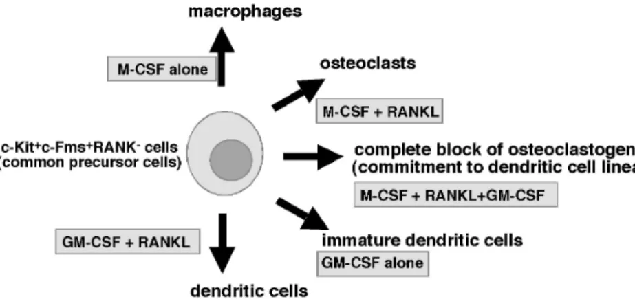
Thus, some other factor might exist inhibiting osteoclast differentiation in the soft tissues. We focused on GM-CSF, and found that osteoclastogenesis induced by M-CSF plus RANKL was completely inhibited by GM-CSF[7]. The osteoclast precursor cells differentiated into TRAP-negative mononuclear cells in the presence of M-CSF, RANKL and GM-CSF. These cells express CD11c, a marker of dendritic cells, indicating that GM-CSF induces the cells to a dendritic cell lineage. The commitment of common precursor cells to macrophages, osteoclasts, and dendritic cells in the presence of specific cytokines are shown in Fig. 1. GM-CSF strongly reduced the expression of c-Fos, an essential transcription factor for osteoclast differentiation, and forced expression of c-Fos rescued the inhibition of osteoclastogenesis by GM-CSF, suggesting that downregulation of c-Fos is critical for the inhibition of osteoclast differentiation by GM-CSF.
Identification of an osteoclast fusion molecule;
DC-STAMP
Since cell-cell fusion is mediated through cell contact, various cell surface molecules such as E-cadherin, Macrophage Fusion Receptor (MFR), CD44, CD9 and CD81 have been implicated in the multinucleation of osteoclasts and macrophages[8~11]. However, defects in multinucleation of osteoclasts and macrophages have not
Fig. 1. Osteoclast, macrophage and dendritic cell differentiation from common precursor cells.
Macrophages are induced by M-CSF alone, while osteoclasts are induced by M-CSF followed by RANKL stimulation. Immature dendritic cells are induced by GM-CSF alone, and GM-CSF plus RANKL induces activated dendritic cells. The osteoclastogenesis induced by M-CSF and RANKL is completely inhibited by GM-CSF, and that cells differentiate into dendritic cell lineage cells.
been shown by gene targeting. In order to isolate osteoclast cell-cell fusion molecules, we undertook DNA subtraction screens between multinuclear osteoclasts and mononuclear macrophages. As above, multinuclear osteoclasts were induced by M-CSF and RANKL, whereas mononuclear macrophages were induced by M-CSF alone.
We have already found that various molecules such as RANK, c-Fms, integrinαvβ3 and Mac-1 are equally expressed in both osteoclasts and macrophages. Thus DNA subtraction screen was considered effective in identifying osteoclast specific molecules. More than 100 molecules were identified by the screen, and DC-STAMP was selected as an osteoclast specific molecule[12].
Characterization of DC-STAMP in osteoclasts
DC-STAMP was identified in dendritic cells as DC -STAMP or IL-4 stimulated macrophages as FIND[13,14].
DC-STAMP was also detected in osteoclasts[15]. In order to understand the role of DC-STAMP in vivo, we generated DC-STAMP deficient mice[12]. The DC-STAMP deficient mice show no growth abnormalities, however, osteoclast cell-cell fusion was completely abrogated in DC-STAMP deficient mice in vivo and in vitro. Osteoclasts in DC-STAMP deficient mice show TRAP-activity, and the osteoclast differentiation markers such as cathepsin K was expressed in DC-STAMP deficient osteoclasts as in wild type osteoclasts. NFATc1 and c-Fos, both of which were essential transcription factors required for osteoclast differentiation, were induced in DC-STAMP deficient osteoclasts at equivalent levels to wild type osteoclasts.
Furthermore, the formation of ruffled boarder, a highly specific cytoskeletal feature seen in wild type osteoclasts, was also detected in DC-STAMP-/- osteoclasts. The osteoclast cell-cell fusion was effectively rescued by retro viral transduction of DC-STAMP into DC-STAMP deficient osteoclast progenitor cells, suggesting that DC-STAMP is specifically required for osteoclast cell-cell fusion. In fact, the mononuclear osteoclasts seen in DC-STAMP deficient mice showed bone resorbing activity, and bone marrow cavity formation and teeth eruption were observed in DC-STAMP deficient mice indicating that the osteoclasts in DC-STAMP deficient mice are mononuclear but bone-resorbing cells. However, the bone resorbing activity evaluated by pit formation assay in DC-STAMP deficient osteoclasts was significantly
low compared with that of wild type osteoclasts, even though the total number of nuclei was equal. In in vivo analysis, an elevated radio-opacity was seen in DC-STAMP-/- mice by Soft X-ray analysis, and the bone mineral density (BMD) and bone volume per tissue volume (BV/TV) was increased in DC-STAMP-/- mice compared with wild type mice, suggesting that multinucleation enhances the bone resorbing efficiency of osteoclasts. Thus loss of osteoclast cell fusion leads to an increased bone mass.
Characterization of DC-STAMP in FBGCs
Foreign body giant cells (FBGCs) are derived from monocyte/macrophage lineage cells like osteoclasts, and are generated in response to foreign bodies at the site of implantation[16]. Both osteoclasts and FBGCs form multinuclear cells by fusion of mononuclear cells.
Interestingly, multinuclear FBGCs formation induced by implantation of foreign bodies was also completely abrogated in DC-STAMP-/- mice. FBGCs are formed in vitro from macrophage and osteoclast common precursor cells by stimulation with several cytokine combinations such as IL-3 plus IL-4[17] or GM-CSF plus IL-4. FBGC formation in vitro was also completely inhibited in DC-STAMP-/- cells, indicating that DC-STAMP is universally required for osteoclast and macrophage cell fusion.
Mechanisms of osteoclast cell fusion via DC-STAMP
DC-STAMP knock-out mice were generated as EGFP knock-in mice, therefore, only mononuclear EGFP-positive cells were induced from DC-STAMP deficient osteoclast precursor cells in the presence of M-CSF plus RANKL.
However, multinuclear EGFP-positive cells were induced when osteoclast precursors isolated from DC-STAMP-/- and wild type mice were mixed and cultured together in the presence of M-CSF plus RANKL. This interesting phenomenon suggests that fusion between EGFP-positive DC-STAMP-/- cells and wild type osteoclast precursor cells was induced. These data indicate that a putative DC -STAMP ligand might be expressed on osteoclasts, and the interaction between DC-STAMP and its putative ligand may induce the cell-cell fusion, and therefore the
expression of DC-STAMP is not required in all the cells participating in multinucleation. Heterogeneous EGFP expression in DC-STAMP-EGFP knock-in cells during the course of osteoclast differentiation supports this possibility.
Thus, DC-STAMP might be involved in cell-cell interactions in a receptor-ligand fashion. Further analysis is required to clarify the mechanisms of cell-cell fusion via DC-STAMP.
References
1. Boyce BF, Yoneda T, Lowe C, Soriano P, Mundy GR: Requirement of pp60c-src expression for osteoclasts to form ruffled borders and resorb bone in mice. J Clin Invest 90:1622-1627, 1992
2. Grigoriadis AE, Wang ZQ, Cecchini MG, Hofstetter W, Felix R, Fleisch HA, Wagner EF: c-Fos: a key regulator of osteoclast-macrophage lineage determination and bone remodeling. Science 266:443-448, 1994 3. Kong YY, Yoshida H, Sarosi I, Tan HL, Timms E,
Capparelli C, Morony S, Oliveira-dos-Santos AJ, Van G, Itie A, Khoo W, Wakeham A, Dunstan CR, Lacey DL, Mak TW, Boyle WJ, Penninger JM: OPGL is a key regulator of osteoclastogenesis, lymphocyte development and lymph-node organogenesis. Nature 397:315-323, 1999
4. Dougall WC, Glaccum M, Charrier K, Rohrbach K, Brasel K, De Smedt T, Daro E, Smith J, Tometsko ME, Maliszewski CR, Armstrong A, Shen V, Bain S, Cosman D, Anderson D, Morrissey PJ, Peschon JJ, Schuh J: RANK isessential for osteoclast and lymph node development. Genes Dev 13:2412-2424, 1999 5. Arai F, Miyamoto T, Ohneda O, Inada T, Sudo T,
Brasel K,Anderson DM, Suda T: Commitment and differentiation of osteoclast precursor cells by the sequential expression of c-Fms and receptor activator of nuclear factor kappaB (RANK) receptors. J Exp Med 190:1741-1754, 1999
6. Miyamoto T, Arai F, Ohneda O, Takagi K, Anderson DM, Suda T: An adherent condition is required for formation of multinuclear osteoclasts in the presence of macrophage colony-stimulating factor and receptor activator of nuclear factor kappa B ligand. Blood 96:4335-4343, 2000
7. Miyamoto T, Ohneda O, Arai F, Iwamoto K, Okada S, Takagi K, Anderson DM, Suda T: Bifurcation of
osteoclasts and dendritic cells from common progenitors. Blood 98:2544-2554, 2001
8. Mbalaviele G, Chen H, Boyce BF, Mundy GR, Yoneda T: The role of cadherin in the generation of multinucleated osteoclasts from mononuclear precursors in murine marrow. J Clin Invest 95:2757- 2765, 1995
9. Saginario C, Sterling H, Beckers C, Kobayashi R, Solimena M, Ullu E, Vignery A: MFR, a putative receptor mediating the fusion of macrophages. Mol Cell Biol 18:6213-6223, 1998
10. de Vries TJ, Schoenmaker T, Beertsen W, van der Neut R, Everts V: Effect of CD44 deficiency on in vitro and in vivo osteoclast formation. J Cell Biochem 94:954-966, 2005
11. Takeda Y, Tachibana I, Miyado K, Kobayashi M, Miyazaki T, Funakoshi T, Kimura H, Yamane H, Saito Y, Goto H, Yoneda T, Yoshida M, Kumagai T, Osaki T, Hayashi S, Kawase I, Mekada E:
Tetraspanins CD9 and CD81 function to prevent the fusion of mononuclear phagocytes. J Cell Biol 161:
945-956, 2003
12. Yagi M, Miyamoto T, Sawatani Y, Iwamoto K, Hosogane N, Fujita N. Morita K, Ninomiya K, Suzuki T, Miyamoto K, Oike Y, Takeya M, Toyama Y, Suda T: DC-STAMP is essential for cell-cell fusion in osteoclasts and foreign body giant cells. J Exp Med 202:345-351, 2005
13. Hartgers FC, Vissers JL, Looman MW, van Zoelen C, Huffine C, Figdor CG, Adema GJ: DC-STAMP, a novel multimembrane-spanning molecule preferentially expressed by dendritic cells. Eur J Immunol 30:3585- 3590, 2000
14. Staege H, Brauchlin A, Schoedon G, Schaffner A:
Two novel genes FIND and LIND differentially expressed in deactivated and Listeria-infected human macrophages. Immunogenetics 53:105-113, 2001 15. Kukita T, Wada N, Kukita A, Kakimoto T, Sandra F,
Toh K, Nagata K, Iijima T, Horiuchi M, Matsusaki H, Hieshima K, Yoshie O, Nomiyama H:
RANKL-induced DC-STAMP is essential for osteoclastogenesis. J Exp Med 200:941-946, 2004 16. Kyriakides TR, Foster MJ, Keeney GE, Tsai A,
Giachelli CM, Clark-Lewis I, Rollins BJ, Bornstein P:
The CC chemokine ligand, CCL2/MCP1, participates in macrophage fusion and foreign body giant cell
formation. Am J Pathol 165:2157-2166, 2004 17. McNally AK, Anderson JM: Interleukin-4 induces
foreign body giant cells from human monocytes/
macrophages. Differential lymphokine regulation of
macrophage fusion leads to morphological variants of multinucleated giant cells. Am J Pathol 147:1487 -1499, 1995