Hip Pelvis 26(3): 178-184, 2014 http://dx.doi.org/10.5371/hp.2014.26.3.178
INTRODUCTION
Adhesive capsulitis of the hip (ACH) has also been referred to as “frozen hip”1)or “capsular constriction”2). It was first described by Caroit et al.3)and since then there have only been few publications on the condition4-6). However, subsequent experience has led authors to speculate that this condition does exist more commonly than was suggested earlier6). There have been descriptions of case reports of the same but there is a lack of description of a case series. The medical
Measurement of Capsular Thickness in Magnetic Resonance Arthrography in Idiopathic Adhesive Capsulitis of Hip
Young-Deuk Joo, MD, Anshul Shyam Sobti, Kwang-Jun Oh, MD, PhD
Department of Orthopedic Surgery, Konkuk University School of Medicine, Seoul, Korea
Purpose: The clinical suspicion of idiopathic adhesive capsulitis of the hip (IACH) involves restricted range of motion and normal hip radiographs. The purpose of this study was to delineate the characteristic findings observed on magnetic resonance arthrography (MRA) by identifying the anatomical structures involved and their significance on clinical presentation of restricted range of motion.
Materials and Methods: We retrospectively evaluated MRA’s of 46 hips (44 patients) who suffered hip pain from September 2006 to August 2012 in our hospital. Of those, 10 cases (8 patients) with clinical suspicion of IACH were compared to 20 normal hip cases (control group). To identify anatomical evidence of adhesive capsulitis in the MRA’s of the IACH group, capsular thickness was measured superiorly, inferiorly, anteriorly and posteriorly, and compared to that of the randomly selected control group.
Results: Comparison of the MRA findings of the control group to that of the IACH group showed that there was a statistically significant increase in the mean thickness of the joint capsule superiorly and posteriorly (P<0.01), while comparison of examination findings revealed a statistically significant decrease in the mean range of motion (flexion 122.5。±5.5。/abduction 28.0。±2.8。/adduction 26.5。±2.4。/external rotation 30.5。±3.8。
/internal rotation 25.5。±2.4。) in the IACH group.
Conclusion: A change in the capsular thickness on MRA is a common finding in IACH patients with the increase more evident in the posterior and superior capsules than the anterior and inferior capsules.
Key Words: Idiopathic adhesive capsulitis of hip, Magnetic resonance arthrography, Capsule thickness
Submitted:July 25, 2014 1st revision:August 8, 2014
2nd revision:August 22, 2014 Final acceptance:August 28, 2014 Address reprint request to
Kwang-Jun Oh, MD, PhD
Department of Orthopedic Surgery, Konkuk University School of Medicine, 120 Neungdong-ro, Gwangjin-gu, Seoul 143-729, Korea TEL:+82-2-2030-7616 FAX:+82-2-2030-7369
E-mail:[email protected]
This is an Open Access article distributed under the terms of the Creative Commons Attribution Non-Commercial License (http://creativecommons.
org/licenses/by-nc/3.0) which permits unrestricted non-commercial use, distribution, and reproduction in any medium, provided the original work is properly cited.
literature has described two kinds of ACH; primary idiopathic adhesive capsulitis of hip (IACH) and secondary ACH2,6). They have defined IACH as adhesive capsulitis that is present without known etiology or concomitant pathology. If adhesive capsulitis was present secondary to pathology it was defined as secondary.
Little has been described in literature concerning IACH.
Its presentations can be challenging and does require further diagnostic testing. The clinical suspicion of IACH involves restricted range of movement and normal hip radiographs. This presentation is similar to that for adhesive capsulitis of the shoulder (ACS)3). ACH is due to retraction of the fibrous joint capsule of the hip and unless IACH is diagnosed through surgery or biopsy, clinicians must rely on the patient’s history and clinical findings and normal radiography to diagnose IACH5). There is a lack of description of an objective evaluation modality that demonstrates direct evidence of this. Thus the purpose of this study was to delineate the characteristic of findings observed on magnetic resonance arthrography (MRA) by identifying the capsular thickness involved and their significance on clinical presentation of restricted range of motion in cases with IACH and to report on the authors experience with the diagnosis of IACH.
MATERIALS AND METHODS
From September 2006 to August 2012, MRA images of all the patients with hip pain or restricted range of movements, with normal plain hip radiographs were retrospectively assessed.
At the time of this reporting, the database consisted of
46 hips (44 patients). We excluded all subjects who have soft tissue abnormal (labral tear or ligament tear 16 hips).
Out of them, 10 hips (8 patients) were selected in the study group (Table 1), who were diagnosed as cases of IACH by the evaluation of a single radiologist on MRA findings. Diagnostic criteria of IACH which we assuming are as 1) reduction of intra-articular capacity of joint, 2) limitation of range of motion, with or without pain. Twenty patients were included in the control group with normal MRA findings and with only hip pain without limitation of range of motion. Rests of the cases were excluded from the study (Fig. 1). The exclusion criteria were abnormal laboratory result (blood cell count, inflammation markers, blood phosphate/calcium balance), in the presence of any sign of osteonecrosis or progressing coxarthrosis by radiological examination, inflammatory arthritis (rheumatoid arthritis, systemic lupus erythematosus etc.) excluded secondary ACH.
The study group included 10 hips (8 patients) with 3 males and 7 females. The average age of the patients with IACH was 44.4 (28-64) years. The average duration of symptoms was 8 (2-24) months. The control group included 20 hips (20 patients) with 7 males and 13 females. The average age of the patients was 47.1 (21-72) years (Table 2). The restriction of motion, which was initially recorded by a single senior surgeon was noted and compared for the study group and control group (Table 3). A single radiologist recorded the MRA finding of IACH. The MRA images of the patients in the study group were assessed; T1 weighted image was chosen where the femur head was the widest and coronal cut was taken at the center of the femur head (Fig. 2A). The capsular thickness was measured in the
F
Fiigg.. 11.. Flow chart of selection of IACH cases.
MRA: magnetic resonance arthrography, ACH: adhesive capsulitis of the hip, IACH: idiopathic ACH.
Table 1.Demographic Data of the Study Group (IACH) PatientAgeVASDuration of AssociatedX-ray ofMRA (mm,Follow-upDuration of SexSidesymptomsTreatmentconservative No.(year)(0-10) (month)diseasepelvis*A/P/S/I)period treatment 0154FRight302DM, HLNormal2.7/2.2/2.1/1.612 monthsMedication 2 weeks (NSAID) 0231FRight312-Normal3.5/2.5/2.8/2.502 monthsMedication 7 weeks (NSAID) 0331FLeft212-Normal3.0/3.3/3.0/2.702 monthsMedication 7 weeks (NSAID) 0441MLeft204HTNNormal2.6/2.5/1.8/2.53 weeksMedication 2 weeks (NSAID) 0550MRight306HTNNormal4.8/3.8/3.2/2.41 monthMedication 4 weeks (NSAID) 0639FLeft202-Normal2.0/2.0/2.5/1.62 weeksMedication 2 weeks (NSAID) Hip arthroscopy 0764FLeft324DM, HTNNormal3.5/2.5/2.8/2.5 06 months(synovectomy,- capsulectomy) 0853FRight308DMNormal3.5/2.8/2.7/1.512 monthsMedication 1 year (NSAID) 0953FLeft208DMNormal2.8/2.0/3.4/2.012 monthsMedication 1 year (NSAID) 1028MRight202-Normal3.0/2.5/3.5/2.01 monthMedication 4 weeks (NSAID) * With both hip joints. �� Capsular thickening location. IACH: idiopathic capsulitis of hip, VAS: visual analog scale for pain, MRA: magnetic resonance arthrography, DM: diabetes mellitus, HTN: hypertension, HL: hyperlipidemia, A: anterior, P: posterior, S: superior, I: inferior, NSAID: non-steroidal anti-inflammatory drug.
anterior and posterior and inferior recess as shown in Fig. 2B. Similarly, T1 weighted images showing femur head as widest was cut in the axial plane at the center of the femoral head (Fig. 3A). The capsular thickness was
measured in the superior and inferior recess as shown in Fig. 3B. And three surgeons independently measured capsule thickness to evaluate the interobserver variability (intraclass correlation coefficient, 0.853). All
F
Fiigg.. 33.. (AA) Magnetic resonance arthrography (MRA) image showing the location of the axial cut of the femoral head. (BB) MRA image showing the superior and inferior capsular recess.
A B
F
Fiigg.. 22.. (AA) Magnetic resonance arthrography (MRA) image showing the location of the coronal cut of the femoral head. (BB) MRA image showing the anterior and posterior capsular recess.
A B
patients with IACH received conservative therapy, but patient who unrelieved the symptoms by conservative treatment, in whom arthroscopic release was performed.
Conservative treatment included lifestyle modifications to avoid pain-provoking activities, supervised physical therapy, and oral anti-inflammatory medications.
The statistical analysis was performed to compare the differences between the two groups. An independent Student t-test and Wilcoxon rank-sum test were using SPSS Statisics software (for Windows Release ver. 17.0;
SPSS Inc., Chicago, IL, USA), and significance was accepted at the 95% level. P-values of less than 0.05 were considered significant.
RESULTS
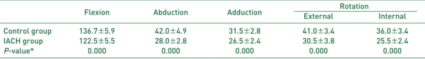
The range of motion possible was recorded in flexion, abduction, adduction, external rotation and internal rotation. In the control group, the mean flexion was 136.7。±5.9。; mean abduction was 42。±4.9。; mean adduction was 31.5。±2.8。; mean external rotation was 41。±3.4。, mean internal rotation was 36。±3.4。. For patients in the IACH group the mean flexion was 122.5。
±5.5。; mean abduction was 28。±2.8。; mean adduction was 26.5。±2.4。; mean external rotation was 30.5。±
3.8。, mean internal rotation was 25.5。±2.4。. There was a statistically significant reduction in the mean range of motion of hip joints in all planes, in patients of the IACH group when compared with the control group.
However, even though there was global restriction of motion in the hip joint, the patient complained of maximum restriction in range of rotation (Table 3).
The capsular thickness was measured on the MRA in millimeters as described earlier, in the anterior, posterior, superior and inferior recess. The mean capsular thickness in the control group was 2.61±0.8 mm anteriorly, 1.94±0.5 mm posteriorly, 1.88±0.5 mm superiorly and 1.84±0.5 mm inferiorly. Similarly, the mean capsular thickness in the IACH group was 3.14±
0.7 mm anteriorly, 2.61±0.5 mm posteriorly, 2.78±0.5 mm superiorly and 2.13±0.4 mm inferiorly. The capsular thickness showed statistically significant difference between the control and IACH groups in the posterior and superior recess (Table 4).
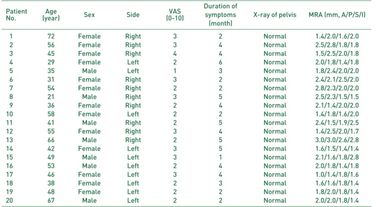
Table 2. Demographic Data of the Control Group
Patient Age VAS Duration of
Sex Side symptoms X-ray of pelvis MRA (mm, A/P/S/I)
No. (year) (0-10)
(month)
01 72 Female Right 3 2 Normal 1.4/2.0/1.6/2.0
02 56 Female Right 3 4 Normal 2.5/2.8/1.8/1.8
03 45 Female Right 4 4 Normal 1.5/2.5/2.0/1.8
04 29 Female Left 2 6 Normal 2.0/1.8/1.4/1.8
05 35 Male Left 1 3 Normal 1.8/2.4/2.0/2.0
06 31 Female Right 3 2 Normal 2.4/2.1/2.5/2.0
07 54 Female Right 2 2 Normal 2.8/2.3/2.0/2.0
08 21 Male Right 3 5 Normal 2.5/2.3/1.5/1.5
09 36 Female Right 2 4 Normal 2.1/1.4/2.0/2.0
10 58 Female Left 2 2 Normal 1.4/1.8/1.6/2.0
11 41 Male Right 2 5 Normal 2.4/1.5/1.9/2.5
12 55 Female Right 3 4 Normal 1.4/2.5/2.0/1.7
13 66 Male Right 2 5 Normal 3.0/3.0/2.6/2.8
14 42 Female Left 3 5 Normal 1.6/1.5/1.4/1.4
15 49 Male Left 3 1 Normal 2.1/1.6/1.8/2.8
16 53 Male Left 2 4 Normal 2.0/1.8/1.4/1.8
17 46 Female Left 3 4 Normal 1.0/1.4/1.8/1.6
18 38 Female Left 2 3 Normal 1.6/1.6/1.8/1.4
19 48 Female Left 2 2 Normal 1.8/2.0/1.8/1.4
20 67 Male Left 2 2 Normal 2.0/2.0/1.8/1.4
* With both hip joints.
�
�Capsular thickening location.
VAS: visual analog scale for pain, MRA: magnetic resonance arthrography, A: anterior, P: posterior, S: superior, I: inferior.
DISCUSSION
Caroit et al.3) introduced the concept of ACH. Since then only a dozen or so articles have been published referring to this diagnosis1-3,5-12). Adhesive capsulitis of a joint has been defined as a condition that begins with synovial inflammation and ends in capsular fibrosis13-16). The fibrosis of joint capsule in IACH is caused by the same cytokines as that for ACS; however, their levels vary. Hsu et al.17) has reported the association between ACS and IACH, but this has not been previously discussed in the literature in details. Presently, there is no study that has examined or that can pinpoint the changes that occur, in otherwise unexplained IACH.
The exact reported incidence of ACH is unknown but it is probably higher than is generally thought. It is said to selectively affect women between the ages of 35 and
501,3,6,18). In our study seven out of ten patients, of the
IACH group were middle-aged women ranging in age from 31 to 61 years. Six out of the ten patients had other co-morbid medical conditions typically associated with adhesive capsulitis like diabetes mellitus, hypertension and hyperlipidemia. Patients suffering from diabetes mellitus have shown to have tendency to develop IACH, associations have been established in similar involvement for the shoulder2,19). However this cohort is too small to draw any reliable conclusions on this aspect.
For the diagnosis of IACH clinicians have mostly relied on their clinical findings, unless it is diagnosed through surgery or biopsy6). Literature shows that in the diagnosis of IACH, hip radiographs often reveal only osteopenia.
The other tests also are most often negative5). Radiographic abnormalities have generally been reported only when there is underlying disease that leads to adhesive capsulitis2,7). In this study the radiographs of the hip were normal in all the 10 hips and this is consistent with the literature. Magnetic resonance imaging (MRI) and computed tomography criteria for ACS have been well known3,11,18,20). Similarly, authors6)also suggest role of MRI of the hip in order to detect potential bone or cartilage pathologies. Although some authors2,7) have described observations of tightness during arthrography as sign of reduced articular capacity and adhesive capsulitis, others have only argued of its relevance for IACH. Studies have also pointed out the need for a contra-lateral injection to compare and validate results6).
And MRI can show evidence of capsular fibrosis where there is thickness of the anterior joint capsule11). But the literature only evaluated axial view in MRI. So best to our knowledge the characteristic of MRA for IACH has not been described in literature yet.
In ACH, there is limitation of motion in all three planes (flexion-extension, internal-external rotation and abduction-adduction)3,6). Some authors suggest that
Table 4. Comparison of the Restriction of Range of Motion of the Hip Joint between Control and Study Group Capsular thickness (mm)
Anterior Posterior Superior Inferior
Control group 2.61±±0.84 1.94±±0.57 1.88±±0.53 1.84±±0.53
IACH group 3.14±±0.75 2.61±±0.57 2.78±±0.54 2.13±±0.45
P-value* 0.112 0.006 0.0009 0.121
Values are presented as mean±±standard deviation.
* By Wilcoxon rank-sum test.
IACH: idiopathic adhesive capsulitis of hip.
Table 3. Comparison of the Restriction of Range of Motion of the Hip Joint between Control and Study Group
Flexion Abduction Adduction Rotation
External Internal
Control group 136.7±±5.9 42.0±±4.9 31.5±±2.8 41.0±±3.4 36.0±±3.4
IACH group 122.5±±5.5 28.0±±2.8 26.5±±2.4 30.5±±3.8 25.5±±2.4
P-value* 0.000 0.000 0.000 0.000 0.000
Values are presented as mean±±standard deviation.
* By t-test.
IACH: idiopathic adhesive capsulitis of hip.
IACH is under-diagnosed as it leads to less functional limitation as compared to the other joints, loss of range of motion is much better tolerated in the hip than other joints, such as the shoulder and knee3,12). Similar results were observed in our study that the range of motion of the hip joint in the patients with IACH was statistically significantly restricted in flexion, abduction, adduction and rotations. However, the patient complained mainly of loss in motion.
The limitation of this study is its retrospective design and small sample size. Future studies are needed with larger sample size and longer follow-up. And we could not compare to contralateral normal hips of the IACH patients due to 2 patients have both hip problem. If we compare contralateral normal hip of the IACH patients, we would have been excluded to differences in individual difference in thickness of the capsule. And we did not distinguish between each stage of IACH like an ACS22), because there was no proven or disproved by definitive diagnostic test like histopathology or arthroscopic finding. So In order to make an accurate diagnosis of IACH need to histopathology or arthroscopic finding.
CONCLUSION
In summary, IACH is a clearly identifiable entity. The clinical presentations are similar to those commonly attributed in the shoulder. The principal clinical finding is painful restricted motion especially rotations. There is a predilection for middle-aged women. The characteristic of MRA is to identify the presence and location of capsular thickening superiorly and posteriorly, which correlates clinically as restricted motions. And there change in the capsular thickness can be helpful in further surgical treatment.
REFERENCES
01. Chard MD, Jenner JR. The frozen hip: an underdiagnosed condition. BMJ. 1988;297:596-7.
02. Lequesne M, Becker J, Bard M, Witvoet J, Postel M.
Capsular constriction of the hip: arthrographic and clinical considerations. Skeletal Radiol. 1981;6:1-10.
03. Caroit M, Djian A, Hubault A, Normandin C, De Seze S. 2 Cases of retractile capsulitis of the hip. Rev Rhum Mal
Osteoartic. 1963;30:784-9.
04. Joassin R, Vandemeulebroucke M, Nisolle JF, Hanson P, Deltombe T. Adhesive capsulitis of the hip: three case reports. Ann Readapt Med Phys. 2008;51:301-14.
05. Lowe R. Adhesive capsulitis of the hip: a case report: an entity in question. Man Ther. 2013;18:594-7.
06. Byrd JW, Jones KS. Adhesive capsulitis of the hip.
Arthroscopy. 2006;22:89-94.
07. Murphy WA, Siegel MJ, Gilula LA. Arthrography in the diagnosis of unexplained chronic hip pain with regional osteopenia. AJR Am J Roentgenol. 1977;129:283-7.
08. Griffiths HJ, Utz R, Burke J, Bonfiglio T. Adhesive capsulitis of the hip and ankle. AJR Am J Roentgenol. 1985;144:101-5.
09. Luukkainen R, Asikainen E. Frozen hip. Scand J Rheumatol.
1992;21:97.
10. Modesto C, Crespo E, Villas C, Aquerreta D. Adhesive capsulitis. Is it possible in childhood? Scand J Rheumatol.
1995;24:255-6.
11. Mont MA, Lindsey JM, Hungerford DS. Adhesive capsulitis of the hip. Orthopedics. 1999;22:343-5.
12. McGrory BJ, Endrizzi DP. Adhesive capsulitis of the hip after bilateral adhesive capsulitis of the shoulder. Am J Orthop (Belle Mead NJ). 2000;29:457-60.
13. Rodeo SA, Hannafin JA, Tom J, Warren RF, Wickiewicz TL. Immunolocalization of cytokines and their receptors in adhesive capsulitis of the shoulder. J Orthop Res. 1997;15:
427-36.
14. Kabbabe B, Ramkumar S, Richardson M. Cytogenetic analysis of the pathology of frozen shoulder. Int J Shoulder Surg. 2010;4:75-8.
15. Neviaser AS, Hannafin JA. Adhesive capsulitis: a review of current treatment. Am J Sports Med. 2010;38:2346-56.
16. Neviaser AS, Neviaser RJ. Adhesive capsulitis of the shoulder. J Am Acad Orthop Surg. 2011;19:536-42.
17. Hsu JE, Anakwenze OA, Warrender WJ, Abboud JA.
Current review of adhesive capsulitis. J Shoulder Elbow Surg. 2011;20:502-14.
18. Lefevre-Colau MM, Drape´ JL, Fayad F, et al. Magnetic resonance imaging of shoulders with idiopathic adhesive capsulitis: reliability of measures. Eur Radiol. 2005;15:
2415-22.
19. Lequesne M, Dang N, Bensasson M, Mery C. Increased association of diabetes mellitus with capsulitis of the shoulder and shoulder-hand syndrome. Scand J Rheumatol.
1977;6:53-6.
20. Jung JY, Jee WH, Chun HJ, Kim YS, Chung YG, Kim JM.
Adhesive capsulitis of the shoulder: evaluation with MR arthrography. Eur Radiol. 2006;16:791-6.
21. Wagner FV, Negra˜o JR, Campos J, et al. Capsular ligaments of the hip: anatomic, histologic, and positional study in cadaveric specimens with MR arthrography.
Radiology. 2012;263:189-98.
22. Looney CG, Raynor B, Lowe R. Adhesive capsulitis of the hip: a review. J Am Acad Orthop Surg. 2013;21:749-55.